Anaes · Airway management & difficult airway
The difficult airway & rapid sequence induction
Also known as Difficult airway · Difficult intubation · Unanticipated difficult airway · Rapid sequence induction · RSI · Can't intubate can't oxygenate
The difficult airway is the single most examined topic in anaesthesia, and the one in which failure kills. The framework rests on four ideas: predict where you can (LEMON, MACOCHA), optimise every attempt (position, preoxygenation, paralysis), and follow a fixed rescue sequence when intubation fails — the Difficult Airway Society Plans A through D, a ladder that ends in emergency front-of-neck access when the patient can no longer be oxygenated. The physiology underpinning it all is the safe apnoea period: preoxygenation denitrogenates the functional residual capacity into an oxygen reservoir, and apnoeic oxygenation (classically transnasal humidified rapid-insufflation ventilatory exchange, THRIVE) can extend that window many-fold — buying the calm, planned best attempt that prevents a spiral into crisis. Built around the landmark guidelines (DAS 2015, ASA 2022, AIDAA, the DAS/ICS critically-ill guideline), the national audit NAP4, the Vortex approach, and the trials that have redrawn practice — the IRIS trial showing cricoid pressure does not reduce aspiration, the Cochrane review and the INTUBE study on videolaryngoscopy, and the MACOCHA score for the predicted-difficult intubation in the ICU.
On this page & tools
Your progress
Saved locally on this device.
8 MCQs with explanations
Target exams
Red flags
Overview & definition
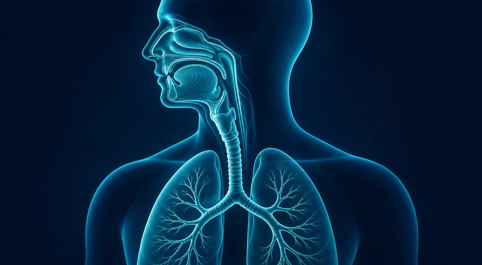
The difficult airway is the circumstance in which a conventionally trained anaesthetist experiences difficulty with mask ventilation, with laryngoscopy, with intubation, or with all three. The 2022 American Society of Anesthesiologists (ASA) guidelines frame it more broadly still, as any clinical situation in which the airway is at risk — the difficult airway is anticipated, encountered unexpectedly, or already lost[3][4]. It is the most examined topic in the fellowship because it is the one in which failure is most rapidly fatal: an unmanageable airway progresses from first-class laryngoscopy to brain injury in the minutes a desaturating apnoeic patient can survive.
The working definitions an examiner expects: [1]
- Difficult mask ventilation — the inability to maintain an oxygen saturation above 90 per cent, or to prevent or reverse inadequate ventilation, despite optimal positioning and a competent mask seal.
- Difficult laryngoscopy — a Cormack–Lehane grade 3 or 4 view (no more than the epiglottis visible) despite optimal external laryngeal manipulation and head position.
- Difficult intubation — more than two attempts at laryngoscopy, or a total procedure time exceeding ten minutes[1].
The whole subject turns on a single principle: the airway is managed by a sequence, not a single act. When intubation fails, the response is a fixed, rehearsed ladder — the Difficult Airway Society (DAS) Plans A through D — that returns the patient to oxygenation at each tier and ends, only at the last, in an emergency surgical airway[1][8]. The physiology that buys time for that ladder is the safe apnoea period: the window after paralysis during which the patient can be left apnoeic without desaturating, prolonged by meticulous preoxygenation and apnoeic oxygenation[9].
Airway anatomy and predicting the difficult airway
The airway is a shared conduit for digestion and respiration, and its geometry is what makes laryngoscopy feasible or impossible. The blade of the laryngoscope must create a straight line of sight from the incisors, over the base of the tongue, around the curve of the pharynx, to the glottis — the angle and the distances between the mouth opening, the hyoid, and the thyroid notch are the anatomical determinants of whether that line can be made straight. A short thyromental distance, a high anterior larynx, and a receding mandible all force the glottis out of the direct line of sight. [1]
Prediction is imperfect but worthwhile. The bedside LEMON assessment scores the external features: Look (facial trauma, beard, overbite), Evaluate the 3-3-2 rule (inter-incisor distance three finger-breadths, hyoid–mentum three, thyroid–floor of mouth two), Mallampati class, Obstruction, and Neck mobility. For the critically ill patient, the MACOCHA score adds the physiological axes that the elective assessment misses — reduced Mallampati, apnoea or obstructive sleep apnoea, reduced cervical mobility, restricted mouth opening, coma, hypoxia, and a non-specialist operator — and it was developed and validated specifically in the intensive care unit, where intubation is both harder and more dangerous[10].
The honest position is that no score predicts the difficult airway with certainty, and a substantial proportion present unanticipated. Prediction is a reason to prepare, not a reason to be reassured: the airway that looked easy on the ward is sometimes the airway that fails in theatre. The trained response is therefore to behave as though every induction may be difficult — preoxygenate fully, position optimally, and have the rescue equipment and the DAS ladder to hand before the first drug is given[1][3].
The unanticipated difficult airway: the DAS management algorithm
When, despite preparation, laryngoscopy fails, the response is the DAS 2015 algorithm — a fixed sequence of four plans, each with a single objective and a defined trigger to move on[1].
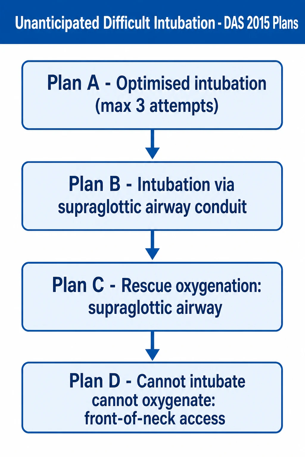
- Plan A — the initial intubation attempt. Optimise every factor that can be optimised: head-elevated ramped position, full preoxygenation, neuromuscular blockade to provide the best intubating conditions, external laryngeal manipulation, videolaryngoscopy, and a bougie or stylet. Limit the attempts to three by the most experienced anaesthetist available, and declare failure early rather than persist[1].
- Plan B — the secondary intubation technique. If Plan A fails, intubate through a second-generation supraglottic airway used as a conduit (with a fibreoptic scope or a dedicated intubating device), or use an alternative videolaryngoscope blade. The aim is still to place the tube[1][15].
- Plan C — rescue oxygenation. If intubation is impossible, abandon the tube and insert a second-generation supraglottic airway to oxygenate and ventilate the patient while expert help is summoned. Supraglottic devices rescue the majority of cannot-intubate situations[7][18].
- Plan D — front-of-neck access. If the patient can no longer be oxygenated — cannot intubate, cannot oxygenate (CICO) — declare it aloud and perform an emergency surgical airway without delay[1][7].
The discipline of the algorithm is to move on. The most common failure of human performance in a crisis is to repeat a failing technique (a fourth, fifth, sixth laryngoscopy) while the saturation falls; the fixed ladder exists precisely to prevent that[8].
Preoxygenation and the physiology of apnoeic oxygenation
The safe apnoea period — the time from the onset of apnoea to the fall of the arterial oxygen saturation below a safe threshold — is the central physiological currency of the difficult airway. It is determined by how much oxygen is stored in the lungs at the moment apnoea begins, and how fast it is removed. [1]
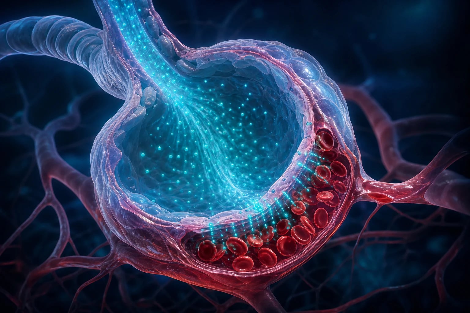
The functional residual capacity (FRC) — the volume of gas left in the lungs at end-expiration — is the reservoir. Breathing 100 per cent oxygen for three minutes, or taking eight vital-capacity breaths, denitrogenates the FRC, replacing its nitrogen with oxygen and storing roughly 2.5 litres of oxygen at the alveolus. Once apnoea begins, this reservoir is consumed only at the rate of the body's metabolic oxygen demand — about 250 mL per minute — so a well-preoxygenated patient of normal weight and cardiac output may not desaturate for five to eight minutes[9].
Two caveats shrink that window sharply, and both are common. First, a reduced functional residual capacity — in obesity, pregnancy, and the supine, anaesthetised patient — empties the reservoir faster and desaturates the patient in under two minutes. Second, high metabolic demand or shunt — sepsis, the critically ill, the foetus — draws the oxygen down faster still[2]. These are precisely the patients in whom a slow exploratory laryngoscopy is intolerable.
Apnoeic oxygenation extends the window. Even with no ventilation, oxygen continues to flow from the pharynx into the alveoli down the partial-pressure gradient created by the alveolar uptake of oxygen, while carbon dioxide rises only slowly (it is highly soluble and tissue-buffered). Delivering high-flow humidified oxygen at the nares — transnasal humidified rapid-insufflation ventilatory exchange (THRIVE) — keeps the upper airway filled with 100 per cent oxygen and can maintain full oxygenation through an apnoea of many minutes, far beyond the classical two-to-three-minute window. THRIVE was first reported in patients with predicted difficult airways and has transformed the practice of the elective difficult airway from a race against the clock into a calm, planned event[9].
Rapid sequence induction and the role of cricoid pressure
The rapid sequence induction (RSI) is the technique for the patient at risk of regurgitation and aspiration — the non-fasted patient, the obstetric emergency, the bowel obstruction, the upper-GI bleed. Its three elements are designed to minimise the interval between loss of consciousness and an inflated, protected cuffed tracheal tube: a fast-acting induction agent, a rapidly acting neuromuscular blocker (succinylcholine or high-dose rocuronium), no mask ventilation between induction and intubation, and cricoid pressure from the moment of loss of consciousness until the tube is confirmed[2][17].
Cricoid pressure — the Sellick manoeuvre — was adopted for half a century on the physiological reasoning that posterior pressure on the cricoid cartilage occludes the oesophagus against the vertebral body, preventing passive regurgitation. The evidence, however, has not supported it. The IRIS randomised trial, a sham-controlled study of cricoid pressure in rapid sequence induction, found that cricoid pressure did not reduce the incidence of pulmonary aspiration, and that it was associated with a worse laryngoscopic view and more intubation difficulty[14]. Modern practice has therefore softened: many anaesthetists use a modified RSI — gentle mask ventilation (to maintain oxygenation, accepting a small theoretical aspiration risk), reduced or relaxed cricoid pressure, and a willingness to release the cricoid pressure entirely if it obstructs the view[3]. The principle — protect the airway swiftly in the patient at risk of aspiration — is unchanged; the sacrosanct status of cricoid pressure is gone.
In the critically ill patient the RSI is more dangerous still. The INTUBE study of peri-intubation events in intensive care units documented a high rate of cardiovascular collapse — hypotension, severe bradycardia and cardiac arrest in the minutes around intubation — driven by the loss of sympathetic tone on induction, the vasodilation of the induction agent, and positive-pressure ventilation against a preload-dependent circulation. The critically ill intubation demands resuscitation before, during and after: preoxygenate, prepare vasopressors, avoid prolonged apnoea, and defend the blood pressure throughout[2][13].
Videolaryngoscopy versus direct laryngoscopy
Videolaryngoscopy — a camera or prism at the tip of the blade, displayed on a screen — improves the view of the glottis, particularly the anterior larynx that defeats a direct line of sight. Whether it improves the outcome of intubation is a subtler question. [1]
The Cochrane systematic review of videolaryngoscopy versus direct laryngoscopy in adults found that videolaryngoscopy improves the glottic view and reduces the rate of failed intubation on the first attempt in the predicted-difficult airway, but did not show a clear reduction in the overall rate of failed intubation, hypoxaemia or airway trauma across all comers[11]. In the critically ill, the INTUBE subanalysis found videolaryngoscopy associated with fewer severe hypoxaemic events but a higher rate of first-attempt failure when used by less experienced operators — the device rewards familiarity[12]. The practical position is that videolaryngoscopy is the first-line instrument for the anticipated difficult airway and a key tool in Plan A, but it does not abolish difficulty, and a tube that cannot be directed through an unseen glottis is no better than a poor view.
Supraglottic airway devices and the rescue role
The second-generation supraglottic airway device — the i-gel, the LMA Supreme — is the workhorse of airway rescue. It seals the pharynx well enough to ventilate, it incorporates an oesophageal drainage port to vent regurgitated matter, and it can be placed rapidly and with a high first-attempt success rate by operators of widely varying experience. In the DAS algorithm it appears in two places: in Plan B, as a conduit through which a tracheal tube can be passed (with a fibreoptic scope), and in Plan C, as the device that re-establishes oxygenation when intubation has failed entirely[1][15].
The evidence for the supraglottic airway extends beyond rescue. In selected low-risk patients it is the primary airway for elective surgery, and a meta-analysis in caesarean delivery has confirmed that a supraglottic airway, in the properly selected patient, is a reasonable alternative to tracheal intubation[18]. The supraglottic airway is not, however, a substitute for a cuffed tracheal tube in the patient at high risk of aspiration or of high airway pressures.
Front-of-neck access: emergency surgical airway
Front-of-neck access (FONA) is the final tier of the algorithm and the one that cannot fail. It is performed for cannot-intubate, cannot-oxygenate (CICO) — the situation in which the patient cannot be intubated by any route and cannot be oxygenated by face mask or supraglottic device. It is rare, and it is the airway event most likely to end in death or brain injury if delayed[7].
NAP4 taught two painful lessons about FONA. First, needle cricothyroidotomy failed repeatedly — in obese patients the standard needle and jet ventilator could not be placed or driven effectively, and rescue was delayed while the needle technique was attempted. Second, anaesthetists were slow to declare CICO and slow to proceed to a surgical airway, having rarely or never performed one. The DAS 2015 algorithm therefore specifies the scalpel–bougie–tube technique as the default for the adult emergency surgical airway: a transverse stab incision through the cricothyroid membrane, a bougie passed into the trachea, and a cuffed tracheal tube railroaded over the bougie. It is fast, it works in the obese neck, and it provides a cuffed airway that protects against aspiration and permits high ventilating pressures — the technique rehearsed so that, on the one occasion it is needed, it can be performed without hesitation[1][7].
Awake tracheal intubation
When the difficult airway is anticipated — the known predicted-difficult patient, the distorted anatomy of surgery or radiation, the obstructing upper-airway lesion — the safest strategy is often to secure the airway before inducing anaesthesia, by awake tracheal intubation. The technique topically anaesthetises the upper airway and sedates the patient lightly enough to maintain spontaneous ventilation and airway tone, while a flexible fibreoptic scope (or a videolaryngoscope used awake) is passed through the nose or mouth and into the trachea, over which the tube is railroaded[15].
A network meta-analysis of airway devices for awake tracheal intubation found that flexible fibreoptic intubation and awake videolaryngoscopy both achieve high success rates, and a more recent network meta-analysis of sedation found that the choice of sedative — typically a low-dose remifentanil or dexmedetomidine-based technique — is less important than the principle of maintaining a cooperative, spontaneously breathing patient with a preserved airway[15][16]. The awake route converts a potentially unmanageable airway into a controlled one, and it is under-used: NAP4 identified a reluctance to perform awake intubation as a recurring contributor to airway disaster[7].
The difficult airway in special contexts: obstetric and the critically ill
Two populations deserve separate mention because they are common and disproportionately dangerous. [1]
In obstetric general anaesthesia, the airway is oedematous and friable, the functional residual capacity is reduced by the gravid uterus, the oxygen consumption is high, and the patient is by definition at high risk of aspiration — the result is a patient who desaturates rapidly and bleeds easily. Maternal airway disaster remains a leading cause of anaesthesia-related maternal death. The anticipated difficult obstetric airway demands a senior operator, full preoxygenation, a low threshold for awake intubation, and the difficult-airway equipment immediately available; where the risk is appropriate, a supraglottic airway may rescue a failed intubation[17][18].
In the critically ill, the difficulties are compounded: hypoxaemia, hypotension, a full stomach, a combative or comatose patient, and an operator who may be outside their area of greatest expertise. The dedicated guideline for tracheal intubation in critically ill adults — a joint product of the Difficult Airway Society, the Intensive Care Society and related bodies — sets out the structured approach: a pre-intubation checklist, a defined team with roles, a plan for the difficult and failed airway, the use of videolaryngoscopy, and the preparation of vasopressors and rescue devices before induction[2]. The AIDAA guidelines extend the same structured approach in the intensive-care and emergency contexts[5].
NAP4: lessons from the national audit
The Fourth National Audit Project (NAP4) of the Royal College of Anaesthetists and the Difficult Airway Society audited every major complication of airway management reported across the United Kingdom over a year, and it remains the defining evidence base for the subject. Its conclusions reshaped practice[7].
- Major airway complications are uncommon but catastrophic. A death or brain injury occurred in a substantial minority of the reported events, concentrated in the obese, in emergency surgery, and in the critically ill.
- Failure was more often of planning and judgement than of technique. Repeated attempts at a failing technique, a reluctance to wake the patient or perform awake intubation, and a delay in declaring CICO and moving to a surgical airway were the recurring human failures.
- Capnography was repeatedly misused or absent. A number of airway deaths were attributed to a failure to apply, or to correctly interpret, continuous waveform capnography — the only reliable confirmation of tracheal intubation. NAP4 mandated its use wherever a tracheal tube or supraglottic airway is in place.
- Needle cricothyroidotomy failed; scalpel techniques succeeded. The audit's finding that needle cricothyroidotomy failed repeatedly in obese patients is the direct reason the modern DAS algorithm specifies the scalpel–bougie–tube technique for emergency FONA. [1]
The Vortex approach
The Vortex is a complementary tool that addresses the team dimension of the airway crisis — not which device to use, but how the team behaves when the saturation is falling[8]. It models the difficult airway as a funnel of three zones of best attempt — green, yellow, red — across the three oxygenation modalities (face mask, supraglottic airway, front-of-neck access). At each zone the team makes a best attempt at each modality, declares success or failure out loud, and descends to the next zone only when all three have failed, ending in the red zone of surgical airway.
The contribution of the Vortex is the discipline of declaring the crisis and moving on. It replaces the silent, escalating attempts of an individual with a shared mental model the whole team can see and track, and it front-loads the decision to proceed to a surgical airway — the decision that is most often delayed. It does not replace the DAS algorithm so much as sit alongside it, giving the team a common language for the descent to Plan D[8].
Extubation of the difficult airway
The difficult airway does not end at intubation. Extubation is its own hazard — around a third of all airway events occur at or after extubation, and the difficult airway that was intubated is the difficult airway that must be extubated, often over a now-oedematous glottis[6]. The dedicated DAS extubation guideline stratifies the patient into low-risk and at-risk extubation, and for the at-risk patient — the known difficult airway, the patient who was difficult to intubate, the obese, the airway surgery case — it mandates a planned strategy: an awake extubation with the patient breathing spontaneously, an airway exchange catheter left in situ as a conduit to reintubation, and the equipment and the personnel to reintubate immediately if extubation fails[6]. The principle is that extubation is performed only when a plan for failed extubation is already in place.
Monitoring, capnography and human factors
The technical and the human sit together in airway safety. On the technical side, continuous waveform capnography is non-negotiable — it confirms tracheal placement from the first breath, it warns of obstruction or disconnection, and it is the single monitoring intervention most strongly endorsed by NAP4[7]. On the human side, the airway crisis is a test of crisis resource management: the early call for help, the allocation of roles, the verbal declaration of plans and of failure, and the discipline to move down the ladder rather than repeat a failing attempt. The Vortex, the DAS algorithm and the WHO-style airway checklist are all instruments of the same underlying discipline — to impose a structure on a crisis that would otherwise collapse into panic[8].
Clinical
- Standard approach
- Evidence-based
Alternative
- Modified technique
- Risk-benefit
Red flags
[1] [1] [1] [1] [1]References
- [1]Frerk C, Mitchell VS, McNarry AF, Mendonca C, Bhagrath R, Patel A, et al. Difficult Airway Society 2015 guidelines for management of unanticipated difficult intubation in adults Br J Anaesth, 2015.PMID 26556848
- [2]Higgs A, McGrath BA, Goddard C, Rangasami J, Suntharalingam G, Gale R, et al. Guidelines for the management of tracheal intubation in critically ill adults Br J Anaesth, 2018.PMID 29406182
- [3]Apfelbaum JL, Hagberg CA, Connis RT, Abdelmalak BB, Agarkar M, Dutton RP, et al. 2022 American Society of Anesthesiologists Practice Guidelines for Management of the Difficult Airway Anesthesiology, 2022.PMID 34762729
- [4]Apfelbaum JL, Hagberg CA, Caplan RA, Blitt CD, Connis RT, Nickinovich DG, et al. Practice guidelines for management of the difficult airway: an updated report by the American Society of Anesthesiologists Task Force on Management of the Difficult Airway Anesthesiology, 2013.PMID 23364566
- [5]Myatra SN, Ahmed SM, Kundra P, Garg R, Ramkumar V, Patwa A, et al. The All India Difficult Airway Association 2016 guidelines for tracheal intubation in the Intensive Care Unit Indian J Anaesth, 2016.PMID 28003694
- [6]Difficult Airway Society Extubation Guidelines Group; Popat M, Mitchell V, Dravid R, Patel A, Swampillai C, Higgs A. Difficult Airway Society Guidelines for the management of tracheal extubation Anaesthesia, 2012.PMID 22321104
- [7]Cook TM, Woodall N, Frerk C; Fourth National Audit Project. Major complications of airway management in the UK: results of the Fourth National Audit Project of the Royal College of Anaesthetists and the Difficult Airway Society. Part 1: anaesthesia Br J Anaesth, 2011.PMID 21447488
- [8]Chrimes N. The Vortex: a universal 'high-acuity implementation tool' for emergency airway management Br J Anaesth, 2016.PMID 27440673
- [9]Patel A, Nouraei SA. Transnasal Humidified Rapid-Insufflation Ventilatory Exchange (THRIVE): a physiological method of increasing apnoea time in patients with difficult airways Anaesthesia, 2015.PMID 25388828
- [10]De Jong A, Molinari N, Terzi N, Mongardon N, Arnal JM, Guitton C, et al. Early identification of patients at risk for difficult intubation in the intensive care unit: development and validation of the MACOCHA score in a multicenter cohort study Am J Respir Crit Care Med, 2013.PMID 23348979
- [11]Lewis SR, Butler AR, Parker J, Cook TM, Schofield-Robinson OJ, Smith AF. Videolaryngoscopy versus direct laryngoscopy for adult patients requiring tracheal intubation: a Cochrane Systematic Review Br J Anaesth, 2017.PMID 28969318
- [12]Russotto V, Lascarrou JB, Tassistro E, Parotto M, Antolini L, Bauer P, et al. Efficacy and adverse events profile of videolaryngoscopy in critically ill patients: subanalysis of the INTUBE study Br J Anaesth, 2023.PMID 37208282
- [13]Russotto V, Tassistro E, Myatra SN, Parotto M, Antolini L, Bauer P, et al. Peri-intubation Cardiovascular Collapse in Patients Who Are Critically Ill: Insights from the INTUBE Study Am J Respir Crit Care Med, 2022.PMID 35536310
- [14]Birenbaum A, Hajage D, Roche S, Ntouba A, Eurin M, Cuvillon P, et al. Effect of Cricoid Pressure Compared With a Sham Procedure in the Rapid Sequence Induction of Anesthesia: The IRIS Randomized Clinical Trial JAMA Surg, 2019.PMID 30347104
- [15]Desai N, Ratnayake G, Onwochei DN, El-Boghdadly K, Ahmad I. Airway devices for awake tracheal intubation in adults: a systematic review and network meta-analysis Br J Anaesth, 2021.PMID 34303493
- [16]El-Boghdadly K, Desai N, Jones JB, Elghazali S, Ahmad I, Sneyd JR. Sedation for awake tracheal intubation: A systematic review and network meta-analysis Anaesthesia, 2025.PMID 39468765
- [17]Mushambi MC, Athanassoglou V, Kinsella SM. Anticipated difficult airway during obstetric general anaesthesia: narrative literature review and management recommendations Anaesthesia, 2020.PMID 32144770
- [18]White LD, Thang C, Hodsdon A, Melhuish TM, Barron FA, Godsall MG, et al. Comparison of Supraglottic Airway Devices With Endotracheal Intubation in Low-Risk Patients for Cesarean Delivery: Systematic Review and Meta-analysis Anesth Analg, 2020.PMID 32925330