Anaes · Airway management
Airway emergencies: recognition and the structured response
Also known as Airway emergencies · The airway crisis · The lost airway · Intraoperative airway emergency · Acute airway obstruction · Crisis resource management in the airway
An airway emergency is any acute failure to oxygenate or ventilate — from laryngospasm and bronchospasm to aspiration, obstruction, the lost airway and cannot-intubate-cannot-oxygenate. Survival hinges less on a single heroic technique than on early recognition, calling for help, basic airway manoeuvres applied well, and a structured, rehearsed escalation — because the time from a falling saturation to a hypoxic cardiac arrest is short.
On this page & tools
Your progress
Saved locally on this device.
Target exams
Red flags
Overview
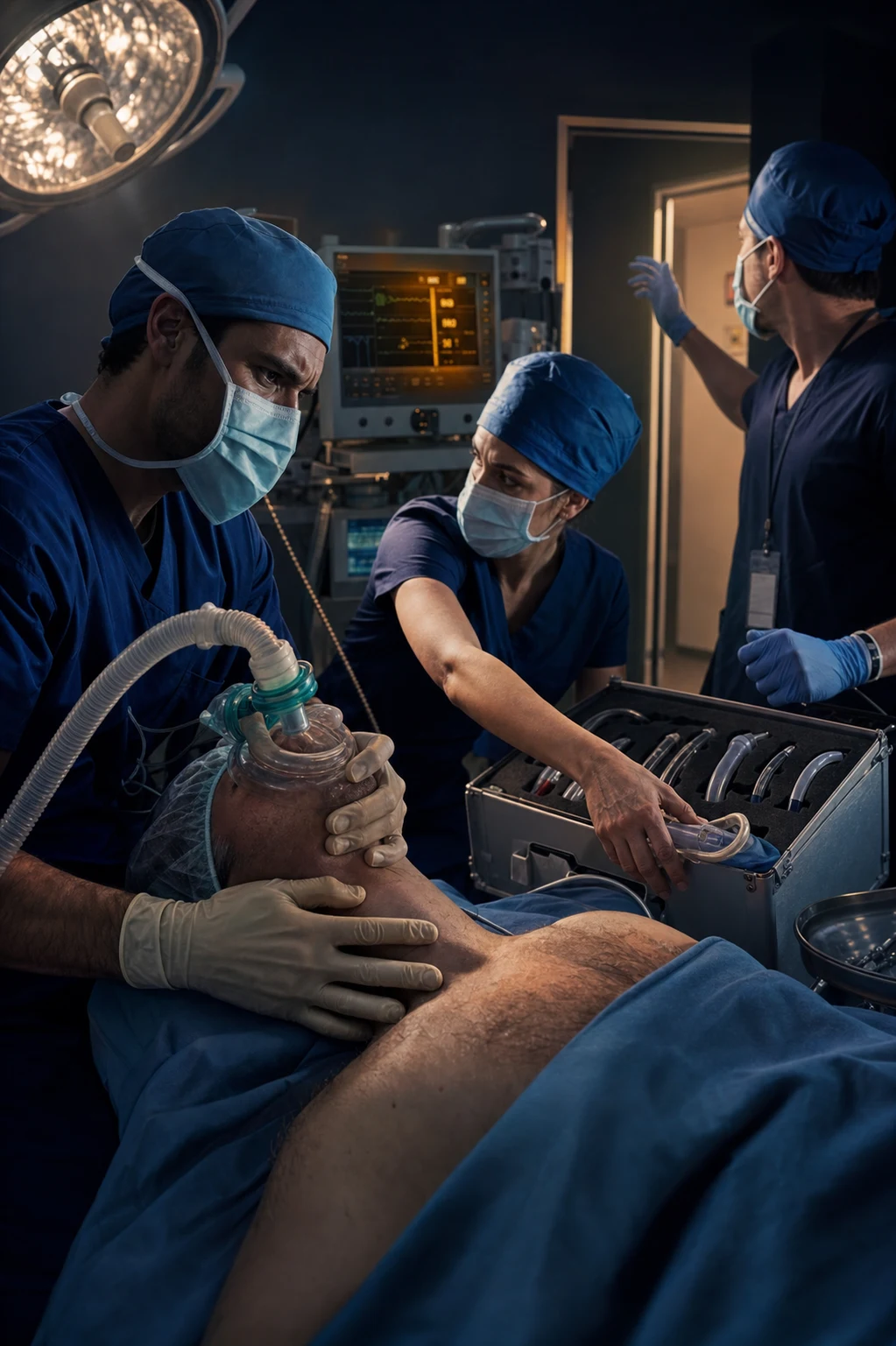
Airway emergencies are the emergencies anaesthetists fear most, because they combine time-critical hypoxia with technical difficulty and high cognitive load[5]. The emergencies span the airway irritant (laryngospasm, bronchospasm), the obstructed airway (oedema, foreign body, epiglottitis), the contaminated airway (aspiration), the lost airway (failed ventilation), and the ultimate emergency cannot-intubate-cannot-oxygenate[6]. The common thread is a falling saturation and a closing window before hypoxic cardiac arrest, and the evidence is consistent that survival depends on early recognition, an immediate call for help, well-practised basic manoeuvres, and a structured escalation rather than on any single device[5][6]. This topic frames the airway emergency as a generic crisis to be recognised and managed systematically, with the specific emergencies as recognisable patterns within it.
Recognition of the airway emergency
The earliest sign of an airway emergency is usually a falling oxygen saturation coupled with a problem ventilating or a high airway pressure, but the saturation lags the event, and the more sensitive real-time signs are the loss of the expired carbon-dioxide trace, a rising or falling airway pressure, audible obstruction, and the loss of chest movement[6]. The disciplined response is to treat any failure to oxygenate or ventilate as an emergency from the first moment, rather than waiting for the saturation to confirm it. Bradycardia in this setting is a pre-terminal sign of hypoxia, not a primary cardiac event, and it must be treated as the airway emergency it represents[1].
The universal first response
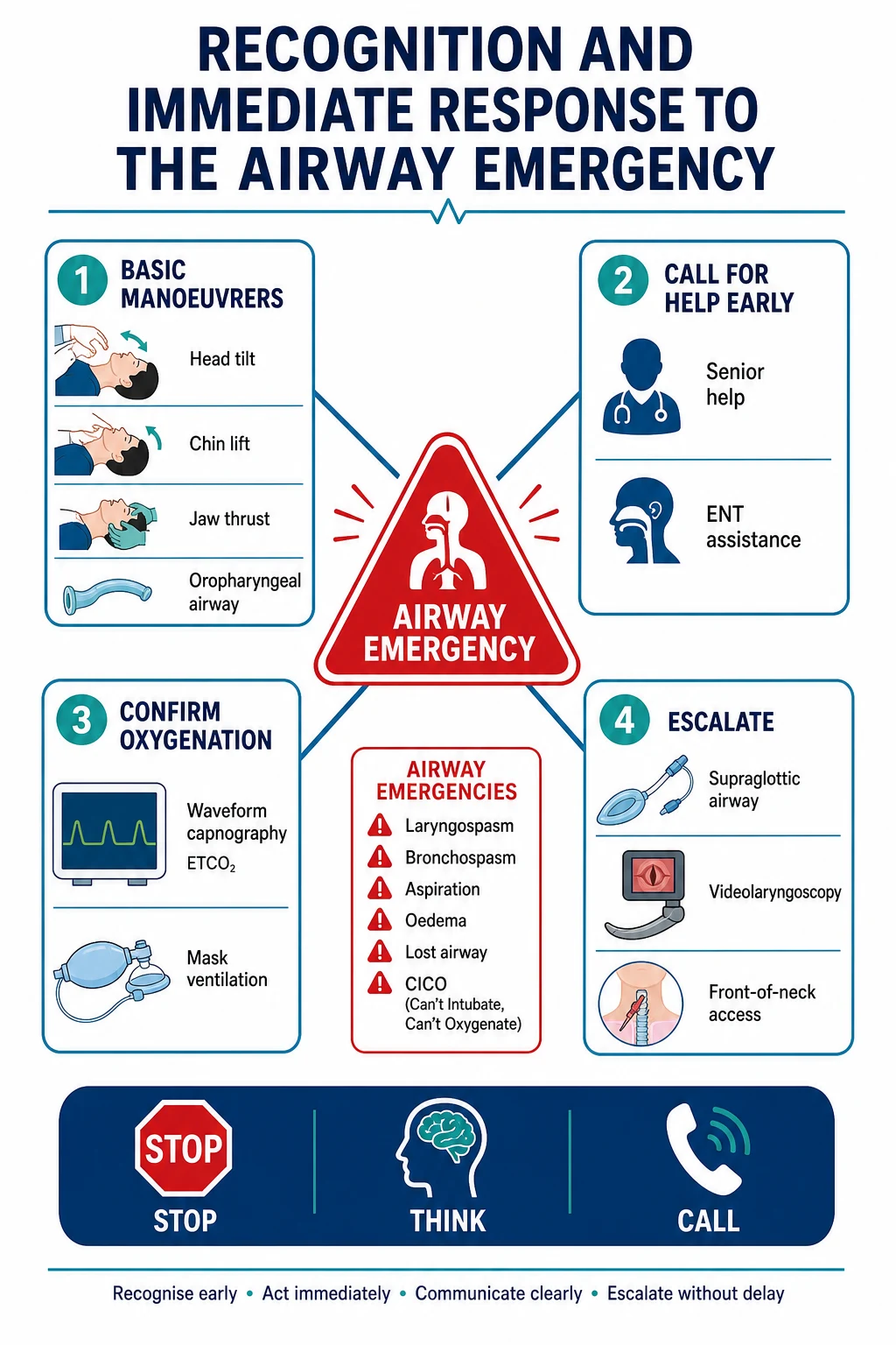
Every airway emergency is met with the same opening sequence, taught because it works under stress[5][6]. Call for help early and explicitly — the single most consistently missed and most consistently beneficial action. Apply one hundred percent oxygen at high flow. Perform basic airway manoeuvres — head tilt, chin lift, jaw thrust, and an oropharyngeal or nasopharyngeal airway — and assess mask ventilation with two-person technique if one-person fails. Confirm oxygenation by watching chest movement and the capnograph. Only after this is the diagnosis refined and the escalation begun: suction if obstruction, deepen anaesthesia for laryngospasm, bronchodilator for bronchospasm, head-down and suction for aspiration, and a supraglottic airway or front-of-neck access for the failed airway[6].
Hypoxic bradycardia and the route to cardiac arrest
The physiological endgame of an unresolved airway emergency is hypoxic bradycardia progressing to cardiac arrest, and the time course is short[1]. A randomised study of atropine in hypoxic bradycardia found that treating the bradycardia with an antimuscarinic does not by itself avert arrest when the underlying cause — hypoxia — continues, confirming the doctrine that the only effective treatment is oxygenation[1]. Cardiopulmonary resuscitation is begun if the patient arrests, but the airway remains the priority: an arrest during an airway emergency is a hypoxic arrest, and it reverses only when oxygenation is restored. Adrenaline is given per the arrested-patient algorithm while the airway is secured by whatever means[1].
Laryngospasm
Laryngospasm — sustained glottic closure in response to a stimulus at a light plane of anaesthesia — is the commonest airway emergency in fit patients and the gateway to negative-pressure pulmonary oedema and hypoxic arrest[4]. Prevention is by avoiding the excitatory light plane on induction and emergence and by clearing the airway of blood and secretions before reflexes return. Treatment is to remove the stimulus, apply one hundred percent oxygen with continuous positive airway pressure and a firm jaw thrust, and, if closure persists, a small dose of propofol or suxamethonium to relax the cords before hypoxia[6]. In the severe case, suxamethonium at a full intubating dose (intravenous or intramuscular if access is lost) breaks the spasm and allows reintroduction of oxygen[1].
Bronchospasm
Intraoperative bronchospasm presents as high airway pressures, wheeze (or, in the severe case, a silent chest with no air movement), and a rising or falling carbon-dioxide trace[4]. The causes are airway irritability (asthma, recent upper respiratory infection, anaphylaxis), mechanical irritation (a tube at the carina), and drug reaction. Treatment is to deepen anaesthesia (a volatile agent or incremental propofol), give an inhaled or intravenous bronchodilator (salbutamol), give adrenaline if anaphylaxis is suspected, and exclude a mechanical cause[4]. Neostigmine and other cholinergics can precipitate bronchospasm on reversal, and cases of neostigmine-associated bronchospasm complicated by pulmonary oedema are described, reminding the operator to consider the drug cause[4].
Aspiration
Pulmonary aspiration of gastric contents presents as hypoxia, wheeze or obstruction after regurgitation around the airway, most often at induction or emergence in the patient with a full stomach, obstruction, or depressed consciousness[6]. The immediate response is a head-down lateral position, suction of the pharynx under direct vision, and intubation to protect and ventilate the lungs; bronchoscopy removes particulate matter, and bronchodilators and positive end-expiratory pressure manage the chemical pneumonitis[6]. Lavage with saline is avoided, as it disperses the aspirate. Prevention — fasting, rapid sequence induction with cricoid pressure, and a protected airway — is far more effective than treatment, which is why aspiration is central to obstetric and emergency airway practice.
Airway obstruction
Acute airway obstruction arises from oedema (after airway manipulation, allergy, or prolonged prone surgery), a foreign body, or infection such as acute epiglottitis[2]. The patient with inspiratory stridor, tracheal tug and recession is partially obstructed and may obstruct completely, and the principle is to preserve spontaneous ventilation, keep the patient upright, and summon senior anaesthetic and surgical help before any intervention[2]. Acute epiglottitis in the adult is a classic example: the case literature describes planned, multidisciplinary decision-making between an inhalational induction preserving spontaneous ventilation and an elective awake tracheostomy, with a rigid bronchoscope and surgical airway immediately available — the opposite of a rushed, instrumented airway that converts partial obstruction into complete[2].
The lost airway
The lost airway is the acute failure to ventilate an anaesthetised patient — mask ventilation impossible, a supraglottic airway failing, intubation failing[6]. It demands the structured escalation: reposition, two-person mask ventilation with an adjunct, insert a supraglottic airway, and if all fail, prepare for front-of-neck access while a best-effort laryngoscopy is attempted. The diagnosis of cannot-intubate-cannot-oxygenate is made when neither intubation nor oxygenation by any non-surgical means is possible, and at that point the scalpel-bougie cricothyroidotomy is performed without delay[6].
Cannot-intubate-cannot-oxygenate
CICO is the rare, terminal airway emergency in which every non-surgical lifeline has failed and the saturation is falling[3]. The recommended technique in the adult is the scalpel-bougie cricothyroidotomy: a transverse stab through the cricothyroid membrane, a bougie passed into the trachea, and a cuffed tube railroaded over it[3]. The reasons it goes wrong are human and organisational rather than technical — delay in declaring CICO, failure to identify the membrane, and the cognitive tunnel-vision of repeated failing intubation attempts — which is why simulation of front-of-neck access on realistic tissue models is increasingly emphasised to build procedural and decisional fluency before the once-in-a-career event[3]. Studies of the anaesthetist's mindset during surgical airway management underline that the decisive act is the decision to act, and that it must be rehearsed[5].
Human factors and crisis resource management
The airway emergency is won or lost on human factors as much as technique[5]. The reproducible failures are: not calling for help early; fixation on a single failing technique; loss of situational awareness; poor communication and role ambiguity in the team; and the failure to declare the emergency and the plan aloud. Crisis resource management counters these with explicit leadership, role allocation, a read-back of the plan, periodic loud summarising of the saturation and time, and a willingness to escalate and to stop a failing action[5]. The structured STOP-THINK-CALL pause borrowed from aviation and resuscitation courses gives the team a moment to reframe when the situation is deteriorating, and is taught precisely because the unstructured response is unreliable under stress[5].
Simulation and preparedness
Because the severe airway emergency is individually rare, the skills to manage it are maintained by simulation[3][5]. High-fidelity and tissue-model simulation of failed intubation, CICO and the multi-disciplinary obstructed airway builds both the procedural skill (the scalpel-bougie technique on realistic tissue) and the non-technical skill (leadership, communication, declaration) that determine outcome[3]. Evaluation of front-of-neck-access simulation materials shows that the realism of the tissue model materially affects the operator's ability to learn the technique, supporting investment in dedicated airway-emergency simulation in every anaesthetic department[3].
Clinical
- Standard approach
- Evidence-based
Alternative
- Modified technique
- Risk-benefit
Red flags
References
- [1]Parizek T, et al. Effect of atropine on time to cardiac arrest in hypoxic bradycardia: a randomised cross-over study in a porcine apnoea model BJA Open, 2026.PMID 42317406
- [2]Wu X, et al. Airway Decision-Making in Acute Epiglottitis: A Case Report of Planned Tracheostomy in a Can Ventilate but Cannot Intubate (CVCI) Scenario Clin Case Rep, 2026.PMID 42290811
- [3]Mullally ME, et al. Evaluation of novel materials for front-of-neck access simulations Anaesth Intensive Care, 2026.PMID 42290043
- [4]Sato M, et al. Suspected Neostigmine-Associated Bronchospasm Complicated by Pulmonary Edema During General Anesthesia: A Case Report Cureus, 2026.PMID 42311723
- [5]Del Tedesco JC, et al. [The mindset of anesthesiologists while performing surgical airway management] Anaesthesiologie, 2026.PMID 42230350
- [6]Karim HMR, et al. Letter to the Editor: Insight into emergency endotracheal intubation in critically ill and strategies beyond equipment World J Crit Care Med, 2026.PMID 42272892