Anaes · Airway management
The paediatric airway: anatomy, physiology and management
Also known as Paediatric airway · Pediatric airway management · Infant airway · Neonatal airway · Paediatric difficult airway
The paediatric airway differs from the adult airway in anatomy, physiology and equipment, and these differences drive every aspect of safe management — from positioning and preoxygenation to the difficult-airway algorithm and emergency front-of-neck access. Children desaturate far faster than adults, so airway crises tolerate no delay.
On this page & tools
Your progress
Saved locally on this device.
Target exams
Red flags

Overview
The paediatric airway is not a small adult airway. Its anatomy, its physiology and the equipment used to manage it differ in ways that are predictable and examinable, and these differences are the reason airway events in children are disproportionately dangerous[1]. A child's oxygen reserve is small and their consumption is high, so the margin between a calm, planned airway and a hypoxic cardiac arrest is measured in seconds rather than minutes[1]. Mastery of the paediatric airway rests on three pillars: knowing how the airway differs anatomically and physiologically, sizing and selecting equipment correctly, and applying a difficult-airway algorithm that is modified for the child's faster trajectory to desaturation[2][4].
Anatomical differences from the adult airway
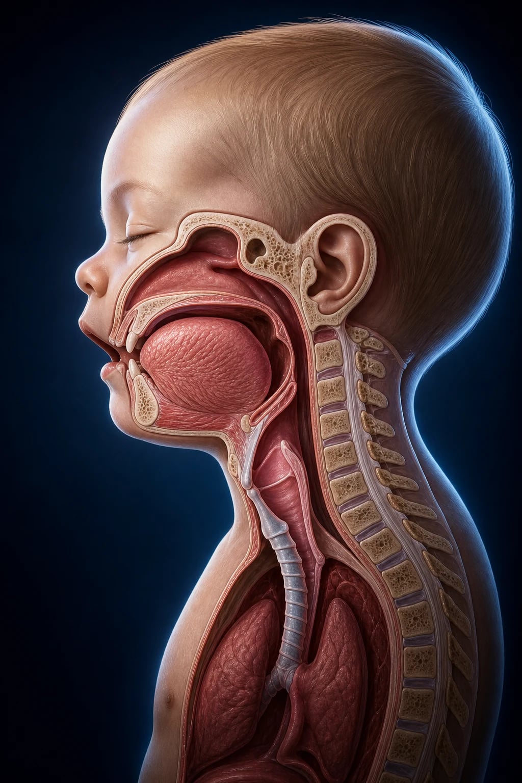
Six anatomical features distinguish the child's airway and each has a practical consequence[4].
- The large occiput. In infants the occiput is proportionally large, so the head naturally flexes forward in the supine position, flexing the neck and obstructing the airway. The corrective manoeuvre is the opposite of the adult: a shoulder roll or a headrest lifts the occiput and aligns the axes, where an adult needs head extension on a pillow[1].
- The large tongue. The tongue fills a disproportionately small oral cavity and falls back against the posterior pharyngeal wall during sedation or anaesthesia, the commonest cause of soft obstruction. A jaw thrust and an oropharyngeal (Guedel) or nasopharyngeal airway relieve it[1].
- The high, anterior larynx. The larynx sits at the level of the third to fourth cervical vertebra in the infant (compared with the sixth in the adult) and is angled anteriorly, so the vocal cords are approached from below and the anterior commissure is easily hidden. A straight (Miller) blade lifted directly onto the epiglottis gives the best view in infants, whereas a curved blade in the vallecula is usual in older children and adults[2].
- The long, floppy, U-shaped epiglottis. The epiglottis is long and angled at roughly forty-five degrees and tends to prolapse over the glottis, so it is often lifted directly with a straight blade rather than indirectly tensioned via the hyoepiglottic ligament[2].
- The funnel-shaped subglottis with the cricoid as the narrowest point. In the adult the vocal cords are the narrowest point and the trachea is cylindrical; in the child the cricoid cartilage — the only complete ring — is the narrowest point and the airway tapers below the cords. This is why a tube that passes the cords may still obstruct, and why an uncuffed tube was historically preferred to avoid subglottic pressure injury[6].
- Short trachea and narrow nasopharynx. The trachea is short (around four to five centimetres in the infant), so mainstem bronchial intubation and accidental extubation are both one centimetre away. The short, delicate nasal mucosa bleeds readily with instrumentation[1].
Physiological differences and the rapid route to desaturation
The physiological reserve of a child is the single most important difference for the practical anaesthetist. Infants and small children have a high metabolic rate and a proportionally large oxygen consumption — roughly six to eight millilitres per kilogram per minute, double the adult rate per kilogram — and a small functional residual capacity[1]. The two together mean that the alveolar oxygen reservoir built up by preoxygenation is small relative to consumption.
In practical terms, a healthy apnoeic adult tolerates several minutes before desaturation, while a small child may fall from full saturation into the seventies within under a minute, and a sick neonate even faster[3]. Preoxygenation is therefore harder and less reliable: the child may cry, refuse the mask, or breathe irregularly. Apnoeic oxygenation with high-flow nasal oxygen extends the window, and a gentle mask ventilation strategy that accepts some gastric insufflation is widely favoured over a strict no-ventilation sequence in children, because hypoxia arrives before paralysis is reliably assured[2][4].
The child also has a relatively small circulating blood volume and a higher baseline heart rate dependent on preload, so bradycardia from hypoxia or vagal stimulation on laryngoscopy signals impending collapse and demands immediate ventilation with one hundred percent oxygen[1].
Airway assessment in children
The adult predictors of difficulty (Mallampati, inter-incisor gap, thyromental distance) are difficult or impossible to apply in the uncooperative child, so assessment is largely historical and observational[2]. The useful questions are: is there a syndrome associated with a difficult airway (Pierre Robin, Treacher Collins, Goldenhar, Down syndrome, mucopolysaccharidoses)? Is there airway obstruction (stridor, stertor, retractions)? Is there a recent upper respiratory tract infection, which multiplies the risk of laryngospasm, bronchospasm and breath-holding? And has the child fed appropriately within the fasting window?[1]
Auscultation of the cry or voice, observation for nasal flaring and the pattern of stridor (inspiratory = extrathoracic, biphasic = glottic or subglottic, expiratory = intrathoracic) localise obstruction. Any child with stridor at rest, tracheal tug or recession is at high risk on induction and the induction technique must preserve spontaneous ventilation[1].
Equipment and sizing
Paediatric airway equipment is sized to weight or age, and the correct size on the first attempt matters because repeated attempts cause oedema in the narrow subglottis[6]. The endotracheal tube internal diameter for a child over two follows the classic formula (age in years divided by four plus four), and the uncuffed tube is taken half a millimetre larger than the cuffed. Modern cuffed tubes with a low-pressure, high-volume cuff and a narrow profile are now used safely in infants and children down to the neonatal period in many centres, with the advantage of fewer repeat laryngoscopies and less leak; the cuff pressure must be checked and kept below approximately twenty centimetres of water in the small child[4].
Laryngoscope blade choice follows anatomy: a straight Miller blade (size zero or one) is preferred in infants up to roughly six months to lift the floppy epiglottis directly, progressing to a size one and then a curved Macintosh as the child grows and the larynx descends. Suction (Yankauer or a rigid paediatric sucker), appropriately sized supraglottic airway devices, and styletted tubes should be ready before induction[2].
The Broselow tape or a weight-based reference gives drug doses and equipment sizes together in an emergency, removing arithmetic from the crisis[1].
Positioning and preoxygenation
Correct positioning aligns the oral, pharyngeal and laryngeal axes. Because of the large occiput, the infant is positioned with a shoulder roll so that the external auditory meatus is level with the sternum — the sniffing position is achieved without a pillow under the head[1]. In the older child a small headrest reproduces the adult sniffing position.
Preoxygenation is attempted with a well-fitting mask for three minutes of tidal breathing or eight vital-capacity breaths where the child cooperates, but in the crying infant it is often partial. Continuous high-flow nasal oxygen during apnoea (paediatric THRIVE) prolongs the safe apnoeic window and is increasingly used during airway instrumentation and difficult intubation[4].
Direct laryngoscopy technique
The laryngoscopist applies a straight blade in the infant to lift the long epiglottis directly, advancing until the epiglottis and posterior cartilages are visualised, then elevating to expose the cords. Because the larynx is high and anterior, external laryngeal manipulation — pressing the larynx posteriorly and cephalad by the free hand or an assistant — often brings the cords into view and should be routine rather than a last resort[2]. The tube is passed under direct vision, watching it pass through the cords and stopping at a depth of roughly three times the internal diameter at the lips, then confirmed by bilateral chest rise, auscultation and, definitively, waveform capnography[1].
Videolaryngoscopy in children
Videolaryngoscopy has become a central tool in paediatric airway management, particularly for the anticipated difficult airway where a blade with an integrated camera and an angled or channelled design exposes an anterior larynx that direct laryngoscopy cannot[4][5]. The percentage of glottic opening (POGO) score quantifies the view and is a more discriminating metric than the adult Cormack and Lehane grade, because even a partial view is adequate if the tube can be passed[5]. The trade-off is that a good view does not guarantee an easy tube pass — anterior angles can make railroading the tube difficult, and a stylet or a bougie is often needed. In the unanticipated difficult intubation, switching early to videolaryngoscopy is now a first escalation rather than a late rescue[2].
Supraglottic airway devices in children
A supraglottic airway device is a rescue device and a conduit, and in children it is arguably even more important than in adults because it can oxygenate when intubation fails and the window is short[2]. A size is chosen by weight, and a second-generation device with a gastric drain port reduces the risk of insufflation and aspiration during rescue ventilation[4]. In the difficult-airway algorithm the supraglottic airway is both the primary oxygenation rescue and, in the intubating variant, a conduit through which a fibreoptic scope and tube can be passed into an otherwise unreachable larynx[2].
The difficult paediatric airway algorithm
The Association of Paediatric Anaesthetists and Difficult Airway Society published an algorithm for the management of the unanticipated difficult intubation in children aged one to eight, structured around the same logic as the adult algorithm but adapted to the child's rapid desaturation[2]. The sequence is: call for help early; limit direct-laryngoscopy attempts and switch to a supraglottic airway to oxygenate; if oxygenation fails, declare cannot-intubate-cannot-oxygenate and move to front-of-neck access without delay[2][3].
Komasawa's paediatric-specific proposal reinforces that the rescue supraglottic airway should be inserted early and that the threshold for escalation is the oxygen saturation, not the number of attempts — the algorithm tolerates no hypoxia[2]. A modified rapid-sequence approach with gentle ventilation and rocuronium (with sugammadex available) rather than suxamethonium is increasingly favoured in children, accepting a small gastric-insufflation risk to avoid the hypoxia of an unventilated stomach[4].
Front-of-neck access in children and the neonate
The cricothyroid membrane is tiny in the child and difficult or impossible to identify in the infant, and the preferred front-of-neck access technique differs from the adult[3]. In older children a narrow-bore cannula (needle) cricothyroidotomy jet-ventilated with a low-pressure device, or in some hands a small surgical cricothyroidotomy, is described, but in the infant and neonate the membrane is so small that a needle technique with careful jet ventilation is preferred over the open scalpel-bougie technique used in adults[3]. Berisha's review of the unexpected difficult airway in neonatal resuscitation emphasises that front-of-neck access is rare, must be rehearsed, and that the best prevention is early use of a supraglottic airway and senior help before the crisis[3].
Specific obstructive conditions
Several conditions present as obstruction and change the induction plan[1][6].
- Croup (viral laryngotracheobronchitis). A barking cough and inspiratory stridor after a viral prodrome; treated with nebulised adrenaline and dexamethasone, with intubation reserved for failure using a tube one size smaller than calculated.
- Epiglottitis. A toxic, febrile, drooling child sitting forward — an emergency managed by senior anaesthetist and ENT together, with induction by sevoflurane in oxygen preserving spontaneous ventilation in the sitting position and a rigid bronchoscope and tracheostomy set ready.
- Foreign body aspiration. Sudden choking in a previously well child; a rigid bronchoscopy under spontaneous-ventilation general anaesthesia is the standard, with the anaesthetist and surgeon ready to convert to an emergency airway[1].
- Subglottic stenosis. Congenital or acquired (prolonged intubation), presenting as unexpected difficult intubation when a tube that should pass instead meets resistance below the cords — a smaller tube or an airway shared with ENT is required, and the diagnosis should be suspected whenever intubation is unexpectedly difficult in a child[6].
- Laryngomalacia. The commonest cause of infant stridor, with floppy supraglottic structures that prolapse on inspiration; usually mild and managed conservatively, but severe cases complicate induction and warrant spontaneous-ventilation techniques.
A recent upper respiratory tract infection increases the risk of every perioperative airway adverse event and is a frequent reason to postpone elective surgery in the small child[1].
Extubation and recovery
Extubation of the child is, as in the adult, a high-risk moment: laryngospasm, breath-holding and post-obstructive pulmonary oedema cluster here[1]. The child is extubated either deep (with a clear airway and an anaesthetist skilled in maintaining the airway) or fully awake, with the stomach emptied if insufflated and all airway equipment ready for reintubation. Laryngospasm is treated with one hundred percent oxygen, continuous positive airway pressure, and if it persists, a small dose of suxamethonium or propofol to relax the cords before hypoxia and bradycardia develop[1]. Recovery in a dedicated, staffed paediatric area with pulse oximetry completes the safe airway pathway, and the integrative safety literature identifies vigilance in recovery, correct equipment sizing and early help as the modifiable factors that reduce harm in paediatric airway management[1].
Clinical
- Standard approach
- Evidence-based
Alternative
- Modified technique
- Risk-benefit
Red flags
References
- [1]Motykiewicz RA, et al. Enhancing Safety in Pediatric Airway Management: An Integrative Review for Perioperative Settings J Perianesth Nurs, 2026.PMID 42319317
- [2]Komasawa N, et al. A proposal for pediatric specific difficult airway management guideline J Clin Anesth, 2020.PMID 31951919
- [3]Berisha G, et al. Management of the Unexpected Difficult Airway in Neonatal Resuscitation Front Pediatr, 2021.PMID 34778121
- [4]Garcia-Marcinkiewicz AG, et al. The future of pediatric airway management Curr Opin Anaesthesiol, 2026.PMID 42013372
- [5]Sasu PB, et al. Diagnostic value of the percentage of glottic opening score for classifying videolaryngoscopy in children: a prospective validation study Anaesthesia, 2026.PMID 42145174
- [6]Aoki S, et al. Unexpected Subglottic Stenosis Detected During Difficult Intubation for the Induction of General Anesthesia: A Case Report Cureus, 2026.PMID 41815624