Anaes · Applied anatomy
Abdominal wall and fascial planes
Also known as Abdominal wall anatomy · Rectus sheath · Transversus abdominis plane · TAP block · Quadratus lumborum block · Fascial plane blocks
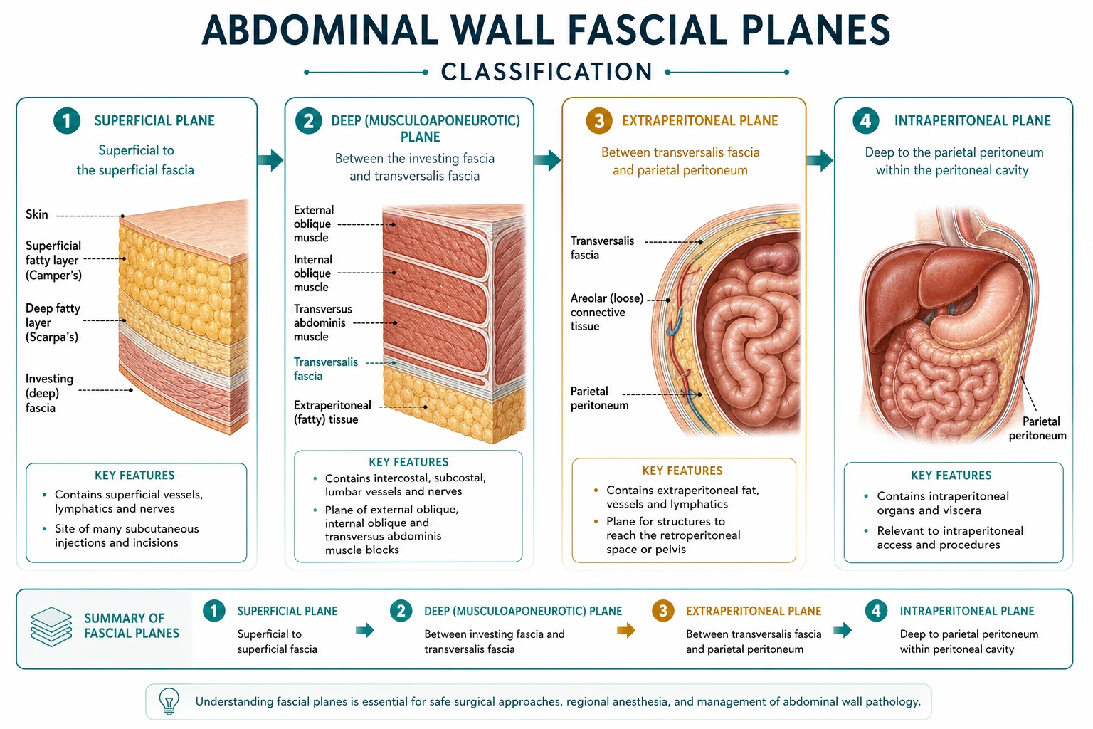
The abdominal wall is the setting for a generation of fascial-plane regional blocks (the TAP, rectus sheath, quadratus lumborum and erector spinae plane blocks) that have transformed analgesia for abdominal surgery, and for the laparoscopic port sites and surgical incisions of the abdomen. The framework rests on six exam-critical ideas. First, the anterolateral abdominal wall is built of nine layers from superficial to deep: skin, subcutaneous fat (Camper's fascia), the membranous layer (Scarpa's fascia), the three flat muscles (external oblique, internal oblique, transversus abdominis) with their aponeuroses, the transversalis fascia, the extraperitoneal fat, and the parietal peritoneum. Second, the midline is closed by the LINEA ALBA, the fusion of the three aponeuroses, into which the rectus abdominis muscles sit either side wrapped in the RECTUS SHEATH; the sheath's composition changes at the ARCUATE LINE (about halfway between the umbilicus and the pubis), below which the posterior wall is transversalis fascia only. Third, the wall is innervated segmentally by the thoracoabdominal nerves (the anterior rami of T7 to T11), the SUBCOSTAL nerve (T12) and the ILIOHYPOGASTRIC and ILIOINGUINAL nerves (L1); the motor and sensory branches of T7-L1 run in the fascial plane between the internal oblique and transversus abdominis — the TRANSVERSUS ABDOMINIS PLANE (TAP). Fourth, the TAP block deposits local anaesthetic in this plane to anaesthetise the anterior abdominal wall (the anterolateral skin and the parietal peritoneum), giving analgesia for lower-abdominal surgery such as caesarean section, hysterectomy and hernia repair. Fifth, the related fascial-plane blocks target planes at different depths and spread — the RECTUS SHEATH block (between rectus and posterior sheath, for midline incisions), the QUADRATUS LUMBORUM block (the pararenal plane, which can spread to the thoracic paravertebral space and cover the visceral as well as the somatic abdominal wall), and the ERECTOR SPINAE PLANE block (the plane over the erector spinae, with cranial-caudal spread for thoracic and abdominal wall analgesia). Sixth, the inguinal canal and the surface landmarks (umbilicus at L4/L5, McBurney's point at the appendix, the semilunar line of the rectus border) complete the applied anatomy for hernia surgery and port placement. Built on the TAP-versus-ilioinguinal-iliohypogastric study (Reda 2026), the hyaluronidase-TAP study (Amin 2026), the erector-spinae-versus-paravertebral study (Turhan 2026), the classical-versus-deep rectus-sheath study (Chooklin 2026), the combined quadratus-lumborum study (Ji 2026), the inguinal-hernia-repair study (Filip 2026), the inguinal-canal spermatic-cord study (Tepelenis 2026), and the umbilical endometriosis study (Huang 2026).
On this page & tools
Your progress
Saved locally on this device.
Target exams
Red flags
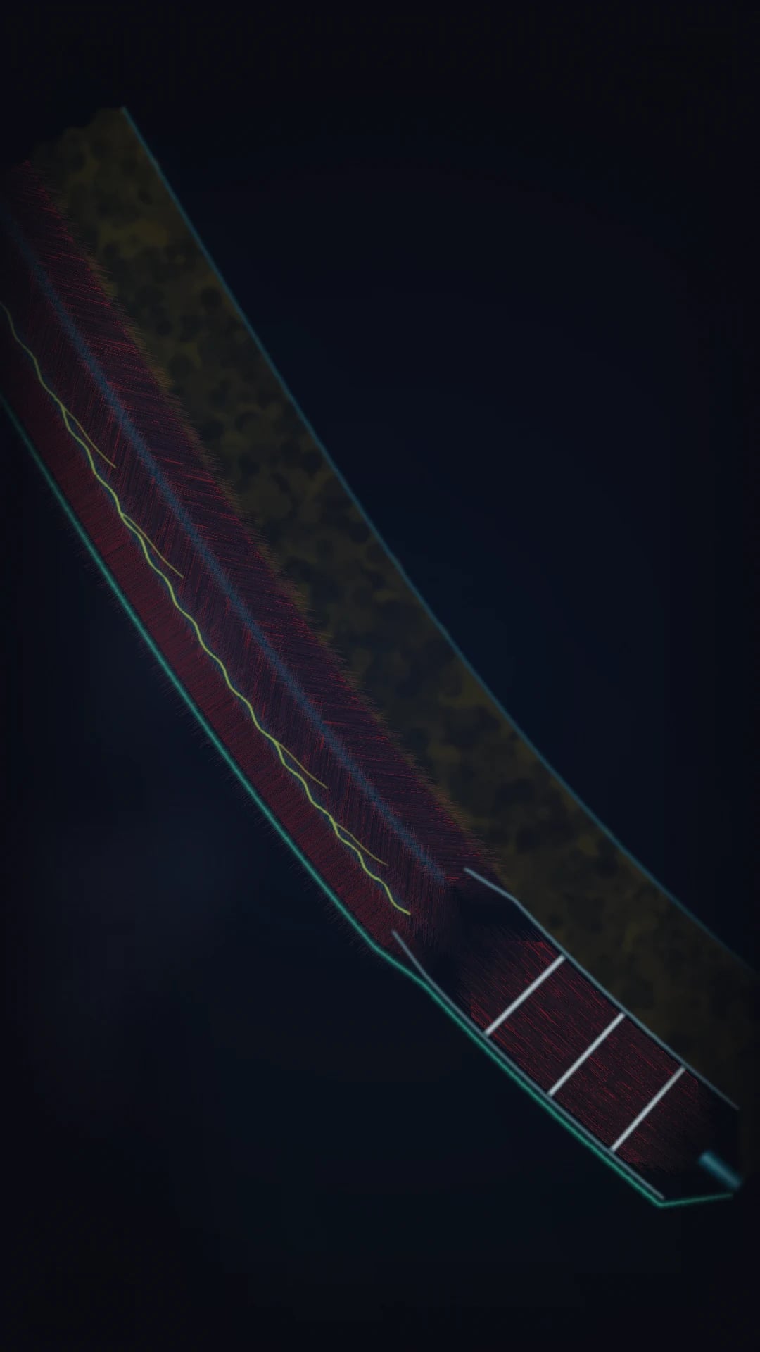
Why this matters to the anaesthetist
Fascial-plane blocks for laparotomy, caesarean section, hernia repair and laparoscopic port-site pain live or die on layer identification. Examiners want the nine layers, the plane that carries T7–L1, how the rectus sheath changes at the arcuate line, and the honest limit that somatic wall blocks do not anaesthetise viscera. [1]
Nine layers (superficial to deep)
- Skin
- Camper fascia (fatty superficial fascia)
- Scarpa fascia (membranous superficial fascia)
- External oblique (+ aponeurosis)
- Internal oblique (+ aponeurosis)
- Transversus abdominis (+ aponeurosis)
- Transversalis fascia
- Extraperitoneal fat
- Parietal peritoneum [1]
The three flat muscles and their aponeuroses form the functional wall; the transversus abdominis plane between internal oblique and transversus is the highway for thoracoabdominal nerves. [1]
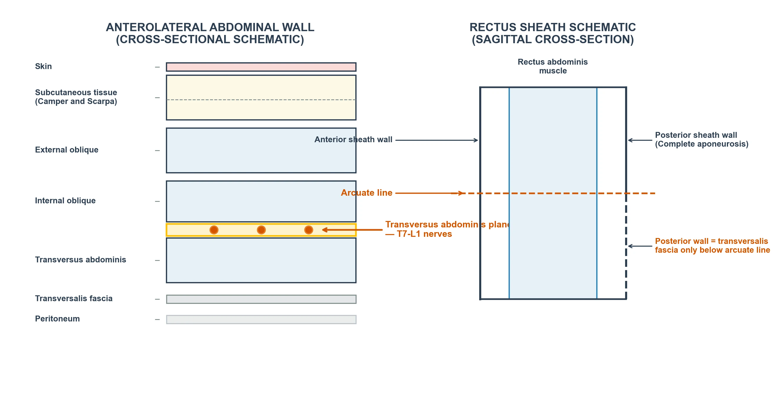
Rectus sheath and arcuate line
| Feature | Above arcuate line | Below arcuate line |
|---|---|---|
| Anterior sheath | External oblique + anterior leaf of internal oblique | All three aponeuroses |
| Posterior sheath | Posterior leaf of internal oblique + transversus | Absent — only transversalis fascia |
| Clinical | Classic rectus sheath block plane | LA can track more freely posteriorly; lower risk/benefit differs |
Linea alba is the midline fusion of aponeuroses. Semilunar (spigelian) line is the lateral rectus border — a hernia and landmark site. [1]
Segmental innervation T7–L1
| Segment | Nerve | Territory |
|---|---|---|
| T7–T11 | Thoracoabdominal | Upper and mid abdomen |
| T12 | Subcostal | Lower abdomen |
| L1 | Iliohypogastric + ilioinguinal | Suprapubic, inguinal, scrotal/labial skin |
Branches run between internal oblique and transversus abdominis before piercing to supply skin. Umbilicus is approximately T10 dermatome and surface marker near L4 vertebra. [1]
Fascial-plane blocks — clinical matrix
| Block | Plane / target | Coverage | Does NOT cover | Typical use | Adult volume guide |
|---|---|---|---|---|---|
| Subcostal TAP | IO–TA plane upper | Upper abdomen wall | Viscera | Upper laparotomy, cholecystectomy | 15–20 mL/side dilute |
| Lateral TAP | IO–TA at mid-axillary | Anterolateral wall T10–L1 | Viscera, midline deeply variable | Lower laparotomy, CS adjunct | 15–20 mL/side |
| Rectus sheath | Posterior to rectus, anterior to posterior sheath (above arcuate line) | Midline wall | Flanks, viscera | Midline incision, umbilical hernia | 10–15 mL/side |
| QL (QL1–3 variants) | Around quadratus lumborum | Wall ± some visceral via paravertebral spread | Variable; technique-dependent | Major abdominal, CS | 20–30 mL/side |
| ESP | Deep to erector spinae at transverse process | Multi-level trunk | Visceral unreliable alone | Thoracic/abdominal adjunct | 20–30 mL |
| Ilioinguinal/iliohypogastric | Near ASIS between IO and TA | Inguinal region | Deep visceral hernia content alone | Open inguinal hernia | 5–10 mL |
Exact surface landmarks
| Landmark | Location | Use |
|---|---|---|
| Umbilicus | ≈ L4 vertebral level; T10 dermatome | Midline orientation |
| ASIS | Anterior superior iliac spine | TAP, ilioinguinal entry reference |
| McBurney point | 1/3 from ASIS to umbilicus | Appendix base / surgical reference |
| Semilunar line | Lateral border of rectus | Spigelian hernia; rectus edge |
| Mid-axillary line | Midway axilla | Lateral TAP probe position |
| Petit's triangle | Iliac crest, latissimus, external oblique | Landmark QL/TAP variants |
Inguinal canal relations (hernia blocks)
Deep ring is lateral to inferior epigastric vessels; superficial ring is above and medial to pubic tubercle. Ilioinguinal nerve runs on the cord (spermatic cord or round ligament). Ultrasound near ASIS identifies IO/TA plane before nerves pierce — safer than blind fan techniques. [1]
Sonoanatomy tips
- TAP: linear or curvilinear probe; three muscle layers like a sandwich; needle in-plane to IO–TA plane; hydrodissection should open a dark lens between IO and TA, not intramuscular.
- Rectus sheath: identify rectus and dual sheath lines; inject deep to muscle, superficial to posterior sheath (above arcuate line).
- QL: curvilinear, lateral abdomen/"shamrock" appearance of QL, psoas, erector spinae around transverse process. [1]
SAQ scaffold
- List nine layers superficial to deep.
- Explain arcuate line change and rectus sheath block implication.
- State TAP coverage limits vs QL.
- Plan analgesia for midline laparotomy vs open inguinal hernia.
- Give ASIS-related landmarks for ilioinguinal block. [1]
Viva phrases
- "Where do T7–L1 nerves run?" → "In the plane between internal oblique and transversus abdominis — the TAP."
- "Will TAP alone do a colectomy?" → "No — somatic wall only; visceral pain needs systemic or neuraxial cover." [1]
Common traps
- Claiming TAP provides surgical anaesthesia of bowel.
- Ignoring arcuate line when teaching rectus sheath.
- Intramuscular injection (failed spread).
- Excessive volume without bilateral total LA dose calculation (LAST risk). [1]
TAP
- IO–TA plane
- Wall somatic
- Easy US
- No reliable visceral cover
Rectus sheath
- Midline incisions
- Arcuate line matters
- Bilateral for laparotomy
- Flank not covered
QL
- Deeper; more visceral potential
- Higher skill
- Larger volume
- Watch LAST and hypotension
Ilioinguinal/IH
- ASIS landmark
- Hernia surgery
- Small volume
- Risk of femoral spread if too deep
Red flags
[1]Primary exam expansion — dense examiner pack
Nine layers of anterolateral abdominal wall (list without hesitation)
- Skin. 2. Subcutaneous fat (Camper). 3. Scarpa fascia (membranous). 4. External oblique. 5. Internal oblique. 6. Transversus abdominis. 7. Transversalis fascia. 8. Extraperitoneal fat. 9. Parietal peritoneum. [1]
Nerves T7–L1 run in the plane between internal oblique and transversus abdominis (TAP plane) giving lateral cutaneous and anterior branches to skin and muscle of the wall — not visceral peritoneum sensation from gut organs (that is autonomic/visceral afferent pathways). [1]
Dermatomes and incision mapping
| Landmark | Dermatome | Block relevance |
|---|---|---|
| Xiphisternum | T6 | High TAP / subcostal TAP / epidural |
| Umbilicus | T10 | Classic TAP coverage aim |
| Inguinal ligament / pubis | L1 | Ilioinguinal/iliohypogastric |
| Midline laparotomy | Bilateral T6–T12 variable | Rectus sheath ± TAP/QL/epidural |
Rectus sheath and arcuate line (critical)
Above arcuate line: rectus has anterior and posterior aponeurotic sheaths. Below arcuate line (~midway umbilicus to pubis): posterior wall deficient — rectus lies on transversalis fascia. Ultrasound rectus sheath block: LA between rectus muscle and posterior sheath (above arcuate line) targeting anterior cutaneous branches of T7–T12. Bilateral for midline. Does not cover lateral flank wounds. [1]
TAP block variants
| Approach | Needle path | Coverage tendency |
|---|---|---|
| Classic mid-axillary | IO–TA plane | Lower quadrants more reliable |
| Subcostal | Along costal margin plane | Upper abdomen better |
| Posterior | Near lumbar triangle | May spread more posterior |
| Dual TAP | Subcostal + lateral | Broader wall cover |
Volume dependent (e.g. 15–20 mL per side teaching ranges — always calculate toxic dose). Ultrasound: flat needle, hydrodissection opens plane, avoid intraperitoneal and intramuscular. [1]
Quadratus lumborum and other plane blocks
QL variants (lateral, posterior, anterior/transmuscular) deposit LA near QL thoracolumbar fascia pathways — potential visceral coverage greater than TAP in some studies/practices, higher technical skill, larger volumes, sympathetic spread hypotension risk. Ilioinguinal/iliohypogastric near ASIS for hernia; risk femoral nerve spread if deep. ESP and midline blocks are alternatives for specific surgeries — mention existence without overclaiming. [1]
Visceral versus somatic pain (must say)
Somatic wall pain: sharp, well localised — plane blocks help. Visceral pain: dull, poorly localised, referred — needs opioids, epidural, or blocks with visceral spread (QL/epidural/paravertebral discussions). Telling examiner TAP equals “complete laparotomy anaesthesia” is a fail. [1]
LAST and dosing
Bilateral multi-plane blocks easily approach maximum lidocaine/bupivacaine/ropivacaine mass. Use lean body weight principles, adrenaline where appropriate, staged injection, monitoring. Have lipid available. [1]
Surgical correlates
Kocher, midline, Pfannenstiel, gridiron/Lanz — map each to dermatomes and choose block/epidural accordingly. Laparoscopy: port sites somatic + pneumoperitoneum visceral/shoulder (phrenic C3–5 referred) — TAP alone insufficient for shoulder tip pain. [1]
SAQ: anatomy of TAP block (8 marks)
Layers to plane (3). Nerves T7–L1 (2). Ultrasound endpoints (1). Indications/limitations visceral (1). Complications (peritoneal puncture, LAST, block failure) (1). [1]
Viva
Q: Between which muscles is TAP? A: Internal oblique and transversus abdominis. Q: What does arcuate line change? A: Posterior rectus sheath ends — anatomy for rectus block and surgical hernias. Q: Will TAP cover bowel pain? A: No reliable visceral cover. [1]
High-yield viva battery and numbers lock-in
Block choice by incision (practical)
| Surgery / incision | Useful wall blocks | Still need for viscera |
|---|---|---|
| Open inguinal hernia | Ilioinguinal/IH ± TAP | Local infiltration / systemic |
| Midline laparotomy | Bilateral rectus ± TAP/QL or epidural | Epidural/opioids for viscera |
| Pfannenstiel | TAP / QL / wound catheters | Visceral cover plan |
| Laparoscopic cholecystectomy | Subcostal TAP ports | Shoulder tip / pneumoperitoneum pain |
| Nephrectomy flank | Posterior TAP/QL/wound | Visceral/renal bed pain |
Ultrasound endpoints quality markers
Visualise three muscle layers; needle tip in TAP plane; anechoic LA lens separating IO from TA; no intramuscular swelling only; no peritoneal puncture (bowel slide loss carefully); aspirate, inject fractionally, watch spread. [1]
Complications list
LAST; intraperitoneal injection; block failure; bowel injury rare; femoral nerve block from deep ilioinguinal; hypotension from extensive QL/epidural spread; haematoma on anticoagulants; infection. [1]
Full viva dialogue (additional)
Examiner: Why is the arcuate line important to the anaesthetist? [1]
Candidate: It marks where the posterior rectus sheath ends, so the anatomy of a rectus sheath block and the integrity of the posterior wall change below that line. Surgeons also care because Spigelian and other hernias relate to these aponeurotic transitions. [1]
Examiner: Give the nine layers of the abdominal wall. [1]
Candidate: Skin, Camper fascia, Scarpa fascia, external oblique, internal oblique, transversus abdominis, transversalis fascia, extraperitoneal fat, parietal peritoneum. The TAP plane is between internal oblique and transversus with nerves T7 to L1. [1]
Exam traps
- TAP as sole anaesthetic for laparotomy.
- Ignoring bilateral dose toxicity.
- Wrong plane (subcutaneous or intramuscular).
- Forgetting umbilicus T10 dermatome. [1]
Examiner synthesis paragraph
Abdominal wall regional anaesthesia is layer counting plus honesty about visceral pain. List nine layers, place the TAP plane between internal oblique and transversus for nerves T7–L1, and respect the arcuate line when teaching rectus sheath blocks. Match blocks to incisions: ilioinguinal for hernia, bilateral rectus for midline, subcostal TAP for upper ports. Always calculate bilateral local anaesthetic mass against LAST limits, confirm an ultrasound lens of local in the correct plane, and state clearly that bowel and shoulder-tip pain need visceral strategies — epidural, opioids or blocks with deeper spread — not TAP alone. [1]
Worked SAQ mark plan — TAP block anatomy (8)
List anterolateral wall layers to the TAP plane (3). Name nerves T7–L1 in the plane (1). Describe ultrasound endpoint as hydrodissection between internal oblique and transversus (1). State somatic not visceral cover (1). Give one indication and one complication including LAST from bilateral volume (2). [1]
Common viva closer: TAP is internal oblique–transversus for T7–L1 somatic wall cover only; always total the bilateral local anaesthetic milligrams against toxic limits before dual-plane wall blocks. [1]
References
- [1]Reda I, et al. Ultrasound-guided transversus abdominis plane block versus ilioinguinal-iliohypogastric nerve block for postoperative analgesia after open total abdominal hysterectomy: a randomised comparative trial Anaesth Rep, 2026.PMID 42311962
- [2]Amin SR, et al. Effect of Two Doses of Hyaluronidase with Bupivacaine in Ultrasound-Guided Transversus Abdominis Plane Block for Post Cesarean Delivery Pain: A Randomized Trial Pain Physician, 2026.PMID 42263311
- [3]Turhan O, et al. Erector Spinae Plane Block Versus Thoracic Paravertebral Block in Laparoscopic Cholecystectomy: A Randomized Controlled Study J Clin Med, 2026.PMID 42355760
- [4]Chooklin S, et al. In Search of Ideal Analgesia: Classical and Deep Rectus Sheath Block in Laparoscopic Cholecystectomy Local Reg Anesth, 2026.PMID 42358227
- [5]Ji J, et al. Effects of multimodal analgesia with combined quadratus lumborum block on enhanced recovery after Kasai portoenterostomy in infants with biliary atresia: a retrospective cohort study Front Pediatr, 2026.PMID 42255912
- [6]Filip SS, et al. Clinical effectiveness of various methods of inguinal hernia repair Wiad Lek, 2026.PMID 42359862
- [7]Tepelenis K, et al. Laparoscopic Removal of a Large Spermatic Cord Lipoma During Bilateral Transabdominal Preperitoneal Inguinal Hernia Repair: A Case Report and Narrative Review of the Literature Cureus, 2026.PMID 42359185
- [8]Huang XN, et al. Primary Cutaneous Endometriosis of the Umbilicus: A Diagnostic Challenge Cureus, 2026.PMID 42326293