Anaes · Applied anatomy
Brachial plexus anatomy
Also known as Brachial plexus · Interscalene block · Supraclavicular block · Infraclavicular block · Axillary block · Erb palsy
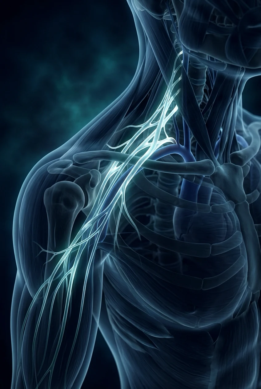
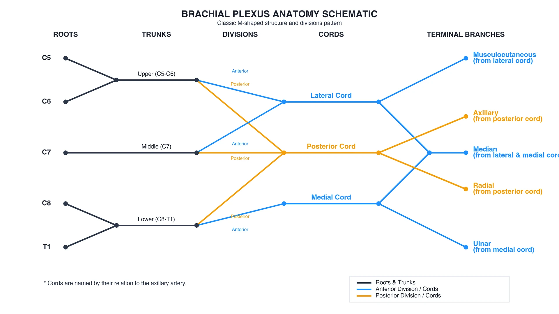
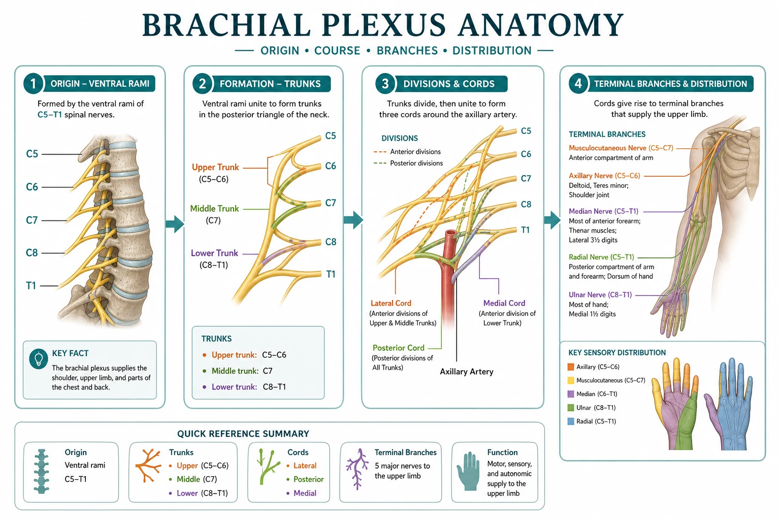
The brachial plexus supplies the entire upper limb and is the most blocked nerve structure in regional anaesthesia. Its anatomy is taught as a five-stage sequence — roots, trunks, divisions, cords and branches — that maps directly onto the four classic approaches to plexus blockade. The framework rests on six exam-critical ideas. First, the plexus is formed by the ventral rami of C5, C6, C7, C8 and T1 (the roots), which unite into three trunks: the upper trunk (C5-C6), the middle trunk (C7) and the lower trunk (C8-T1). Second, each trunk splits into an anterior and a posterior division; the six divisions regroup into three cords named by their relationship to the axillary artery — the lateral, medial and posterior cords. Third, the cords give rise to five terminal branches: the musculocutaneous nerve (lateral cord, elbow flexors and lateral forearm), the axillary nerve (posterior cord, deltoid and teres minor), the radial nerve (posterior cord, all the extensor compartment of arm and forearm), the median nerve (lateral and medial cords, most of the thenar muscles and lateral palm) and the ulnar nerve (medial cord, the intrinsic hand muscles and medial one and a half digits). Fourth, the plexus runs a recognisable course — between the scalenus anterior and medius (the interscalene groove), over the first rib, behind the clavicle, and into the axilla around the axillary artery — and each location is the site of a named block. Fifth, the four classic ultrasound-guided approaches are interscalene (roots/trunks, shoulder surgery but with a high rate of phrenic-nerve block), supraclavicular (divisions, the whole upper limb, near the pleura), infraclavicular/costoclavicular (cords around the axillary artery, hand and forearm surgery) and axillary (branches, forearm and hand). Sixth, two birth-injury patterns localise damage: Erb's palsy (upper trunk C5-C6, the waiter's-tip posture) and Klumpke's palsy (lower trunk C8-T1, a claw hand and sometimes Horner syndrome). Built on the ultrasound-guided interscalene block study (Soor 2026), the diaphragm-sparing low-volume interscalene study (Verbeke 2026), the selective-trunk and supraclavicular block study (Rani 2026), the costoclavicular versus infraclavicular block study (Yazar 2026), the axillary-plexus-blockade study (Solomos 2025), the obstetric brachial-plexus-palsy study (Cimilli 2026), the musculocutaneous-nerve-variation study (Lv 2026), and the greater-auricular-nerve imaging study (Kabra 2026).
On this page & tools
Your progress
Saved locally on this device.
8 MCQs with explanations
Target exams
Red flags
Why this matters to the anaesthetist
The brachial plexus is the single most frequently blocked nerve structure in regional anaesthesia. Every upper-limb block — shoulder, arm, forearm, hand — is an exercise in knowing which stage of the five-stage sequence sits at which anatomical location, which nerves will and will not be covered, and which vital structures sit next door (phrenic nerve, pleura, subclavian artery, vertebral artery). The viva rewards exact relations and block-choice logic, not generic "block the plexus" answers [1][2].
The five-stage organisation (Read That Damn Cadaver)
| Stage | Elements | Location | Block target |
|---|---|---|---|
| Roots | C5, C6, C7, C8, T1 ventral rami | Interscalene groove (between scalenus anterior and medius) | Interscalene |
| Trunks | Upper (C5–6), middle (C7), lower (C8–T1) | Posterior triangle; cross over first rib | Interscalene / selective trunk |
| Divisions | Anterior + posterior from each trunk (six total) | Behind the clavicle | Supraclavicular |
| Cords | Lateral, medial, posterior — named by relation to axillary artery | Infraclavicular / costoclavicular region | Infraclavicular, costoclavicular |
| Branches | Musculocutaneous, axillary, radial, median, ulnar | Axilla around axillary artery | Axillary |
Prefixed plexus = C4 contribution; postfixed = T2 contribution. Both alter dermatome maps slightly and are best recognised under ultrasound [7].
Exact relations along the course
| Level | Key relations | Clinical hazard |
|---|---|---|
| Interscalene groove | Roots between scalenus anterior (anterior) and scalenus medius (posterior); phrenic nerve on anterior surface of scalenus anterior; vertebral artery medial/deep | Phrenic block (hemidiaphragm); vertebral artery injection → seizure |
| Supraclavicular | Divisions as "cluster of grapes" superolateral to subclavian artery; pleural dome immediately inferior/medial; first rib is deep landmark | Pneumothorax; arterial puncture |
| Infraclavicular / costoclavicular | Cords around axillary artery deep to pectoralis minor (infraclavicular) or between clavicle and second rib (costoclavicular) | Pneumothorax (lower risk than supra); vessel puncture |
| Axillary | Median, ulnar, radial around axillary artery; musculocutaneous usually in coracobrachialis (separate) | Incomplete block if musculocutaneous missed |
Terminal branches — origin, motor, sensory
| Branch | Cord | Motor | Sensory | Block implication |
|---|---|---|---|---|
| Musculocutaneous | Lateral | Biceps, brachialis, coracobrachialis (elbow flexion, supination) | Lateral forearm (lateral cutaneous n. of forearm) | Must be found separately in axillary block |
| Axillary | Posterior | Deltoid, teres minor | Regimental-badge patch over deltoid | Covered by interscalene / supra; missed by pure axillary |
| Radial | Posterior | Triceps + all wrist/finger extensors | Posterior arm/forearm, dorsolateral hand | Posterior cord coverage critical for tourniquet pain and extensors |
| Median | Lateral + medial roots | LOAF thenar muscles, most forearm flexors | Lateral palm, lateral 3½ digits | The "M" of the plexus — dual cord origin |
| Ulnar | Medial | Most intrinsic hand muscles, FCU, medial FDP | Medial 1½ digits, medial palm | Lower trunk / medial cord — often missed by interscalene |
Read That Damn Cadaver
RTDC
C5–T1 ventral rami
Upper, middle, lower
Anterior and posterior
Lateral, medial, posterior
Five terminal nerves
LOAF — median motor hand
LOAF
Median motor
Thenar
Thenar
Thenar (superficial head)
The four classic ultrasound-guided blocks
| Block | Target level | Surgical coverage | Landmark / sono | Main caveat | Typical volume (adult) |
|---|---|---|---|---|---|
| Interscalene | Roots / upper trunk (C5–C6) | Shoulder, proximal humerus | Short-axis: 2–3 hypoechoic roots between scalenes | Phrenic block; misses C8–T1 (ulnar) | 7–15 mL (low-volume preferred) [1][2] |
| Supraclavicular | Divisions | Whole upper limb distal to shoulder | Cluster of grapes superolateral to subclavian a.; pleura deep | Pneumothorax | 15–25 mL [3] |
| Infraclavicular / costoclavicular | Cords | Forearm, hand; good catheter site | Cords around axillary a.; costoclavicular more compact | Less phrenic risk; vessel puncture | 20–30 mL [4] |
| Axillary | Terminal branches | Forearm, hand (not shoulder) | Nerves around axillary a.; locate MC in coracobrachialis | Missed musculocutaneous; no shoulder cover | 15–25 mL + MC top-up [5] |
Interscalene
- Shoulder surgery first choice
- High phrenic-block rate
- Misses ulnar (C8–T1)
- Avoid severe respiratory disease
Supraclavicular
- Whole-limb from one injection
- Fast onset
- Pleural risk
- Good when shoulder not needed
Infraclavicular / costoclavicular
- Cords around artery
- Reliable catheters
- Costoclavicular more compact spread
- Lower phrenic risk
Axillary
- Safest for respiratory risk
- Forearm/hand only
- Must block musculocutaneous separately
- Add intercostobrachial for tourniquet
Ultrasound sonoanatomy — exam points
- Interscalene: linear probe, short-axis, mid-neck. Scalenus anterior and medius form a "stoplight" of 2–3 hypoechoic roots. Identify carotid artery medially; keep needle tip lateral to vessels. Greater auricular nerve may be seen superficial over scalenus medius [8].
- Supraclavicular: probe in supraclavicular fossa parallel to clavicle. Subclavian artery is the landmark; plexus sits superolateral ("grapes"); first rib and pleural line are deep — never inject below the rib line.
- Axillary: probe high in axilla short-axis to artery. Median (superficial), ulnar (medial), radial (deep/posterior) around artery; musculocutaneous as hyperechoic oval in coracobrachialis — find it deliberately or the lateral forearm wakes up [7].
Injuries examiners love
- Erb's palsy (upper trunk C5–C6): waiter's tip — arm adducted, internally rotated, elbow extended, forearm pronated, wrist flexed. Birth traction or motorcycle fall.
- Klumpke's palsy (lower trunk C8–T1): claw hand ± ipsilateral Horner syndrome if T1 sympathetics involved [6].
- Inadvertent intraneural / arterial injection and local anaesthetic systemic toxicity are procedure hazards, not plexus anatomy — but the vertebral artery proximity at interscalene makes seizure risk real.
Variation that changes the block
Musculocutaneous nerve may pierce coracobrachialis late, run with the median nerve, or send a communicating branch — the commonest reason an axillary block misses the lateral forearm [7]. Prefixed/postfixed plexuses and anomalous cord formation are recognised under ultrasound before injection.
SAQ answer scaffold
- Draw/list the five stages (roots → trunks → divisions → cords → branches) with root contributions.
- Name the five terminal branches with cord of origin, motor and sensory.
- For a given operation (e.g. shoulder arthroscopy vs wrist ORIF), choose the block and justify coverage and risks.
- Explain why interscalene causes hemidiaphragmatic paresis and how to mitigate.
- State how you would complete an axillary block if the lateral forearm remains intact. [1]
Viva stem bank and model phrases
- "Describe the course of the brachial plexus from foramina to axilla." → "C5–T1 roots unite in the interscalene groove between scalenus anterior and medius; trunks cross the posterior triangle over the first rib; divisions form behind the clavicle; cords surround the axillary artery; terminal branches arise in the axilla."
- "Why prefer costoclavicular over classic infraclavicular?" → "Cords are more tightly clustered between clavicle and second rib, improving single-injection spread and catheter stability [4]."
- "Patient with severe COPD needs shoulder surgery — what do you avoid?" → "Classical interscalene because of near-universal phrenic block; discuss alternative (superior trunk low-volume, general alone, or carefully counselled low-volume techniques)."
Common traps
- Thinking interscalene covers the whole arm including ulnar — it often spares C8–T1.
- Forgetting to block musculocutaneous separately at the axilla.
- Ignoring phrenic risk in COPD / obesity / contralateral phrenic palsy.
- Confusing Erb (upper trunk, waiter's tip) with Klumpke (lower trunk, claw ± Horner).
- Injecting deep to the first rib at the supraclavicular level (pleura). [1]
Red flags
[1]References
- [1]Soor B, et al. Ultrasound-guided Interscalene Block as a Safer Alternative to General Anesthesia in a Patient with Ankylosing Spondylitis Undergoing Proximal Humerus Fracture Fixation Ann Afr Med, 2026.PMID 42318963
- [2]Verbeke AL, et al. The diaphragm-sparing effect of interscalene block with a low-volume of ropivacaine 0.1% vs. 0.5%: A double-blind, controlled, randomised trial Eur J Anaesthesiol Intensive Care, 2026.PMID 42244823
- [3]Rani N, et al. Ultrasound-Guided Selective Trunk Block Combined With Supraclavicular Nerve Block for Awake Shoulder Arthroscopy: A Case Series Cureus, 2026.PMID 42220761
- [4]Yazar V, et al. Comparison of Costoclavicular Block and Infraclavicular Block Effects on Tissue Oxygen Saturation in Upper Extremity Surgery: A Randomized, Assessor-Blinded Controlled Trial Diagnostics (Basel), 2026.PMID 42279582
- [5]Solomos D, et al. Effect of Ketamine Supplementation in Axillary Plexus Blockade: A Comparative Study Cureus, 2025.PMID 41583169
- [6]Cimilli E, et al. Cognitive and Executive Function Profiles in School‑Age Children with Obstetric Brachial Plexus Injury Phys Occup Ther Pediatr, 2026.PMID 42322142
- [7]Lv Y, et al. An anatomical variation of the musculocutaneous nerve featuring early bifurcation and a transient common trunk with the median nerve: a case report Folia Morphol (Warsz), 2026.PMID 42345218
- [8]Kabra N, et al. Asymmetrical Brachial Plexus Involvement with Greater Auricular Nerve Involvement in Borderline Tuberculoid Leprosy: An Electrophysiological and Radiological Correlation Indian J Radiol Imaging, 2026.PMID 42344197