Anaes · Applied anatomy
Lumbosacral plexus anatomy
Also known as Lumbar plexus · Sacral plexus · Sciatic nerve anatomy · Femoral nerve · Obturator nerve · Lower limb nerve blocks
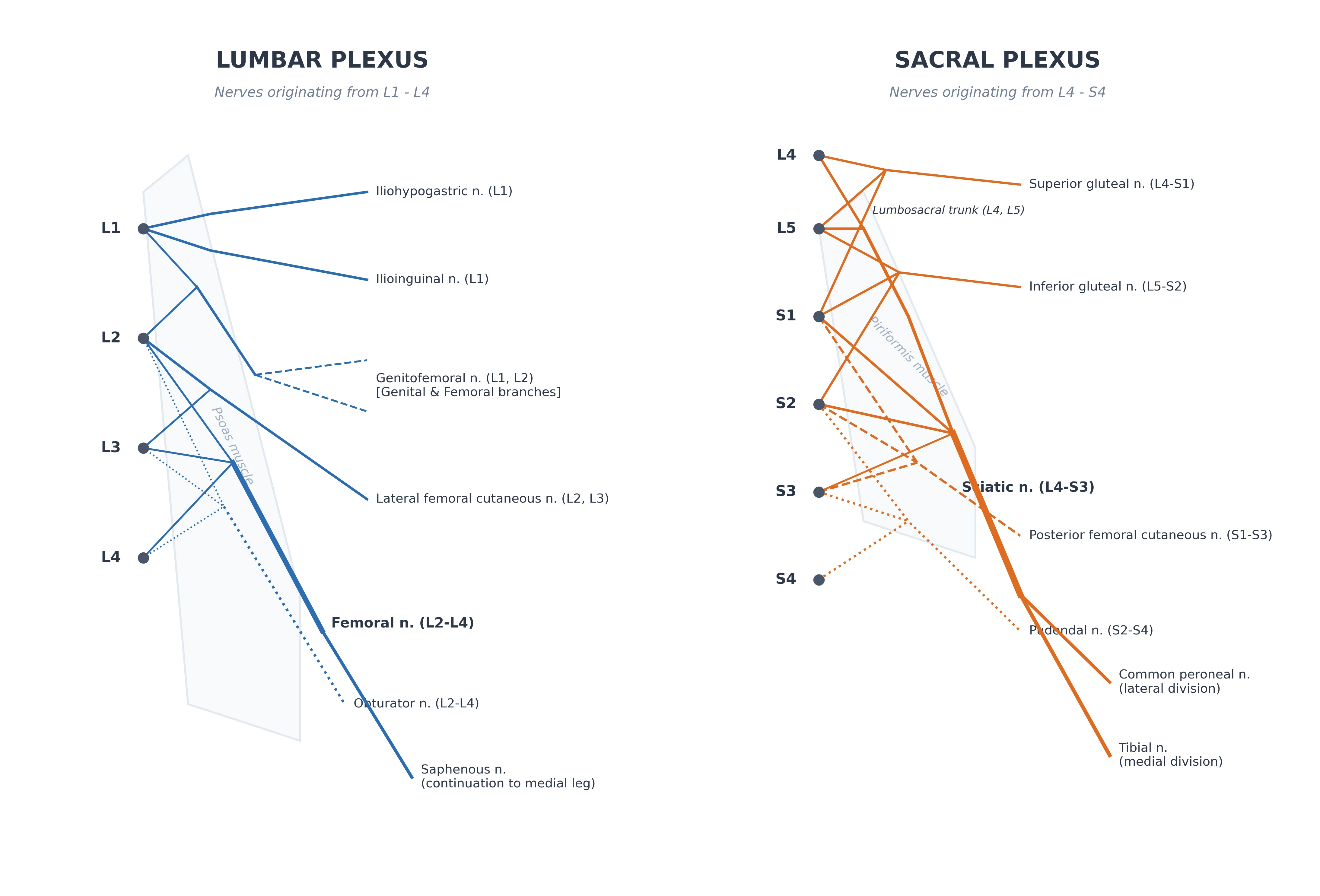
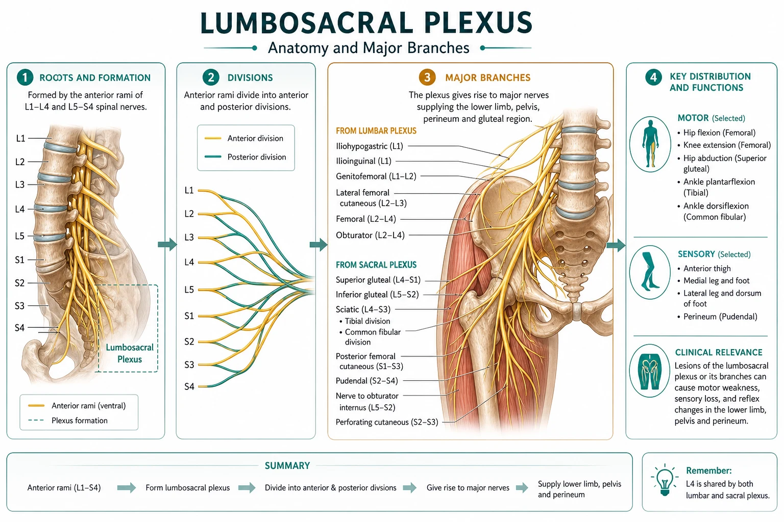
The lumbosacral plexus supplies the entire lower limb, the pelvic floor and the perineum, and its branches are the targets of the lower-limb regional blocks used for hip, knee, ankle and foot surgery. The framework rests on six exam-critical ideas. First, the lumbosacral plexus is actually two plexuses: the LUMBAR plexus, formed by the ventral rami of L1-L4 (with T12 and L5 contributions) and lying within the substance of the psoas major muscle; and the SACRAL plexus, formed by the lumbosacral trunk (L4-L5) and the ventral rami of S1-S4, lying on the anterior surface of the piriformis muscle. Second, the principal lumbar-plexus branches are the iliohypogastric and ilioinguinal (L1, the abdominal wall), the genitofemoral (L1-L2, the genital branch and the femoral triangle), the lateral femoral cutaneous nerve (L2-L3, the lateral thigh — compressed in meralgia paraesthetica), the FEMORAL nerve (L2-L4, the anterior thigh quadriceps and the medial leg via the saphenous nerve) and the OBTURATOR nerve (L2-L4, the medial-thigh adductors). Third, the principal sacral-plexus branch is the SCIATIC nerve (L4-S3), the largest nerve in the body, which leaves the pelvis through the greater sciatic foramen below the piriformis and runs down the posterior thigh to the popliteal fossa, where it divides into the TIBIAL nerve (posterior compartment, plantar foot) and the COMMON PERONEAL nerve (lateral/anterior compartment, dorsum of foot). Fourth, other sacral-plexus branches are the superior and inferior gluteal nerves (gluteal muscles), the pudendal nerve (S2-S4, the perineum) and the posterior femoral cutaneous nerve (posterior thigh). Fifth, the lower-limb blocks target these branches: the femoral and fascia-iliaca blocks (anterior thigh and knee), the obturator block (adductor spasm and knee), the pericapsular nerve group (PENG) block (hip capsule), and the sciatic block by posterior, anterior or popliteal approaches (the leg and foot below the knee). Sixth, two injury patterns localise damage: foot drop from a common-peroneal (or L5) lesion, and sciatica from L4-S1 root irritation. Built on the lumbar-plexus shamrock-view ultrasound study (Cui 2026), the lumbar-plexus intra-psoas study (Gagliardi 2026), the sciatic-nerve radiofrequency study (Gonzalez Godoy 2026), the piriformis-syndrome ultrasound study (Sudhakar 2026), the PENG and lateral-femoral-cutaneous-nerve block study (Cunha 2026), the obturator-nerve-block study (Uchino 2026), the CT-guided pudendal-nerve-block study (Battle 2026), and the peroneal-nerve-injury case report (Li 2026).
On this page & tools
Your progress
Saved locally on this device.
8 MCQs with explanations
Target exams
Red flags
Why this matters to the anaesthetist
Every lower-limb regional technique — fascia iliaca, femoral, PENG, obturator, sciatic, popliteal, ankle — is a map of the lumbosacral plexus. Examiners test which nerves cover which operation, where each nerve sits relative to muscle and vessel landmarks, and classic injury patterns (foot drop, meralgia, sciatica) [3][5][8].
Two plexuses, one limb
| Plexus | Roots | Bed | Link |
|---|---|---|---|
| Lumbar | Ventral rami L1–L4 (± T12, L5) | Within substance of psoas major | Gives femoral, obturator, LFCN, iliohypogastric/ilioinguinal, genitofemoral |
| Sacral | Lumbosacral trunk (L4–L5) + S1–S4 | Anterior surface of piriformis | Gives sciatic, gluteals, pudendal, posterior femoral cutaneous |
The lumbosacral trunk (L4–L5) is the anatomical bridge. Ultrasound "shamrock" views and MRI mapping of the lumbar plexus within psoas are modern teaching aids for deep plexus work [1][2].
Lumbar plexus branches — exact supply and block target
| Nerve | Roots | Course / landmark | Motor | Sensory | Clinical block |
|---|---|---|---|---|---|
| Iliohypogastric / ilioinguinal | L1 | Between internal oblique and transversus; near ASIS | Abdominal wall | Suprapubic / inguinal / scrotal or labial | Hernia, lower abdominal wall |
| Genitofemoral | L1–2 | On psoas; genital + femoral branches | Cremaster | Genital skin, upper anterior thigh | Hernia, orchidectomy analgesia |
| Lateral femoral cutaneous | L2–3 | Under inguinal ligament near ASIS | None (pure sensory) | Lateral thigh | Meralgia paraesthetica; add to hip blocks [5] |
| Femoral | L2–4 | Lateral to femoral artery in femoral triangle, deep to fascia iliaca | Quadriceps (knee extension), iliopsoas contribution | Anterior thigh; saphenous = medial leg to medial malleolus | Femoral / fascia iliaca; knee and hip analgesia |
| Obturator | L2–4 | Through obturator canal into medial thigh | Adductors | Variable medial knee patch | Interadductor approach for spasm / total knee [6] |
Sacral plexus branches
| Nerve | Roots | Landmark | Function | Block / clinical |
|---|---|---|---|---|
| Sciatic | L4–S3 | Greater sciatic foramen below piriformis; posterior thigh; splits in popliteal fossa | Hamstrings; then tibial + common peroneal | Posterior, anterior, subgluteal, or popliteal approaches [3][4] |
| Tibial | (sciatic) | Mid popliteal fossa deep to vein/artery | Plantar flexion, intrinsic foot (most) | Plantar foot sensation and motor |
| Common peroneal (fibular) | (sciatic) | Winds around fibular neck | Dorsiflexion, eversion | Foot drop if injured [8] |
| Superior / inferior gluteal | L4–S2 | Greater sciatic foramen | Gluteus medius/minimus; gluteus maximus | Rarely blocked alone |
| Pudendal | S2–4 | Ischial spine / Alcock's canal | Perineal muscles | Labour, perineal, chronic pelvic pain blocks [7] |
| Posterior femoral cutaneous | S1–3 | With sciatic under gluteus maximus | None significant motor | Posterior thigh skin |
Relations that decide the needle path
- Femoral triangle (lateral → medial): femoral nerve → artery → vein → lymphatics (NAVY reversed: nerve lateral). Fascia iliaca is the plane that spreads local anaesthetic over the femoral and often LFCN with sufficient volume.
- Sciatic exit: below piriformis through greater sciatic foramen; piriformis syndrome and deep gluteal pain relate to this relationship [4].
- Popliteal fossa: tibial nerve medial/deeper near vessels; common peroneal lateral toward biceps femoris tendon — bifurcation level varies; ultrasound finds the split before it occurs.
- Fibular neck: common peroneal is subcutaneous and vulnerable to stirrups, compression, and misplaced needles [8].
Lower-limb block decision matrix
| Surgery / goal | Primary blocks | Notes |
|---|---|---|
| Hip fracture analgesia | Fascia iliaca or PENG ± LFCN | PENG targets articular branches of femoral and accessory obturator to anterior capsule [5] |
| Total knee arthroplasty | Femoral or adductor canal + obturator ± IPACK / sciatic sparingly | Balance analgesia vs quadriceps weakness (falls) |
| Below-knee / ankle / foot | Sciatic (popliteal) + saphenous (adductor canal or ankle) | Dual innervation is mandatory for complete cover |
| Adductor spasm (TURP, TKA) | Obturator (interadductor US) | US vs nerve stimulation both used [6] |
| Perineum / labour adjuvant | Pudendal | Landmark (ischial spine) or image-guided [7] |
Typical adult single-shot volumes (guide only; titrate and use US): fascia iliaca 30–40 mL dilute LA; femoral 15–20 mL; adductor canal 15–20 mL; popliteal sciatic 15–25 mL; obturator 5–10 mL per branch. [1]
Injury patterns examiners expect
- Foot drop: common peroneal at fibular neck or L5 root — loss of ankle/toe dorsiflexion and eversion; sensory dorsum of foot [8].
- Meralgia paraesthetica: LFCN compression under inguinal ligament near ASIS — burning lateral thigh, no motor deficit.
- Sciatica / piriformis-related deep gluteal pain: sciatic irritation at the greater sciatic foramen [4].
SAQ answer scaffold
- Distinguish lumbar vs sacral plexus (roots, muscle bed, major branches).
- Draw femoral triangle relations and explain fascia iliaca spread.
- Describe sciatic course from piriformis to tibial/common peroneal split.
- Plan regional analgesia for NOF fracture vs ankle ORIF with nerve territories.
- Explain foot drop localising features. [1]
Viva stem bank
- "Where does the lumbar plexus form?" → "Within psoas major from L1–L4 ventral rami."
- "What does a femoral block miss for foot surgery?" → "Lateral leg, dorsum and sole of foot, posterior leg — needs sciatic and often lateral sural contributions addressed."
- "Why is the common peroneal the most injured lower-limb nerve?" → "Subcutaneous winding around the fibular neck — compression and trauma." [1]
Common traps
- Assuming femoral block covers the whole leg below the knee.
- Forgetting saphenous (femoral terminal sensory) for medial ankle/foot.
- Blocking only sciatic for TKA and expecting anterior knee analgesia.
- Confusing LFCN (sensory only) with femoral motor block.
- Ignoring fibular neck padding in the lithotomy position. [1]
Femoral / fascia iliaca
- Anterior thigh + saphenous
- Knee and hip analgesia
- Quadriceps weakness risk
- Misses posterior and most foot
Sciatic / popliteal
- Posterior thigh and below-knee majority
- Essential for foot/ankle
- Foot drop if nerve injury
- Combine with saphenous for complete distal cover
Obturator
- Adductors + medial knee patch
- TKA / TURP spasm
- Interadductor US approach
- Variable cutaneous territory
PENG + LFCN
- Anterior hip capsule focus
- Less quadriceps weakness than high femoral
- Hip fracture niche
- Still not complete surgical anaesthesia alone
Red flags
[1]References
- [1]Cui H, et al. DMRNet: a dynamic multi-scale residual network for Shamrock view and lumbar plexus segmentation Comput Assist Surg (Abingdon), 2026.PMID 42284483
- [2]Gagliardi F, et al. Posterolateral approach for lumbar plexus intra-psoas schwannomas: systematic review and comparative anthropometric analysis of surgical corridors with an illustrative case Eur Spine J, 2026.PMID 42289522
- [3]Gonzalez Godoy E, et al. Ultrasound-Guided Pulsed Radiofrequency of the Sciatic Nerve for Chronic Lower Limb Pain: A Real-World Cohort Experience Cureus, 2026.PMID 42326120
- [4]Sudhakar P, et al. Integrated Role of Musculoskeletal Ultrasound in Piriformis Syndrome: A Case Series Cureus, 2026.PMID 42317917
- [5]Cunha DFD, et al. Pericapsular nerve group block combined with lateral femoral cutaneous nerve block versus fascia iliaca compartment block for hip surgery: a systematic review, meta-analysis, and trial sequential analysis Korean J Pain, 2026.PMID 42337250
- [6]Uchino T, et al. Ultrasound versus nerve stimulation-guided interadductor approach for obturator nerve block: evaluation of injectate spread into the obturator canal in a randomized controlled trial BMC Anesthesiol, 2026.PMID 42286464
- [7]Battle W, et al. Clinical Criteria for Improved Outcomes in Patients who Undergo Percutaneous CT-Guided Pudendal Nerve Cryoablation: A Retrospective Analysis J Vasc Interv Radiol, 2026.PMID 42276242
- [8]Li M, et al. [A case report of peroneal nerve injury caused by peripheral nerve block anaesthesia of knee joint] Zhongguo Gu Shang, 2026.PMID 42338211