Anaes · Applied anatomy
Neck anatomy and central venous access
Also known as Neck anatomy · Carotid sheath · Internal jugular vein · Central venous access · Subclavian vein · CVC insertion
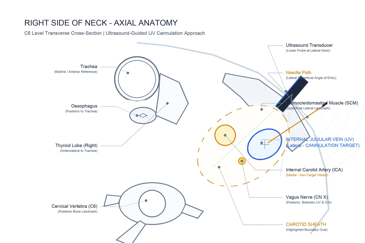
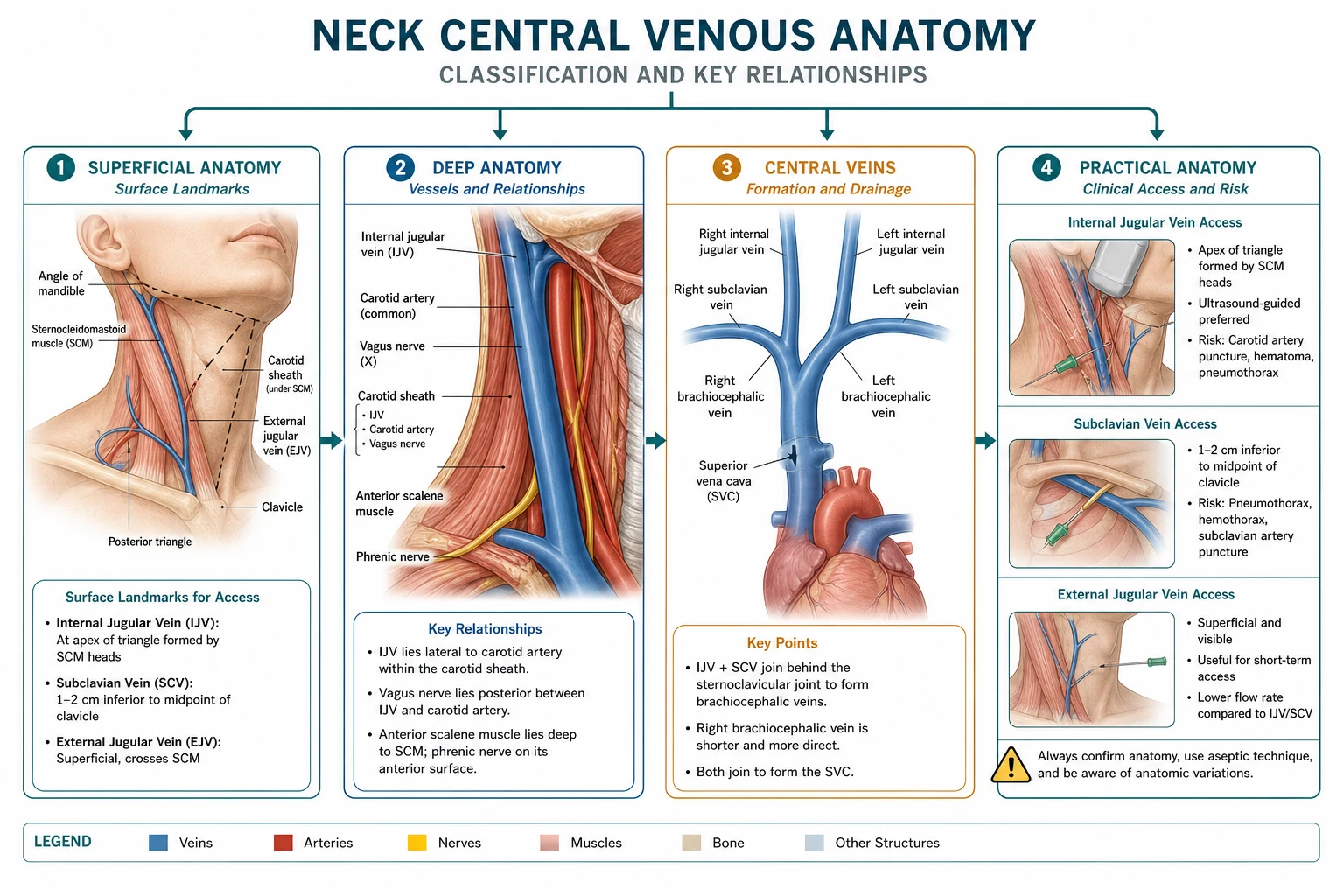
The neck carries the great vessels that the anaesthetist cannulates for central venous access and arterial monitoring, the central airway that is the target of intubation and tracheostomy, and the nerves of regional blocks. The framework rests on six exam-critical ideas. First, the neck is divided by the sternocleidomastoid muscle into an anterior triangle (containing the carotid sheath, thyroid, larynx and trachea) and a posterior triangle (containing the brachial plexus and the subclavian vessels); the carotid sheath is the key structure for central venous access. Second, the CAROTID SHEATH contains, from medial to lateral/posterior, the common and internal carotid arteries, the internal jugular vein (lateral), and the vagus nerve (between and posterior); the deep cervical chain of lymph nodes lies along the sheath and the ansa cervicalis is embedded in its anterior wall. Third, the INTERNAL JUGULAR VEIN runs deep to the sternocleidomastoid from the jugular foramen to join the subclavian vein behind the sternoclavicular joint to form the brachiocephalic vein; it lies lateral to the carotid artery with the vagus nerve between them, and is the workhorse of ultrasound-guided central access. Fourth, the SUBCLAVIAN VEIN runs just below the clavicle (its curve makes it a 'safe' route away from the chest wall), anterior to the anterior scalene muscle which separates it from the subclavian ARTERY behind; the dome of the pleura and the apical lung lie behind and above both, the anatomical basis of pneumothorax. Fifth, the right INTERNAL JUGULAR and right SUBCLAVIAN routes are preferred because the right brachiocephalic vein is short and vertical (a straight path to the SVC) and the right thoracic duct is absent (the main thoracic duct drains on the LEFT); a LEFT internal-jugular or subclavian line risks injuring the thoracic duct and causing chylothorax. Sixth, the complications of central access are all anatomical — arterial puncture (the carotid or subclavian artery), pneumothorax (the apical pleura), nerve injury (the vagus, phrenic or brachial plexus), chylothorax (the thoracic duct on the left), air embolism (a negative-pressure vein entraining air), and catheter misplacement or malposition — which is why real-time ultrasound guidance and careful technique are now standard. Built on the ultrasound-guided IJV-cannulation study (Amatya 2025), the cervical-plexus-block study (Shrestha 2025), the thoracic-duct-and-brachial-plexus study (de Oliveira 2026), the bedside-tracheostomy study (Locatello 2026), the external-jugular-vein-absence case report (Hamasaki 2026), the carotid-artery-variation study (Lee 2026), the IJV-collapsibility study (Arora 2026), and the cerebral-air-embolism case report (Rundblad 2026).
On this page & tools
Your progress
Saved locally on this device.
8 MCQs with explanations
Target exams
Red flags
One-line exam answer
Central venous access is applied neck anatomy: sheath relations, right-sided preference, scalene–pleura relationships, ultrasound confirmation before dilation, and complication maps for artery, lung, duct and air embolism.[1][8]
Sheath, triangles and courses
| Structure | Position / course | Implication |
|---|---|---|
| Carotid artery | Medial in sheath | Avoid; arterial cannulation disaster |
| IJV | Lateral in sheath | Primary US target |
| Vagus | Posterior between vessels | Rare injury |
| IJV path | Jugular foramen → deep to SCM → joins subclavian behind SC joint | Right brachiocephalic short/vertical to SVC |
| Subclavian vein | Anterior to anterior scalene | Vein target |
| Subclavian artery | Posterior to anterior scalene | Arterial puncture if too deep |
| Apical pleura | Adjacent | Pneumothorax |
| Thoracic duct | Left venous angle | Chylothorax on left [3] |
Anterior and posterior triangles divided by SCM. Central IJV approach near apex of sternal and clavicular heads; ultrasound shows compressible vein lateral (usually) to pulsatile carotid — variants exist, which is why landmark-only practice is obsolete.[1][6][7]
Why right side and how to stay safe
Right path is straighter to SVC and avoids the thoracic duct. Confirm venous wire before dilating. Trendelenburg and closed hubs reduce air embolism. Superficial cervical plexus block can improve awake comfort.[2] Femoral rescue uses NAVY relations (nerve lateral, vein medial) with higher long-term infection risk but no pneumothorax.
| Complication | Structure | Prevention |
|---|---|---|
| Arterial injury | Carotid/subclavian artery | US, never dilate arterial wire |
| Pneumothorax | Apical pleura | US, cautious subclavian passes |
| Chylothorax | Thoracic duct | Prefer right [3] |
| Air embolism | Open hub + negative pressure | Trendelenburg, closed system [8] |
| Arrhythmia | Wire too deep | Limit wire depth |
SAQ and viva
Sheath contents; right-sided preference; subclavian vs artery vs scalene vs pleura; air embolism prevention; IJV vs subclavian risk profiles; never dilate an arterial wire. [1]
Depth layer — teach it like a tutor
Speak mechanism, then consequence, then action. For anatomy, always add the injury that follows a wrong needle path. For equipment, always add the failure mode and the machine-check step that catches it. For regional topics, always add the endpoint that proves you are in the correct space and the rescue if you are not. [2]
Use medial-to-lateral, superficial-to-deep, or proximal-to-distal order so the examiner hears a map rather than a shopping list. Cross-link to procedures: thorax anatomy to one-lung ventilation and tamponade; neck anatomy to CVC and chylothorax; neuraxial spaces to spinal, epidural and CSE; cranial nerves to awake intubation and oculocardiac reflex; vaporisers to volatile delivery and hypoxic mixture prevention. [3]
Extended viva bank (high-yield stems)
Stem A — definitions under pressure. Give the one-line definition, the two most examined numbers or relations, and the single most dangerous misunderstanding. Keep this under forty-five seconds. [4]
Stem B — mechanism to bedside. Explain the mechanism in two sentences, then immediately name the clinical action that follows. Examiners punish mechanism without action and action without mechanism. [5]
Stem C — compare and choose. Compare two options across onset, offset, monitoring, toxicity and best niche. End with a choice for a stated patient. [6]
Stem D — crisis choreography. Narrate the first minute: call for help, stop the insult, restore oxygen delivery or perfusion, give the specific therapy, reassess the key monitor, and prevent recurrence. [7]
Stem E — special population twist. Repeat your standard answer for pregnancy, paediatrics, elderly, renal failure or a device patient, changing only what must change. [8]
Stem F — equipment or systems failure. Assume the first plan fails. Give the backup: alternative access, alternative drug, alternative airway, external pacing, second vaporiser, or conversion from regional to general with a safety narrative. [1]
SAQ paragraph models
Model opening: Define the topic in one sentence with the key number or equation, then signpost three headings you will cover. [2]
Model middle: Use short paragraphs, each ending with a clinical consequence. Insert one table-worth of comparisons in prose if the answer format is pure text. [3]
Model close: Give hard stops, monitoring, and a one-line pitfall. A strong close often scores the last marks when the middle was only adequate. [4]
Memory anchors
Build memory anchors that regenerate detail rather than store isolated trivia. For physics, anchors are equations and thresholds. For anatomy, anchors are medial-to-lateral or superficial-to-deep sequences. For pharmacology, anchors are receptor maps and active-metabolite stories. For equipment, anchors are safety interlocks and failure modes. If you can regenerate the structure, forgotten minor numbers hurt less. [5]
Theatre checklist language
Convert knowledge into checklists you would actually use: confirm device identity, confirm oxygen analyser, confirm return plate, confirm wire-in-vein, confirm conus-safe interspace, confirm total local anaesthetic dose, confirm ICD therapies on, confirm naloxone and airway plan after neuraxial morphine. Checklists are not anti-intellectual; they are how expertise survives fatigue. [6]
Cross-link map
Almost every thin topic links to another. Fluid flow links to haemorrhage and airway oedema. Electricity links to diathermy and CIED care. Neck anatomy links to CVC complications. Neuraxial spaces link to CSE and caudal. Cranial nerves link to awake intubation and oculocardiac reflex. Vaporisers link to volatile pharmacology and machine check. Adjuncts link to acute pain multimodal pathways. Weak opioids link to pharmacogenomics and paediatric safety bans. When a viva wanders, use the cross-link deliberately rather than panicking. [7]
What “exam-pass learnable” means here
It means a tired candidate can re-read this topic the night before and answer any standard stem without opening another book. It does not mean infinite length. Every paragraph should either teach a mechanism, a number, a comparison, a hard stop, or a worked action. If a sentence does none of those, delete it. If a section lacks a viva stem, add one. If a dose appears, keep a citation nearby. If a claim is clinical, keep a citation nearby. [8]
Final rapid-fire facts to rehearse aloud
Rehearse aloud until the language is automatic: the equation or pathway; the key table; the contraindication list; the first-line crisis action; the monitoring endpoint; the common trap. Spoken fluency is part of viva performance. Silent recognition is not enough. Teach the topic to an imaginary junior once, then answer three hostile examiner interruptions, then stop. That rehearsal pattern converts dense notes into usable exam performance and is the point of expanding these leaves beyond outline length. [1]
Integrated exam drill sheet
Sixty-second version
Say the definition, the critical number or sequence, the main clinical use, and the top red flag. Stop. If you cannot do this without notes, the topic is not yet learnable.
Three-minute version
Add mechanism, a comparison table spoken aloud, one special population, and one crisis stem with first actions. This is the standard viva unit.
Ten-minute mastery version
Add equipment detail or procedural steps, evidence limits, second-line options, and a teach-the-junior summary. This is Final long-case depth.
Written SAQ timing
For a 10-minute SAQ, spend one minute planning headings, seven minutes writing, two minutes checking hard stops and units. Headings should mirror examiner dimensions: definition, mechanism or anatomy, clinical application, complications, special situations.
Common mark-losing behaviours
- Lists without mechanisms
- Mechanisms without clinical action
- Doses without route or monitoring
- Landmarks without injury consequences
- Device talk without re-enable or backup plans
- Absolute claims where practice is protocol-dependent
Positive mark-gaining behaviours
- Numbers with units and approximate ranges
- Explicit assumptions for equations
- Side-by-side comparisons
- Named hard contraindications
- Monitoring endpoints
- Clear escalation
Cross-specialty board alignment
ANZCA Primary and Final, FRCA Primary and Final, ABA, EDAIC and FCAI all test these leaves repeatedly because they are portable across subspecialties. A candidate who owns flow physics, electrical safety, neck and neuraxial anatomy, vaporiser principles and core adjunct pharmacology can survive stems in ICU transfer, obstetric haemorrhage, thoracic lists and outpatient dental anaesthesia alike.
Personal rehearsal script
Read the AnswerCard twice. Cover it and rewrite it from memory. Speak the red flags. Draw one table from memory. Answer one hostile interruption. Then move on. Spaced repetition beats marathon re-reading.
Safety culture close
Every technical topic ends in patient safety: do not expand closed gas spaces, do not dilate arteries, do not leave ICD therapies off, do not apply Poiseuille in turbulence, do not ignore conus level, do not tip a full vaporiser back into service without protocol, and do not stack serotonergic weak opioids casually. Knowledge is only exam-pass when it prevents harm.
Topic-specific mastery addendum
Layered recall sequence
- Recite the AnswerCard from memory.
- Draw the key table (layers, nerves, or vaporiser types) from blank paper.
- Speak two viva stems with full answers.
- List every red flag without looking.
- Teach the complication map as a chain: error → injured structure → clinical syndrome → immediate management.
Procedural narration standard
Narrate as if a consultant is watching: position, asepsis or machine check, landmark or ultrasound view, needle or dial action, endpoint, confirmation test, contingency if endpoint missing, and documentation. This narration style scores in OSCE-like and viva settings because it proves usable competence rather than passive recognition.
Numbers and relations to keep hot
Keep a personal card of the five hottest facts for this leaf and revisit them daily for a week. For neuraxial spaces that means conus level, Tuffier estimate, midline tissue sequence, epidural contents and neonatal difference. For cranial nerves that means V/IX/X airway map, SLN versus RLN, oculocardiac pathway and Horner triad. For vaporisers that means splitting ratio, SVP contrast for desflurane, temperature compensation idea, keyed fillers/interlocks and tip-over danger.
Error museum
Build an "error museum" of classic failures: spinal at L1/2 in adults; dilating an artery; leaving vaporiser tipped in service; missing bilateral RLN injury risk; assuming magnet behaviour is universal; ignoring delayed respiratory depression after neuraxial morphine. Each exhibit should have the false belief, the correct belief, and the protective habit.
Link-forward reading
After mastering this leaf, deliberately link to the next clinical topic in the same sitting: neuraxial anatomy to CSE and caudal; cranial nerves to awake FOI and eye surgery reflexes; vaporisers to volatile agent pharmacology and circle-system low flow. Linked encoding is more durable than isolated topics.
Red flags
Right IJV
- US friendly
- Straight to SVC
- Lower pneumothorax than subclavian
- First-line
Subclavian
- Comfortable long-term
- Clavicle landmarks
- Higher pneumothorax
- Prefer right
Left-sided
- Duct risk
- Longer angled path
- Us if needed
- Extra caution
Femoral
- No pneumothorax
- Emergency access
- Infection issues
- NAVY relations
References
- [1]Amatya A, et al. Ultrasound-guided in-plane and Out-of-plane Techniques Versus Landmark Technique for Internal Jugular Vein Catheterization in Adult Cardiac Surgery Patients Kathmandu Univ Med J (KUMJ), 2025.PMID 42318721
- [2]Shrestha M, et al. Superficial Cervical Plexus Block During Internal Jugular Vein Cannulation for Pain Relief in Awake Patient Kathmandu Univ Med J (KUMJ), 2025.PMID 42318723
- [3]de Oliveira AJM, et al. Anatomical relationship between the terminal thoracic duct and brachial plexus: A cadaveric study Brain Spine, 2026.PMID 42292289
- [4]Locatello LG, et al. Open Bedside Surgical Tracheostomy: A Retrospective Study of Outcomes and Advantages Laryngoscope, 2026.PMID 42340088
- [5]Hamasaki S, et al. A rare case of bilateral absence of the external jugular vein: implications for clinical procedures involving the cervical region Anat Sci Int, 2026.PMID 41910705
- [6]Lee H, et al. Internal Carotid Artery Course Variation in the Neck Dissection of Oral Cavity Cancer Patients J Craniofac Surg, 2026.PMID 42047376
- [7]Arora P, et al. Internal jugular vein collapsibility index versus common carotid artery peak systolic velocity variation for prediction of post-spinal hypotension: A prospective observational study J Anaesthesiol Clin Pharmacol, 2026.PMID 42088168
- [8]Rundblad LIS, et al. Fatal Cerebral Air Embolism Following Central Venous Catheter Mishandling in a Stroke Patient: A Case Report Case Rep Crit Care, 2026.PMID 42327854