Anaes · Applied anatomy
Thorax, mediastinum and heart anatomy
Also known as Thoracic anatomy · Mediastinum · Heart anatomy · Coronary arteries · Great vessels · Intercostal block
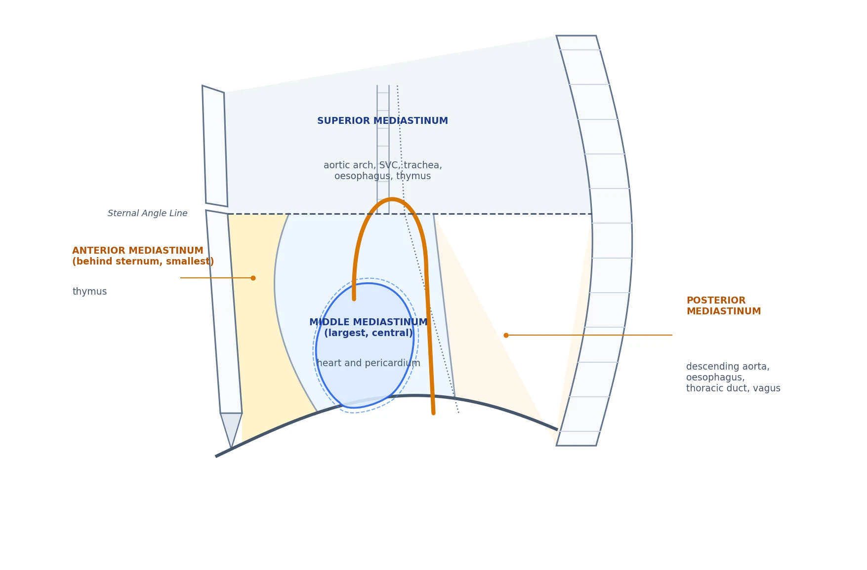
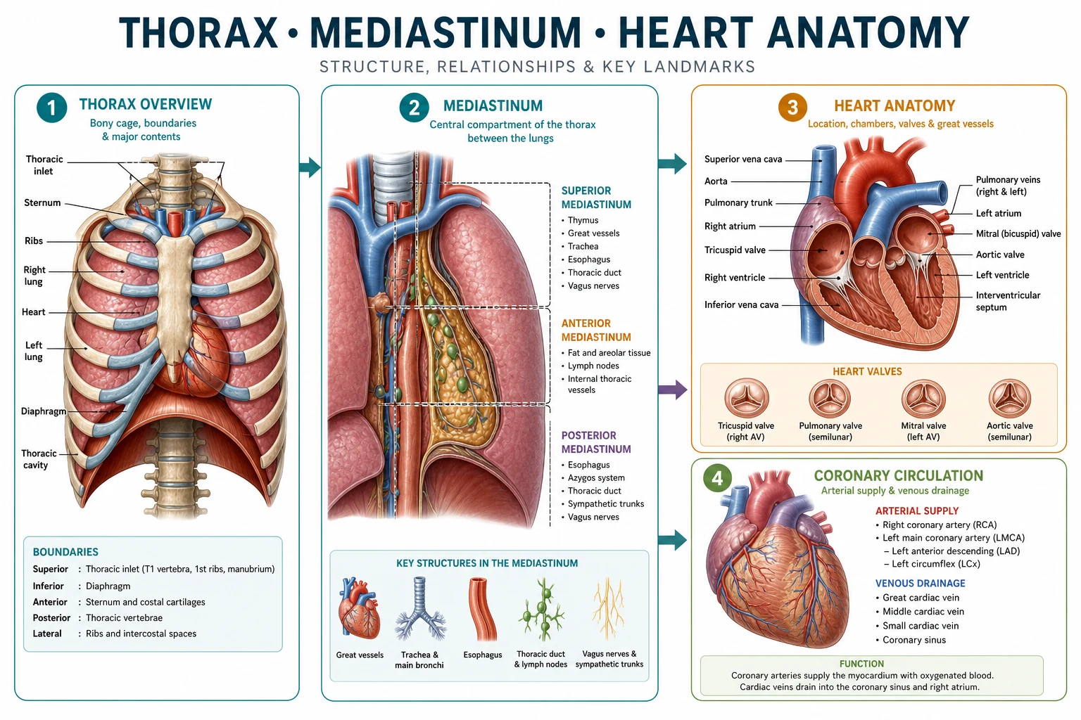
The thorax houses the heart and great vessels, the lungs and the central airway, and is the setting for central venous cannulation, intercostal and paravertebral block, pericardiocentesis and cardiac surgery. The framework rests on six exam-critical ideas. First, the thoracic cage is built of 12 thoracic vertebrae, 12 pairs of ribs and the sternum (manubrium, body and xiphoid); the intercostal space carries the nerve-artery-vein triad in the costal groove in the order V-A-N (vein superior, artery, nerve inferior) running along the lower border of the rib above — the basis for intercostal block and for the rule to needle just above the upper border of the rib below. Second, the mediastinum is the central compartment between the pleural cavities; it is divided into the SUPERIOR mediastinum (above the sternal angle) and the INFERIOR mediastinum, the latter split into ANTERIOR (thymus, behind the sternum), MIDDLE (the heart and pericardium) and POSTERIOR (oesophagus, thoracic aorta, thoracic duct, azygos, vagus). Third, the heart has four chambers (right and left atria, right and left ventricles), an apex (left fifth intercostal space, midclavicular line), three surfaces (anterior/sternocostal, inferior/diaphragmatic, posterior/base) and borders (right atrial, left ventricular), and sits in the middle mediastinum within the pericardium. Fourth, the coronary arteries are the right coronary artery (RCA, supplying the inferior wall, the AV node in 90 percent and usually the SA node), the left anterior descending (LAD, the anterior wall and interventricular septum — the 'widow-maker' territory) and the circumflex (the lateral wall); coronary dominance is determined by which artery supplies the posterior descending artery (the RCA in about 85 percent — right dominant). Fifth, the cardiac conduction system runs from the SA node (high right atrium, the pacemaker) through the atria to the AV node (lower right atrium, near the coronary sinus), then to the bundle of His, its branches and the Purkinje fibres; the SA and AV nodes are both supplied by the RCA in most people, which is why inferior myocardial infarction causes bradycardia and heart block. Sixth, the thoracic duct ascends from the cisterna chyli through the aortic hiatus, crosses from right to left at the T4-T5 level, and empties into the left venous angle — the reason a right internal-jugular line rarely causes chylothorax but can injure the duct, and a left one can. Built on the sternal-healing study (Sarrazin 2026), the mediastinal-imaging study (Dogan 2026), the chylothorax-after-IJV-cannulation report (Alsadeh 2026), the coronary-anastomosis imaging study (Soto Diaz 2026), the sinus-of-Valsalva study (Majadla 2026), the ultrasound intercostal-block study (Yang 2026), the intercostal-cryoanalgesia study (Drake 2026), and the sinoatrial-node report (Tang 2026).
On this page & tools
Your progress
Saved locally on this device.
8 MCQs with explanations
Target exams
Red flags
One-line exam answer
Thoracic anaesthesia anatomy is compartmental: sternal-angle plane at T4/5 organises carina and arch landmarks; middle mediastinum holds the heart; bronchial asymmetry drives tube misplacement; anterior mediastinal masses threaten the airway at induction.[1][2]
Mediastinal compartments
| Compartment | Boundaries | Anaesthetic contents |
|---|---|---|
| Superior | Inlet to sternal angle (T4/5) | Trachea, oesophagus, arch branches, upper SVC, thoracic duct |
| Anterior inferior | Sternum to pericardium | Thymus, fat, nodes — mass effect when supine |
| Middle inferior | Pericardial sac | Heart, ascending aorta, main PA, cava junctions, phrenic nerves |
| Posterior inferior | Pericardium to spine | Descending aorta, oesophagus, azygos, duct, vagus |
Sternal angle / T4–5 plane: tracheal bifurcation, aortic arch start/end, azygos into SVC. [2]
Airway, heart, coronaries, vessels
| Structure | Key feature | Implication |
|---|---|---|
| Right main bronchus | Shorter, wider, more vertical | Endobronchial intubation; foreign body |
| Left main bronchus | Longer, more horizontal | Left DLT common |
| Carina | About T4/5 | ETT depth landmark |
| Phrenic | On pericardium, anterior to roots | Hemidiaphragm palsy if injured |
| Vagus | Posterior to roots | Cardiac and laryngeal branches |
| Left RLN | Loops under aortic arch | Voice/airway after left chest or arch surgery |
| Thoracic duct | To left venous angle | Left neck line chyle risk |
| Coronary dominance | Usually RCA gives PDA | Inferior MI / node blood supply patterns |
Heart surfaces: sternocostal mostly RV; diaphragmatic LV+RV; apex LV. Chambers and valves orient TOE windows through the oesophagus behind the left atrium. [4]
Clinical applications
- One-lung ventilation: bronchial anatomy for DLT/blocker; right upper lobe takeoff near carina complicates right-sided DLT.
- Mediastinal mass: anterior mass + supine + GA can collapse airway or vessels — spontaneous ventilation strategies and rigid readiness.
- Tamponade: pericardial constraint, equalised diastolics, pulsus paradoxus, Beck triad.
- CVC tip: cavoatrial junction in SVC territory.
- Drains/needles: know lung and vessel relations for decompression. [5]
SAQ and viva
Divide mediastinum; compare bronchi; CVC tip anatomy; why anterior mass is dangerous; coronary territories; carina level; left RLN course. [6]
Depth layer — teach it like a tutor
Speak mechanism, then consequence, then action. For anatomy, always add the injury that follows a wrong needle path. For equipment, always add the failure mode and the machine-check step that catches it. For regional topics, always add the endpoint that proves you are in the correct space and the rescue if you are not. [7]
Use medial-to-lateral, superficial-to-deep, or proximal-to-distal order so the examiner hears a map rather than a shopping list. Cross-link to procedures: thorax anatomy to one-lung ventilation and tamponade; neck anatomy to CVC and chylothorax; neuraxial spaces to spinal, epidural and CSE; cranial nerves to awake intubation and oculocardiac reflex; vaporisers to volatile delivery and hypoxic mixture prevention. [8]
Extended viva bank (high-yield stems)
Stem A — definitions under pressure. Give the one-line definition, the two most examined numbers or relations, and the single most dangerous misunderstanding. Keep this under forty-five seconds. [1]
Stem B — mechanism to bedside. Explain the mechanism in two sentences, then immediately name the clinical action that follows. Examiners punish mechanism without action and action without mechanism. [2]
Stem C — compare and choose. Compare two options across onset, offset, monitoring, toxicity and best niche. End with a choice for a stated patient. [3]
Stem D — crisis choreography. Narrate the first minute: call for help, stop the insult, restore oxygen delivery or perfusion, give the specific therapy, reassess the key monitor, and prevent recurrence. [4]
Stem E — special population twist. Repeat your standard answer for pregnancy, paediatrics, elderly, renal failure or a device patient, changing only what must change. [5]
Stem F — equipment or systems failure. Assume the first plan fails. Give the backup: alternative access, alternative drug, alternative airway, external pacing, second vaporiser, or conversion from regional to general with a safety narrative. [6]
SAQ paragraph models
Model opening: Define the topic in one sentence with the key number or equation, then signpost three headings you will cover. [7]
Model middle: Use short paragraphs, each ending with a clinical consequence. Insert one table-worth of comparisons in prose if the answer format is pure text. [8]
Model close: Give hard stops, monitoring, and a one-line pitfall. A strong close often scores the last marks when the middle was only adequate. [1]
Memory anchors
Build memory anchors that regenerate detail rather than store isolated trivia. For physics, anchors are equations and thresholds. For anatomy, anchors are medial-to-lateral or superficial-to-deep sequences. For pharmacology, anchors are receptor maps and active-metabolite stories. For equipment, anchors are safety interlocks and failure modes. If you can regenerate the structure, forgotten minor numbers hurt less. [2]
Theatre checklist language
Convert knowledge into checklists you would actually use: confirm device identity, confirm oxygen analyser, confirm return plate, confirm wire-in-vein, confirm conus-safe interspace, confirm total local anaesthetic dose, confirm ICD therapies on, confirm naloxone and airway plan after neuraxial morphine. Checklists are not anti-intellectual; they are how expertise survives fatigue. [3]
Cross-link map
Almost every thin topic links to another. Fluid flow links to haemorrhage and airway oedema. Electricity links to diathermy and CIED care. Neck anatomy links to CVC complications. Neuraxial spaces link to CSE and caudal. Cranial nerves link to awake intubation and oculocardiac reflex. Vaporisers link to volatile pharmacology and machine check. Adjuncts link to acute pain multimodal pathways. Weak opioids link to pharmacogenomics and paediatric safety bans. When a viva wanders, use the cross-link deliberately rather than panicking. [4]
What “exam-pass learnable” means here
It means a tired candidate can re-read this topic the night before and answer any standard stem without opening another book. It does not mean infinite length. Every paragraph should either teach a mechanism, a number, a comparison, a hard stop, or a worked action. If a sentence does none of those, delete it. If a section lacks a viva stem, add one. If a dose appears, keep a citation nearby. If a claim is clinical, keep a citation nearby. [5]
Final rapid-fire facts to rehearse aloud
Rehearse aloud until the language is automatic: the equation or pathway; the key table; the contraindication list; the first-line crisis action; the monitoring endpoint; the common trap. Spoken fluency is part of viva performance. Silent recognition is not enough. Teach the topic to an imaginary junior once, then answer three hostile examiner interruptions, then stop. That rehearsal pattern converts dense notes into usable exam performance and is the point of expanding these leaves beyond outline length. [6]
Integrated exam drill sheet
Sixty-second version
Say the definition, the critical number or sequence, the main clinical use, and the top red flag. Stop. If you cannot do this without notes, the topic is not yet learnable.
Three-minute version
Add mechanism, a comparison table spoken aloud, one special population, and one crisis stem with first actions. This is the standard viva unit.
Ten-minute mastery version
Add equipment detail or procedural steps, evidence limits, second-line options, and a teach-the-junior summary. This is Final long-case depth.
Written SAQ timing
For a 10-minute SAQ, spend one minute planning headings, seven minutes writing, two minutes checking hard stops and units. Headings should mirror examiner dimensions: definition, mechanism or anatomy, clinical application, complications, special situations.
Common mark-losing behaviours
- Lists without mechanisms
- Mechanisms without clinical action
- Doses without route or monitoring
- Landmarks without injury consequences
- Device talk without re-enable or backup plans
- Absolute claims where practice is protocol-dependent
Positive mark-gaining behaviours
- Numbers with units and approximate ranges
- Explicit assumptions for equations
- Side-by-side comparisons
- Named hard contraindications
- Monitoring endpoints
- Clear escalation
Cross-specialty board alignment
ANZCA Primary and Final, FRCA Primary and Final, ABA, EDAIC and FCAI all test these leaves repeatedly because they are portable across subspecialties. A candidate who owns flow physics, electrical safety, neck and neuraxial anatomy, vaporiser principles and core adjunct pharmacology can survive stems in ICU transfer, obstetric haemorrhage, thoracic lists and outpatient dental anaesthesia alike.
Personal rehearsal script
Read the AnswerCard twice. Cover it and rewrite it from memory. Speak the red flags. Draw one table from memory. Answer one hostile interruption. Then move on. Spaced repetition beats marathon re-reading.
Safety culture close
Every technical topic ends in patient safety: do not expand closed gas spaces, do not dilate arteries, do not leave ICD therapies off, do not apply Poiseuille in turbulence, do not ignore conus level, do not tip a full vaporiser back into service without protocol, and do not stack serotonergic weak opioids casually. Knowledge is only exam-pass when it prevents harm.
Topic-specific mastery addendum
Layered recall sequence
- Recite the AnswerCard from memory.
- Draw the key table (layers, nerves, or vaporiser types) from blank paper.
- Speak two viva stems with full answers.
- List every red flag without looking.
- Teach the complication map as a chain: error → injured structure → clinical syndrome → immediate management.
Procedural narration standard
Narrate as if a consultant is watching: position, asepsis or machine check, landmark or ultrasound view, needle or dial action, endpoint, confirmation test, contingency if endpoint missing, and documentation. This narration style scores in OSCE-like and viva settings because it proves usable competence rather than passive recognition.
Numbers and relations to keep hot
Keep a personal card of the five hottest facts for this leaf and revisit them daily for a week. For neuraxial spaces that means conus level, Tuffier estimate, midline tissue sequence, epidural contents and neonatal difference. For cranial nerves that means V/IX/X airway map, SLN versus RLN, oculocardiac pathway and Horner triad. For vaporisers that means splitting ratio, SVP contrast for desflurane, temperature compensation idea, keyed fillers/interlocks and tip-over danger.
Error museum
Build an "error museum" of classic failures: spinal at L1/2 in adults; dilating an artery; leaving vaporiser tipped in service; missing bilateral RLN injury risk; assuming magnet behaviour is universal; ignoring delayed respiratory depression after neuraxial morphine. Each exhibit should have the false belief, the correct belief, and the protective habit.
Link-forward reading
After mastering this leaf, deliberately link to the next clinical topic in the same sitting: neuraxial anatomy to CSE and caudal; cranial nerves to awake FOI and eye surgery reflexes; vaporisers to volatile agent pharmacology and circle-system low flow. Linked encoding is more durable than isolated topics.
Red flags
Anterior mediastinum
- Thymus/nodes
- Mass airway risk
- Supine danger
- Spontaneous vent strategies
Middle mediastinum
- Heart
- Phrenic on pericardium
- Tamponade space
- TOE targets
Right main bronchus
- Short wide vertical
- Easy endobronchial ETT
- Foreign body path
- RUL near carina
Left main bronchus
- Longer horizontal
- Left DLT common
- Arch relations
- Harder blind entry
References
- [1]Sarrazin BD, et al. Temporal Evolution of Sternal Healing on Chest CT Following Median Sternotomy: A Retrospective Analysis of Patterns of Fat Stranding, Lymphadenopathy, and Callus Formation J Comput Assist Tomogr, 2026.PMID 42335078
- [2]Dogan D, et al. CT-Derived Radiomic Features for the Non-Invasive Differentiation of Mediastinal Lymphadenopathy in Lung Cancer and Sarcoidosis Biomedicines, 2026.PMID 42351755
- [3]Alsadeh G, et al. Right chylothorax following right internal jugular vein cannulation BMJ Case Rep, 2026.PMID 42362338
- [4]Soto Diaz FP, et al. Imaging-Based Diagnosis of a Right Coronary Artery-to-Left Anterior Descending Artery Fistula Associated With a Double Right Coronary Artery Presenting With Angina Cureus, 2026.PMID 42326121
- [5]Majadla S, et al. Unruptured congenital sinus of Valsalva aneurysm causing right ventricular outflow tract obstruction associated with hidden ventricular septal defect: a case report BMC Cardiovasc Disord, 2026.PMID 42350997
- [6]Yang MW, et al. Noninferiority of Ultrasound-Guided Modified Intercostal Block to Traditional Approach for Analgesia After Minimally Invasive Repair of Pectus Excavatum in Children: A Randomized Trial Pain Res Manag, 2026.PMID 41830195
- [7]Drake L, et al. Cryoanalgesia in Thoracic Surgery Innovations (Phila), 2026.PMID 42087707
- [8]Tang E, et al. Anti-NMDA receptor encephalitis associated with severe sinoatrial node dysfunction: three case reports and a review of the literature Neurol Sci, 2026.PMID 42265428