Anaes · Applied anatomy
Vertebral column and neuraxial spaces
Also known as Vertebral column anatomy · Neuraxial anatomy · Epidural space · Subarachnoid space · Ligamentum flavum · Meninges
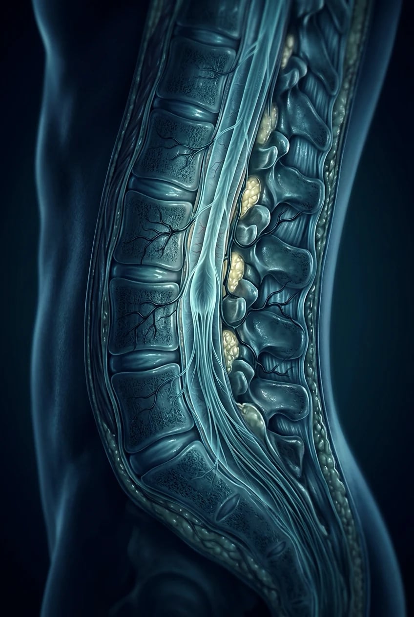
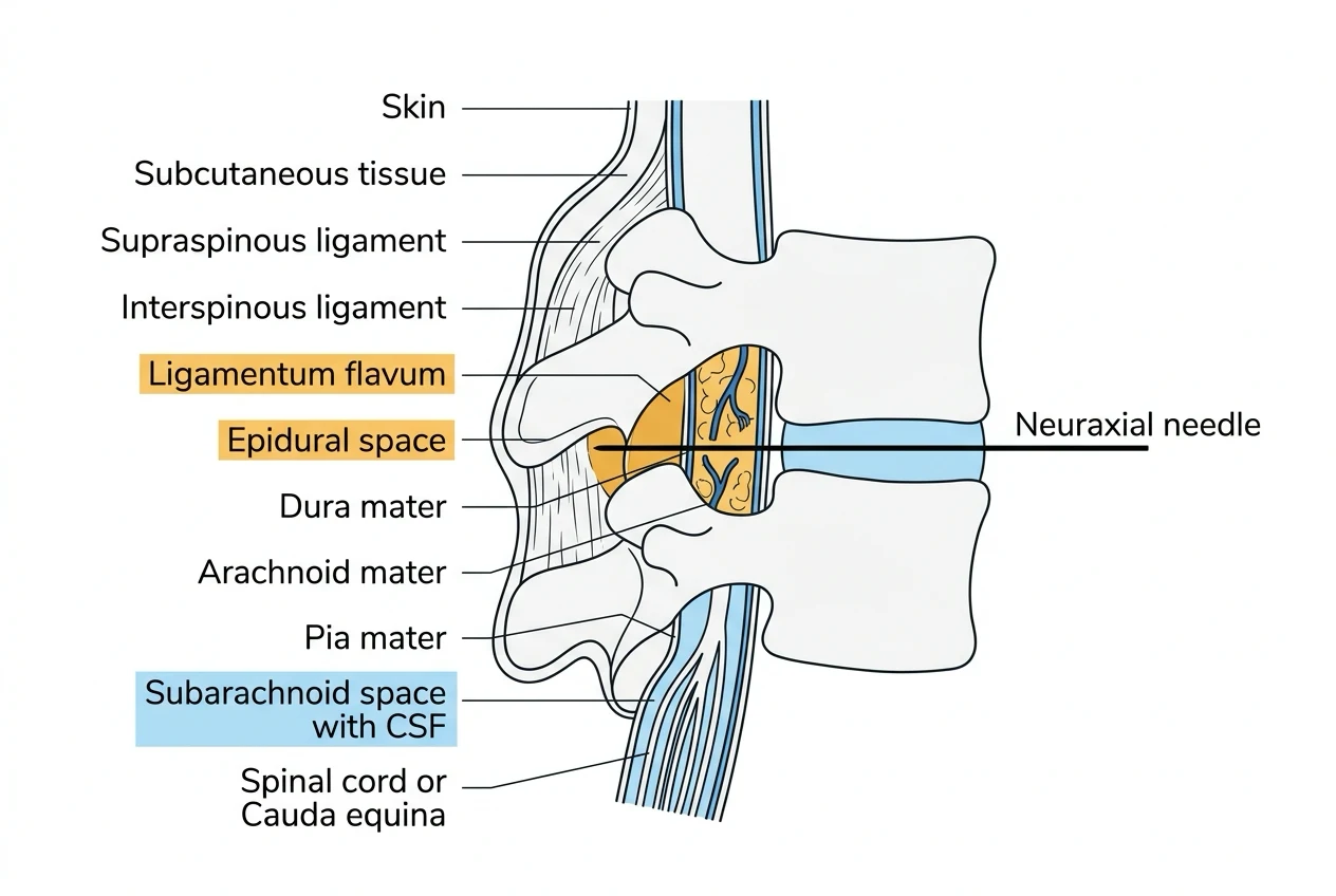
Neuraxial anaesthesia — spinal, epidural and combined spinal-epidural — depends entirely on knowing the layered anatomy from skin to cerebrospinal fluid. The framework rests on six exam-critical ideas. First, the vertebral column is built of 33 vertebrae (7 cervical, 12 thoracic, 5 lumbar, 5 sacral fused, 4 coccygeal) with four curvatures (cervical and lumbar lordotic, thoracic and sacral kyphotic); a typical vertebra has a body anteriorly, a vertebral arch posteriorly forming the vertebral canal, and seven processes (two transverse, four articular, one spinous). Second, the spinal cord is shorter than the vertebral canal: it ends as the conus medullaris at the L1/L2 disc in the adult (lower, around L3, in the neonate), and below it the lumbar and sacral nerve roots form the cauda equina — which is why a lumbar puncture or spinal needle is inserted at or below L3/L4 to avoid the cord. Third, three meningeal layers invest the cord: the tough dura mater outermost, the delicate arachnoid mater in the middle, and the pia mater adherent to the cord; CSF lies in the subarachnoid space between arachnoid and pia. Fourth, the epidural space lies outside the dura and contains fat, lymphatics, the internal vertebral venous plexus (Batson's valveless plexus) and the spinal nerve roots as they exit; its depth from the skin varies with body habitus and is measurable by ultrasound or MRI. Fifth, the ligamentum flavum (yellow ligament) is the tough elastic ligament joining the laminae of adjacent vertebrae and is the key resistance landmark for loss-of-resistance in epidural placement. Sixth, a midline neuraxial needle passes in order through skin, subcutaneous tissue, the supraspinous ligament, the interspinous ligament, the ligamentum flavum (the epidural endpoint), and then — if a spinal is intended — the dura mater and arachnoid mater into the subarachnoid space. Built on the MRI epidural-space-depth study (Alsaati 2026), the ligamentum-flavum study (Gu 2026), the tethered-spinal-cord anaesthetic-management report (Alessi 2026), the loss-of-resistance technique study (Goksu 2026), the inadvertent-dural-puncture report (Greenspon 2026), the ultrasound combined-spinal-epidural study (Sethi 2026), the myodural-bridge meningeal histology study (Rodriguez-Vazquez 2026), and the lumbar-puncture simulation-training study (Lopez-Brotons 2026).
On this page & tools
Your progress
Saved locally on this device.
8 MCQs with explanations
Target exams
Red flags
Why this matters
Spinal, epidural and CSE succeed when the layered anatomy is a mental model, not a hope. Conus level, ligamentum flavum feel, Tuffier's line limits, and neonatal differences are pure Primary gold. [1]
Vertebral column facts
| Region | Count | Curve | Anaesthetic note |
|---|---|---|---|
| Cervical | 7 | Lordosis | High epidural rare; C7 vertebra prominens |
| Thoracic | 12 | Kyphosis | Steep spinous processes → paramedian often easier |
| Lumbar | 5 | Lordosis | Standard spinal/epidural site |
| Sacral | 5 fused | Kyphosis | Caudal hiatus / sacral cornua |
| Coccygeal | 4 | — | Rarely relevant |
Typical vertebra: body + arch (laminae, pedicles) + 7 processes (1 spinous, 2 transverse, 4 articular). Vertebral canal contains cord/meninges/roots/epidural fat and veins. [3]
Cord termination and safe interspace
| Age | Conus level | Safe LP/spinal |
|---|---|---|
| Adult | L1/L2 disc (range T12–L3) | At or below L3/4 |
| Neonate / infant | Around L3 | Lower interspaces; careful technique |
| Tethered cord | Variable low | Imaging / avoid neuraxial if known [3] |
Tuffier's line: line joining highest points of iliac crests crosses L4 spinous process or L4/5 interspace — a surface estimate, not MRI truth. Ultrasound improves interspace identification, especially in obesity and pregnancy [1][6].
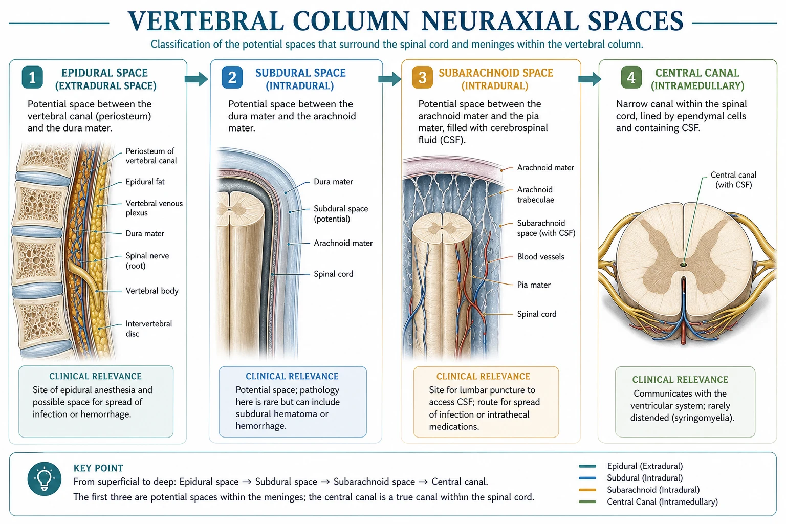
Meninges and spaces
| Layer / space | Content / role |
|---|---|
| Epidural space | Fat, lymphatics, Batson valveless venous plexus, exiting roots — outside dura |
| Dura mater | Tough outer meningeal layer |
| Subdural (potential) | Rare accidental injection plane — patchy block |
| Arachnoid mater | Delicate middle layer |
| Subarachnoid space | CSF — spinal target |
| Pia mater | Adherent to cord |
Midline needle path (commit this order)
- Skin
- Subcutaneous tissue
- Supraspinous ligament
- Interspinous ligament
- Ligamentum flavum → loss of resistance = epidural space
- Dura mater
- Arachnoid mater
- Subarachnoid space (CSF) [5]
Ligamentum flavum is the elastic "yellow ligament" joining laminae — the classic firm resistance then give of LOR to saline/air [2][4]. Inadvertent dural puncture → PDPH risk; air in canal (pneumorrhachis) is rare but reported [5].
Epidural depth and contents
Skin-to-epidural depth varies with habitus (often ≈ 4–6 cm in average adults; wider range in obesity) and correlates imperfectly with anthropometry — ultrasound/MRI refine estimates [1]. Batson's plexus engorges in pregnancy → higher bloody tap risk; avoid intravascular injection with aspiration and test dose discipline.
Surface landmarks
| Landmark | Meaning |
|---|---|
| Tuffier's line | ≈ L4 / L4–5 |
| Vertebra prominens | C7 |
| Inferior scapular angle | ≈ T7 |
| Iliac crest highest point | Tuffier |
| Sacral cornua + hiatus | Caudal entry |
| Intercristal line pitfalls | Often higher than believed in pregnancy |
Paramedian versus midline
Thoracic spinous processes angulate sharply caudad — paramedian approach walks off lamina under the ligamentum flavum more reliably. Lumbar midline is standard; ultrasound can choose midline vs paramedian for CSE [6].
SAQ scaffold
- List midline tissue sequence skin to CSF.
- State adult vs neonatal conus levels with safe interspace.
- Define Tuffier's line and its limitation.
- Contents of epidural space and why veins matter in pregnancy.
- Difference between epidural LOR endpoint and spinal CSF endpoint. [7]
Viva phrases
- "Why not L1/2 spinal in adults?" → "Conus may reach that level — risk of cord injury; use L3/4 or below."
- "What is the ligamentum flavum?" → "Elastic ligament between laminae; LOR landmark for epidural." [8]
Common traps
- Relying on Tuffier's line as ground truth in obese/pregnant patients.
- Confusing epidural fat/veins with subarachnoid CSF.
- Neonatal cord assumed at adult level.
- Ignoring total LA dose and catheter migration risks (clinical, not pure anatomy). [1]
Spinal
- Pierce dura+arachnoid
- CSF confirms
- L3/4 or below adult
- Single-shot or CSE
Epidural
- Stop after flavum LOR
- Catheter possible
- Segmental, volume-dependent
- Test dose / aspiration
Midline
- Standard lumbar
- Through interspinous
- Easier sitting midline feel
- Hard if calcified ligaments
Paramedian
- Thoracic favourite
- Laminar walk-off
- Avoids calcified interspinous
- Different needle angle
Integrated exam drill sheet
Sixty-second version
Say the definition, the critical number or sequence, the main clinical use, and the top red flag. Stop. If you cannot do this without notes, the topic is not yet learnable.
Three-minute version
Add mechanism, a comparison table spoken aloud, one special population, and one crisis stem with first actions. This is the standard viva unit.
Ten-minute mastery version
Add equipment detail or procedural steps, evidence limits, second-line options, and a teach-the-junior summary. This is Final long-case depth.
Written SAQ timing
For a 10-minute SAQ, spend one minute planning headings, seven minutes writing, two minutes checking hard stops and units. Headings should mirror examiner dimensions: definition, mechanism or anatomy, clinical application, complications, special situations.
Common mark-losing behaviours
- Lists without mechanisms
- Mechanisms without clinical action
- Doses without route or monitoring
- Landmarks without injury consequences
- Device talk without re-enable or backup plans
- Absolute claims where practice is protocol-dependent
Positive mark-gaining behaviours
- Numbers with units and approximate ranges
- Explicit assumptions for equations
- Side-by-side comparisons
- Named hard contraindications
- Monitoring endpoints
- Clear escalation
Cross-specialty board alignment
ANZCA Primary and Final, FRCA Primary and Final, ABA, EDAIC and FCAI all test these leaves repeatedly because they are portable across subspecialties. A candidate who owns flow physics, electrical safety, neck and neuraxial anatomy, vaporiser principles and core adjunct pharmacology can survive stems in ICU transfer, obstetric haemorrhage, thoracic lists and outpatient dental anaesthesia alike.
Personal rehearsal script
Read the AnswerCard twice. Cover it and rewrite it from memory. Speak the red flags. Draw one table from memory. Answer one hostile interruption. Then move on. Spaced repetition beats marathon re-reading.
Safety culture close
Every technical topic ends in patient safety: do not expand closed gas spaces, do not dilate arteries, do not leave ICD therapies off, do not apply Poiseuille in turbulence, do not ignore conus level, do not tip a full vaporiser back into service without protocol, and do not stack serotonergic weak opioids casually. Knowledge is only exam-pass when it prevents harm.
Topic-specific mastery addendum
Layered recall sequence
- Recite the AnswerCard from memory.
- Draw the key table (layers, nerves, or vaporiser types) from blank paper.
- Speak two viva stems with full answers.
- List every red flag without looking.
- Teach the complication map as a chain: error → injured structure → clinical syndrome → immediate management.
Procedural narration standard
Narrate as if a consultant is watching: position, asepsis or machine check, landmark or ultrasound view, needle or dial action, endpoint, confirmation test, contingency if endpoint missing, and documentation. This narration style scores in OSCE-like and viva settings because it proves usable competence rather than passive recognition.
Numbers and relations to keep hot
Keep a personal card of the five hottest facts for this leaf and revisit them daily for a week. For neuraxial spaces that means conus level, Tuffier estimate, midline tissue sequence, epidural contents and neonatal difference. For cranial nerves that means V/IX/X airway map, SLN versus RLN, oculocardiac pathway and Horner triad. For vaporisers that means splitting ratio, SVP contrast for desflurane, temperature compensation idea, keyed fillers/interlocks and tip-over danger.
Error museum
Build an "error museum" of classic failures: spinal at L1/2 in adults; dilating an artery; leaving vaporiser tipped in service; missing bilateral RLN injury risk; assuming magnet behaviour is universal; ignoring delayed respiratory depression after neuraxial morphine. Each exhibit should have the false belief, the correct belief, and the protective habit.
Link-forward reading
After mastering this leaf, deliberately link to the next clinical topic in the same sitting: neuraxial anatomy to CSE and caudal; cranial nerves to awake FOI and eye surgery reflexes; vaporisers to volatile agent pharmacology and circle-system low flow. Linked encoding is more durable than isolated topics.
Worked neuraxial stems
Stem — adult spinal for TURP. Choose L3/4 or L4/5 because adult conus usually ends at L1/L2. Confirm free CSF flow, inject, then watch for high block and hypotension. Do not use Tuffier’s line as absolute truth in the obese patient; ultrasound helps when landmarks are poor.[1][6]
Stem — intended epidural becomes wet tap. You have crossed dura. Do not inject the epidural dose into CSF. Options include converting to a deliberate spinal with an appropriate spinal dose, or resiting at another level with meticulous total local anaesthetic accounting and PDPH counselling.[2][5]
Stem — neonatal lumbar puncture. Cord ends lower (around L3), so the safe interspace is lower than adult habit. Position and experience matter; force is not a technique.[3]
Stem — pregnant patient for CSE. Engorged Batson veins raise bloody-tap risk; estimate of interspace by Tuffier may be high; lateral or sitting position must protect aortocaval flow after block. Ultrasound can refine level and midline.[1][6]
Tissue sequence chant: skin, fat, supraspinous, interspinous, ligamentum flavum (epidural LOR), dura, arachnoid, CSF. If you can chant it under stress, you can narrate both spinal and epidural endpoints. [2]
Red flags
References
- [1]Alsaati I, et al. MRI-Based Evaluation of Lumbar Epidural Space Depth and Its Correlation with Anthropometric Factors in Saudi Adults Tomography, 2026.PMID 42042941
- [2]Gu C, et al. Full Endoscopic Piecemeal Resection of Lumbar Ligamentum Flavum Cyst With Uniaxial Large Working Channel Spinal Endoscope: Technical Report and Case Series Orthop Surg, 2026.PMID 42062771
- [3]Alessi L, et al. Anesthetic Management in a Patient With Tethered Spinal Cord Syndrome Undergoing Knee Replacement Surgery Case Rep Anesthesiol, 2026.PMID 42339046
- [4]Goksu H, et al. Evaluation of loss of resistance technique using an air-filled injector to enhance accuracy of landmark-guided knee joint injections Turk J Phys Med Rehabil, 2026.PMID 42291381
- [5]Greenspon NH, et al. Pneumorrhachis After Inadvertent Dural Puncture as a Cause of Severe Neck Pain Limiting Labor Participation and Necessitating Operative Vaginal Delivery: A Case Report A A Pract, 2026.PMID 42257607
- [6]Sethi D, et al. Ultrasound-guided midline versus paramedian approach for combined spinal-epidural anesthesia: A randomized controlled study J Anaesthesiol Clin Pharmacol, 2026.PMID 42088167
- [7]Rodriguez-Vazquez JF, et al. Morphogenesis of Myodural Bridges: A Histological Study in Human Fetuses Cells Tissues Organs, 2026.PMID 41166506
- [8]Lopez-Brotons M, et al. Evaluation of a peer-assisted, simulation-based clinical skills training program in Spain: a prospective single-group before-and-after study J Educ Eval Health Prof, 2026.PMID 42272202