Anaes · Airway management
Airway assessment: the LEMON tool, Mallampati classification & prediction of difficult direct laryngoscopy
Also known as LEMON assessment · Mallampati classification · Mallampati score · Cormack-Lehane grade · Difficult airway prediction · 3-3-2 rule · Wilson score · El-Ganzouri index
Pre-operative airway assessment is the single bedside exercise most consistently examined in the airway viva and short-answer question, because it is the one step that converts an unanticipated crisis into a planned event. The framework rests on four ideas: a small set of anatomical axes — mouth opening, mandibular space, laryngeal position, neck mobility — determines whether a direct line of sight from the incisors to the glottis can be made; the LEMON tool (Look externally, Evaluate the 3-3-2, Mallampati, Obstruction, Neck mobility) packages those axes into a reproducible screen; the Mallampati classification grades the oropharyngeal view and correlates, imperfectly, with the Cormack-Lehane laryngoscopic grade that defines a difficult intubation; and the honest limitation — individual bedside tests have poor positive predictive value, so combined multi-factor scores are used and the assessment exists to inform preparation, not to guarantee prediction. Anchored to contemporary evidence spanning syndromic and physiological difficult airways, the optimisation of video-laryngoscopic intubating conditions, the bronchoscopic management of dynamic airway obstruction, and the airway compromise of anaphylaxis.
On this page & tools
Your progress
Saved locally on this device.
8 MCQs with explanations
Target exams
Red flags

Overview & definition
Airway assessment is the structured bedside evaluation performed before every induction of anaesthesia, the aim of which is to predict the difficult direct laryngoscopy and the difficult mask ventilation before they are encountered, and so to convert a potential crisis into a planned event. It is the most consistently examined element of the airway viva and short-answer question because it is cheap, reproducible, and the one step whose omission is most clearly linked to harm in the national audits. [1]
The exercise turns on a single anatomical fact: direct laryngoscopy requires a straight line of sight from the upper incisors, over the base of the tongue, around the curve of the pharynx, to the glottic opening. Whether that line can be made straight is determined by a small set of distances and ranges — the mouth opening, the mandibular space (the room into which the tongue can be displaced), the position of the larynx relative to the base of the tongue, and the mobility of the cervical spine. A receding mandible, a high anterior larynx, a restricted mouth opening, or a stiff neck each force the glottis out of the direct line of sight and predict a poor laryngoscopic view[1].
The standard assessment packages these anatomical axes into a memorable screen — the LEMON tool — and grades the oropharyngeal view with the Mallampati classification. The outcome that the assessment tries to predict is the Cormack-Lehane grade seen at direct laryngoscopy: a grade 3 or 4 view (epiglottis only, or epiglottis not visible) is the operational definition of the difficult intubation. The honest position, examined below, is that no single bedside test predicts this outcome with certainty; the assessment exists to inform preparation, not to replace vigilance[1][3].
The LEMON assessment tool
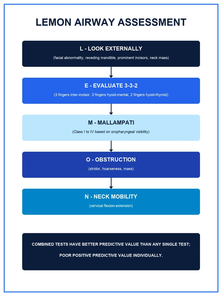
The LEMON tool is the most widely taught bedside screen for the prediction of difficult direct laryngoscopy, originally described in the emergency-medicine literature and adopted across anaesthesia, emergency and critical-care practice. Each letter names one axis of difficulty, and the presence of two or more predicts a higher probability of a poor laryngoscopic view[2].
- L — Look externally. Inspect the face and neck for the features that experience associates with difficulty: facial trauma or congenital abnormality, a receding (retrognathic) mandible, prominent ("buck") upper incisors, a short muscular neck, a neck mass, oedema, beard (which interferes with the mask seal), and the syndromic facies of conditions such as Pierre Robin sequence, Treacher Collins, Down syndrome, or spinal muscular atrophy[1][2].
- E — Evaluate the 3-3-2 rule. Three measured distances, in finger-breadths, that map the line of sight (detailed below).
- M — Mallampati score. The oropharyngeal view, class I to IV (detailed below).
- O — Obstruction. Signs of upper-airway obstruction — stridor, hoarseness, a muffled voice, drooling, a neck mass, or infection (peritonsillar or retropharyngeal abscess, Ludwig angina)[4][6].
- N — Neck mobility. The range of cervical flexion and extension, reduced by arthritis, cervical spine injury, previous surgery or radiotherapy, or the rigid collar.
The tool is a screen, not a score: it does not produce a single number, and its strength is that it forces the operator to consider the anatomical, obstructive and mobility axes together before induction. Its weakness, shared with every bedside test, is a high false-positive rate — it flags many airways as potentially difficult that prove easy at laryngoscopy[2].
L — Look externally
The external inspection is the least quantified and the most experience-dependent element of the assessment, but it captures the patients the bedside tests miss. The operator inspects the face and neck for the features that distort the line of sight or the mask seal. [1]
- Mandibular recession (retrognathia) — a receding chin shortens the thyromental distance and crowds the tongue into the pharynx, the central anatomical cause of the difficult anterior larynx.
- Prominent upper incisors ("buck teeth") — obstruct the blade and force a levering, damaging technique.
- Facial trauma, oedema, or congenital abnormality — distort the anatomy and bleed easily.
- A beard — defeats the mask seal and predicts difficult mask ventilation as much as difficult intubation.
- A short, thick, muscular neck and a neck mass — predict both a poor view and difficult mask ventilation.
- Syndromic and neuromuscular features — the congenital craniofacial syndromes (Pierre Robin sequence, Treacher Collins, Goldenhar, Down syndrome) and the neuromuscular disorders (spinal muscular atrophy, muscular dystrophy) carry a high incidence of difficult intubation that the conventional distances under-represent[1].
The teaching point is that the external look is the screen for the airway that is anatomically difficult for reasons the tape-measure cannot capture. A patient who looks difficult should be treated as difficult even when the Mallampati score is low[1][2].
E — Evaluate the 3-3-2 rule
The 3-3-2 rule measures the three distances that together determine whether a straight line of sight from the incisors to the glottis is geometrically possible. Each is expressed in finger-breadths of the patient's own hand, which scales the measurement to the patient. [1]
- 3 — Mouth opening (inter-incisor distance). The patient should open the mouth to the width of three of their own fingers laid sideways. An inter-incisor distance of less than three finger-breadths (roughly under 4 cm) predicts difficult laryngoscopy, because the blade and the tube cannot be introduced through a narrow gap.
- 3 — Hyoid-to-mental distance (the mandibular space). The distance from the hyoid bone to the mentum (the tip of the chin) should accommodate three fingers. This measures the mandibular space — the room into which the tongue can be displaced by the laryngoscope blade. A short hyomental distance (under three fingers, or under 6 cm) means the tongue has nowhere to go and obstructs the line of sight.
- 2 — Hyoid-to-thyroid distance (laryngeal position). The distance from the hyoid to the thyroid notch should be two fingers. This measures the position of the larynx — a high, anterior larynx (a distance under two fingers) is the classic anatomical cause of the difficult anterior glottis, because the larynx sits out of the direct line of sight. [1]
The rule is anatomically literate: it asks whether there is room to introduce the blade (3), room to displace the tongue (3), and a larynx that lies within the line of sight (2). A failure at any of the three predicts difficulty, and the rule is the most quantitatively taught element of the LEMON screen[2].
M — Mallampati classification
The Mallampati classification grades the view of the oropharynx with the mouth open and the tongue protruded, and it is the single bedside test most commonly examined. It is performed with the patient sitting upright, the mouth open wide, and the tongue protruded without phonation (saying "aah" falsely improves the view by elevating the soft palate). The class is determined by which posterior structures are visible. [1]
- Class I — the soft palate, fauces, uvula and pillars are all visible.
- Class II — the soft palate, fauces and uvula are visible, but the uvula is partly obscured by the base of the tongue.
- Class III — the soft palate and the base of the uvula only are visible; the pillars and the fauces are obscured.
- Class IV — the soft palate only is visible; the uvula is wholly obscured. [1]
The grade correlates, imperfectly, with the Cormack-Lehane laryngoscopic view: a Mallampati class III or IV predicts a Cormack-Lehane grade III or IV view (the difficult intubation). The correlation is anatomical — both measure how much the tongue obscures the posterior pharyngeal structures — but it is loose, because the Mallampati view is obtained awake and upright with an active tongue, while the Cormack-Lehane view is obtained in the anaesthetised, supine, paralysed patient with the tongue displaced by a blade. A modified Mallampati, performed without phonation in the supine position, is often used in the pre-operative clinic[1].
The central teaching, returned to below, is that the Mallampati score in isolation has a high false-positive rate: the majority of class III airways prove easy to intubate, and a substantial minority of difficult intubations occur in patients with a low Mallampati class. It is a screen, not a verdict[1][3].
O — Obstruction
The O of LEMON asks whether there is an obstructing lesion anywhere between the lips and the carina, and it is the element most often under-weighted by the candidate who recites the mnemonic without thinking clinically. Signs of upper-airway obstruction — stridor, a muffled ("hot-potato") voice, hoarseness, drooling, and trismus — mark a threatened airway that may collapse completely with the loss of tone on induction, and they change the plan from a rapid sequence induction to an awake fibreoptic intubation[2].
The causes span the intrinsic and the extrinsic, the acute and the chronic: [1]
- Infection — peritonsillar abscess, retropharyngeal abscess, and Ludwig angina distort and narrow the pharynx, and the tissues bleed and distort further under the blade.
- Tumour and mass — a pharyngeal or laryngeal tumour, a large thyroid goitre, or a neck haematoma (after surgery or trauma) compress or distort the airway.
- Oedema — anaphylaxis, angio-oedema, burns, and post-radiation oedema narrow the lumen; the airway compromise of Kounis syndrome (the acute coronary syndrome of an allergic reaction) is an example of how rapidly an allergic insult can threaten the airway[6].
- Dynamic obstruction — tracheobronchomalacia, in which the tracheal and bronchial walls are weak and collapse on expiration, is a cause of dynamic, position-dependent obstruction that is managed by bronchoscopic and interventional techniques rather than by a standard intubation, and it illustrates how obstruction is not always a fixed stenosis[4].
- Foreign body — an inhaled object that partially obstructs the larynx or trachea.
The practical point is that obstruction is the one axis of LEMON that should rarely be managed by a blind rapid sequence induction: a patient with stridor at rest has a critical airway, and the technique of choice is usually an awake topically-anaesthetised fibreoptic intubation, with a surgical airway plan agreed and prepared[2][6].
N — Neck mobility
The final letter measures the range of cervical flexion and extension, because the "sniffing the morning air" position — flexion of the lower cervical spine and extension at the atlanto-occipital joint — is the position that aligns the oral, pharyngeal and laryngeal axes into the single line of sight. A neck that cannot be positioned cannot provide that line, however favourable the other distances. [1]
The causes of reduced neck mobility are the common and the surgical: [1]
- Cervical spine arthritis (osteoarthritis, rheumatoid arthritis, ankylosing spondylitis) — reduces the atlanto-occipital extension most critically; rheumatoid disease also threatens the unstable atlanto-axial joint.
- Cervical spine injury — the patient in a rigid collar or on a spinal board cannot be positioned, and manual in-line stabilisation is required; these patients are the classical indication for an awake or a video-laryngoscopic technique.
- Previous cervical spine surgery — fusion reduces the range and the "give" of the tissues.
- Radiotherapy and scarring — the irradiated or scarred neck is immobile and indurated.
- Obesity and the short muscular neck — limit extension practically, even when the joints are normal. [1]
Neck mobility is the element most often compromised in the emergency and trauma context, and it is the axis that most often forces the move to a video laryngoscope or an awake technique when the other distances are favourable[2].
The Cormack-Lehane grade: the outcome being predicted
Every bedside airway test is, ultimately, trying to predict the view that will be obtained at direct laryngoscopy, and that view is graded by the Cormack-Lehane classification — the four-point scale described by Cormack and Lehane in 1984 and the operational definition of the difficult intubation. [1]
- Grade I — most of the glottis is visible.
- Grade II — the posterior portion of the glottis, including the arytenoid cartilages, is visible; the anterior glottis is not.
- Grade III — only the epiglottis is visible; no part of the glottis can be seen.
- Grade IV — neither the glottis nor the epiglottis is visible; only the soft palate. [1]
A grade III or IV view is the definition of the difficult direct laryngoscopy. Grade I and II are "easy", grade III and IV are "difficult", and the boundary is the line an examiner draws. A modified three-point scale (the percentage-of-glottic-opening, or POGO, score) refines the grade II to III boundary, but the four-point Cormack-Lehane remains the universal language[3].
The link to the assessment is direct: the Mallampati class III or IV predicts the Cormack-Lehane grade III or IV, and the other LEMON axes predict the same view by different anatomical routes. The grade is the outcome because it predicts both the difficulty of passing the tube and the risk of hypoxaemia, oesophageal intubation and airway trauma on repeated attempts[3].
Additional predictors and the multi-factor scores
Beyond the LEMON screen, a set of measurements and multi-factor scores refine the prediction by combining tests. The consistent finding across them is that combination improves on any single test. [1]
- Thyromental distance — the distance from the thyroid notch to the mentum with the neck extended; under 6.5 cm (under three finger-breadths) is abnormal and predicts difficulty. It is the same axis as the second and third measurements of the 3-3-2 rule.
- Neck circumference — measured at the level of the thyroid cartilage; a circumference above 43 cm increases the risk of both difficult laryngoscopy and difficult mask ventilation, and it is the single best predictor of difficulty in obesity.
- Sternomental distance — from the sternal notch to the mentum with the neck extended; under 12.5 cm is abnormal.
- Inter-incisor distance — mouth opening under 4 cm (under three fingers) predicts difficulty.
- Upper lip bite test — the patient bites the upper lip; class III (cannot reach the vermillion border with the lower incisors) correlates with a difficult intubation and adds a functional axis the static measurements miss.
- Wilson score — a five-component, weighted score (weight, head and neck movement, mouth opening, receding mandible, "buck teeth") that sums to a probability of difficulty; it was the original multi-factor index.
- El-Ganzouri multi-factor risk index — combines seven variables into a single score, and it outperforms any single test because it integrates the anatomical, the obstructive and the mobility axes.
- MACOCHA score — the validated tool for the predicted-difficult intubation in the intensive care unit, adding the physiological axes (apnoea, hypoxia, coma) that the elective assessment misses. [1]
The principle is that no single test is sufficient, and the multi-factor scores exist precisely because combining tests improves the positive predictive value. The El-Ganzouri and Wilson scores are the exam-standard examples of combination, and a candidate is expected to know the components rather than only the name[1][2].
Limitations: the poor positive predictive value of bedside tests
The honest, examined position on airway assessment is that every individual bedside test has a poor positive predictive value and a high false-positive rate. A high Mallampati class flags many airways that prove easy at laryngoscopy; a short thyromental distance does the same; and the majority of "predicted-difficult" patients are intubated without difficulty. The tests are sensitive enough to be worth performing and specific enough that a normal result is reassuring, but they are not accurate enough to predict the difficult airway with certainty[1].
The implications shape the whole subject. First, combination improves prediction — the multi-factor scores (Wilson, El-Ganzouri, MACOCHA) outperform any single test, and the LEMON tool itself is a combination. Second, a substantial proportion of difficult airways remain unanticipated — they present for the first time at induction, in a patient whose assessment was normal, and this is the irreducible residual that the Difficult Airway Society algorithm exists to rescue. The unanticipated difficult airway remains a significant cause of anaesthesia-related morbidity and a recurring theme of the national audits[1][3].
Third, the difficulty that is encountered is sometimes not anatomical at all but physiological. The physiological difficult airway — the patient who is hypoxaemic, hypotensive, acidotic, or in shock from any cause — is dangerous independently of the geometry, and the same patient who is anatomically easy can be physiologically impossible: the safe apnoea period is short, the cardiovascular reserve is gone, and the intubation must be both technically successful and haemodynamically kind. The physiological difficult airway is increasingly recognised in the emergency and critical-care literature as a distinct entity that the anatomical LEMON screen does not capture, and it demands resuscitation before, during and after the intubation[2].
The teaching, examined below, is therefore that the assessment informs preparation but does not abolish risk. Even with the best available video-laryngoscopic equipment and optimal intubating conditions, a proportion of intubations remain difficult, and the prepared operator expects it[3].
How assessment informs preparation
The purpose of the assessment is not to predict with certainty but to inform preparation — to determine, before induction, the equipment, the technique, the team and the threshold for senior help that the predicted degree of difficulty warrants. A structured assessment converts an unanticipated crisis into a planned event, and the plan is built from four components[2].
- Equipment. A predicted difficult airway warrants a video laryngoscope (the standard first-line for the anticipated difficult direct laryngoscopy), a range of alternative blades, a bougie or stylet, a second-generation supraglottic airway device, and a front-of-neck access kit, all prepared and checked before induction. The optimisation of the intubating conditions — full neuromuscular blockade, optimal position, external laryngeal manipulation — is itself a determinant of success[3].
- Technique. The predicted difficult airway, especially with obstruction or limited neck mobility, is often best managed by an awake fibreoptic intubation — securing the airway before the loss of tone — rather than by a rapid sequence induction. The choice of induction agent and the depth of sedation affect the conditions; remimazolam, for example, has been compared with propofol for the conditions it provides, and the principle is that a planned, unhurried technique with good conditions outperforms a rushed one[5].
- Team readiness. A predicted difficult airway warrants a senior anaesthetist present from the outset, a trained assistant, and the roles for the difficult-airway algorithm allocated before induction. The single most consistent finding of the airway audits is that senior help called early changes the outcome; the early call is the action most reliably within the operator's control[2].
- A plan for failure. Every induction that flags a difficult airway on assessment should begin with a rehearsed plan for the failed intubation — the Difficult Airway Society ladder of Plans A through D — agreed with the team before the first drug is given[2].
The assessment, in short, is the trigger for the preparation, and the preparation is what prevents the spiral from a difficult intubation into an airway disaster. A reassuring assessment is never a reason to skip the preparation: the disciplined operator behaves as though every induction may be difficult, because a proportion of them will be[2][3].
Special contexts: the predicted-difficult and the physiological difficult airway
Two contexts deserve separate mention because they are common, disproportionately dangerous, and reshape the assessment. [1]
In the predicted-difficult airway — the syndromic, the post-radiation, the obstructed, the known cervical-spine-injured — the assessment is positive on multiple axes, and the plan is an awake technique with a surgical airway plan agreed. The syndromic and neuromuscular disorders, exemplified by series such as spinal muscular atrophy, carry a high incidence of difficult intubation that combines anatomical distortion with physiological fragility, and they are the patients in whom an under-prepared induction is most likely to end in harm[1].
In the physiological difficult airway, seen most often in the emergency department and the intensive care unit, the anatomy may be favourable but the physiology is hostile: hypoxaemia shortens the safe apnoea period, hypotension and shock remove the cardiovascular reserve that induction erodes, and the patient may be agitated, combative, or unable to cooperate with the bedside tests. The assessment here must extend beyond LEMON to the MACOCHA axes, and the management must interleave resuscitation with the intubation — preoxygenation, vasopressor readiness, and an unhurried best attempt[2]. The recognition of the physiological difficult airway as a distinct entity is the major conceptual advance of the recent emergency and critical-care airway literature, and it is the element most often missing from the candidate who recites LEMON mechanically[2].
Clinical
- Standard approach
- Evidence-based
Alternative
- Modified technique
- Risk-benefit
Red flags
[1] [1] [1] [1] [1]References
- [1]Black KM, et al. Anesthesia Care, Complications, and Airway Management for Patients With Spinal Muscular Atrophy: A Retrospective Chart Review From a Quaternary Children's Hospital Anesth Analg, 2026.PMID 42363899
- [2]Ghaffar S, et al. Physiological difficult airway management in the emergency department J Pak Med Assoc, 2026.PMID 42363338
- [3]Ipsen EO, et al. Remifentanil Versus Rocuronium for Optimising Video Laryngoscopy Assisted Tracheal Intubation-The ROCVIDEO Trial Protocol Acta Anaesthesiol Scand, 2026.PMID 42304626
- [4]Schramm D, et al. Bronchoscopic and interventional management of tracheobronchomalacia in children with bronchopulmonary dysplasia: a review of evidence Paediatr Respir Rev, 2026.PMID 42364941
- [5]Shionoya M, et al. Comparison of the Effects of Remimazolam and Propofol, With Epinephrine-containing Lidocaine, on Rocuronium-induced Muscle Relaxation Anesth Prog, 2026.PMID 42307548
- [6]Baba T, et al. A Case of Clinically Suspected Kounis Syndrome Associated with Platelet Transfusion during Thoracoscopic Right Lower Lobectomy Surg Case Rep, 2026.PMID 42359366