Anaes · Airway management
The anticipated difficult airway and the awake fibreoptic intubation
Also known as Awake fibreoptic intubation · AFOI · Awake intubation · Anticipated difficult airway · Topicalisation of the airway · Awake video laryngoscopy · Conscious intubation
The anticipated difficult airway is the airway that the preoperative assessment — the LEMON tool, the Mallampati class, the multi-factor scores — has flagged as likely to defeat direct or video laryngoscopy before induction is ever attempted. When the assessment is positive, the single safest strategy is usually to secure the airway while the patient is still awake, breathing, and protecting their own reflexes: the awake fibreoptic intubation (AFOI), the long-standing gold standard. The technique rests on four ideas examined here: the indication — predicted difficult laryngoscopy, aspiration risk where a rapid sequence is also unsafe, and the critically ill patient who cannot tolerate apnoea; the principle that the patient must remain cooperative and breathing throughout, so the pharmacology is built around topical anaesthesia and dexmedetomidine sedation rather than apnoea and paralysis; the six-step sequence of preparation, topicalisation, sedation, scope insertion, railroading of the tube, and confirmation before induction; and the honest framing of the alternatives — the awake video laryngoscope is faster and increasingly preferred, and in selected cases the airway can be avoided entirely with a regional technique. Anchored to contemporary evidence on the physiological difficult airway, the syndromic and neuromuscular difficult airway, the cerebral oxygenation and haemodynamic monitoring of the sedated patient, and the preoxygenation and oxygenation that make the awake technique safe.
On this page & tools
Your progress
Saved locally on this device.
8 MCQs with explanations
Target exams
Red flags
Overview & definition
The anticipated difficult airway is the airway that the preoperative assessment has flagged, before any drug is given, as likely to defeat direct or video laryngoscopy. It is the predicted difficult airway, and its management is the single largest subject in the airway viva because the whole point of the assessment is to convert an unanticipated crisis into a planned event. Once the assessment is positive, the central decision is not which blade to use but whether to induce general anaesthesia at all before the airway is secured[5].
The safest answer, in most anticipated difficult airways, is to secure the airway while the patient is still awake, spontaneously breathing, and protecting their own reflexes — the awake intubation. Of the awake techniques, the awake fibreoptic intubation (AFOI) has been the long-standing gold standard: a flexible bronchoscope carrying a preloaded endotracheal tube is passed through the nose or the mouth under topical anaesthesia and conscious sedation, and guided through the vocal cords under direct vision[2][5]. The principle that governs every step is that the patient must remain cooperative and breathing throughout — the pharmacology is therefore built around topical local anaesthesia and arousable sedation, never around apnoea and paralysis.
The honest position, examined below, is that AFOI is no longer the only awake option. The awake video laryngoscope is faster, requires less topicalisation, and is increasingly preferred where the difficulty is anatomical rather than obstructing; in selected patients the airway can be avoided altogether with a regional technique; and the AFOI retains the gold-standard role where the airway is distorted, obstructed, or inaccessible to a rigid blade[5].
The anticipated difficult airway: the decision to stay awake
The decision to perform an awake intubation rests on a simple risk judgement: in this patient, on this assessment, is the probability of failed direct or video laryngoscopy high enough that the loss of tone and the apnoea of induction are unacceptable? When the answer is yes, the airway must be secured before induction, and the awake technique is the means[1][5].
The anticipated difficult airway is identified at the bedside by the LEMON tool, the Mallampati class, and the multi-factor scores (Wilson, El-Ganzouri). A Mallampati class of III or IV, a limited mouth opening of fewer than three finger-breadths, a retrognathic mandible with a short thyromental distance, a high anterior larynx, restricted cervical mobility, a neck mass or radiation change, or a known history of difficult intubation each raise the probability of a poor laryngoscopic view; combined across several axes they convert an isolated risk factor into a planned awake technique[2].
The decision is reinforced by three features the anatomical assessment does not capture but the modern teaching insists on. First, the aspiration risk in whom a rapid sequence induction is also contraindicated — the patient with a full stomach and a known difficult airway, in whom the two hazards compound. Second, the critically ill patient who cannot tolerate apnoea — the hypoxaemic, the shocked, the acidotic — in whom the safe apnoea period after induction is measured in seconds, and in whom the preservation of spontaneous ventilation through an awake technique buys time that an apnoeic induction cannot[1]. Third, the patient in whom a definitive awake airway assessment is required before surgery, because the predicted difficulty is uncertain and the operator needs to see the cords before committing[2][5].
Indications for awake intubation
The indications group into four families, each examined in the viva as a separate reason to stay awake[1][2][5].
- Predicted difficult direct or video laryngoscopy. A Mallampati class of III or IV, a mouth opening of fewer than three fingers, a retrognathic mandible, a short thyromental distance, a high anterior larynx, restricted neck mobility, or a previous difficult intubation documented in the record. The syndromic and neuromuscular disorders — Pierre Robin sequence, Treacher Collins, Down syndrome, and the neuromuscular disorders exemplified by spinal muscular atrophy — combine anatomical distortion with physiological fragility and carry a high incidence of difficult intubation that warrants an awake plan from the outset[2].
- Anticipated difficult mask ventilation. The bearded, edentulous, obese, or stiff-lung patient in whom the mask seal or the ventilation will fail, and in whom the loss of the airway on induction is therefore unrecoverable. The rule is that difficult mask ventilation compounds difficult intubation, and the combination is an awake indication.
- Aspiration risk in whom a rapid sequence is contraindicated. The patient with a full stomach, a bowel obstruction, or delayed gastric emptying who also has a difficult airway — the rapid sequence that would protect from aspiration is itself unsafe because the airway cannot be rescued if it fails, so the airway is secured awake with the cuff inflated before induction[5].
- The critically ill patient who cannot tolerate apnoea. The hypoxaemic, the shocked, the acidotic patient in whom the safe apnoea period is exhausted before the tube is placed, and in whom the preservation of spontaneous ventilation through an awake technique maintains oxygenation while the airway is secured[1].
The unifying principle is that the awake technique is chosen when the risk of inducing anaesthesia before the airway is secured exceeds the risk of securing it awake. In every other case — a normal assessment, a reassuring second attempt, a rescue device available — induction followed by laryngoscopy remains the default[1][5].
Awake fibreoptic intubation: the gold standard
Of the awake techniques, the awake fibreoptic intubation is the long-standing gold standard for the anticipated difficult airway, and the technique against which the alternatives are still measured. A flexible bronchoscope with a preloaded endotracheal tube is passed through the nose or the mouth under topical anaesthesia, the airway anatomy is identified under direct vision, the scope is advanced through the vocal cords into the trachea, and the tube is railroaded over it[2][5].
Its strengths are the qualities that a rigid blade cannot offer. It is flexible, so it navigates a distorted, irradiated, or oedematous airway that a rigid blade cannot reach. It is performed under direct vision, so the operator sees the cords, confirms the tracheal rings and the carina, and excludes an oesophageal or endobronchial placement before the tube is railroaded. It tolerates a limited mouth opening, a fixed cervical spine, and an anterior larynx that defeat direct laryngoscopy, because it does not require a straight line of sight. And it leaves the patient awake, breathing, and protecting their airway until the tube is confirmed[2][5].
Its weaknesses are the time it takes, the dependence on operator skill, the need for a cooperative patient and a still field, and the difficulty of performing it in the presence of blood, secretions, or an obstructing mass that blocks the view. These weaknesses are precisely those addressed by the awake video laryngoscope, examined below as the leading alternative[5].
Step 1: preparation and premedication
The awake technique succeeds or fails on the preparation, and the disciplined preparation is the same six-step sequence that the viva expects recited in order[2][5].
The first step is preparation. The operator explains the procedure to the patient in plain terms, obtains consent, and establishes that the patient can cooperate — cooperation is a prerequisite, not a variable. The patient is positioned sitting upright or semi-recumbent, which offsets the tongue, drains secretions, and preserves the functional residual capacity that keeps the patient oxygenated during the procedure. An anti-sialogogue is given to dry the mucosa, because secretions obscure the lens and wash the topical local anaesthetic away; glycopyrrolate is the agent of choice because it does not cross the blood-brain barrier and causes less sedation and tachycardia than atropine. Standard monitoring is applied — electrocardiography, non-invasive blood pressure, pulse oximetry, and capnography — and supplemental oxygen is delivered by nasal specs or a modified face mask throughout[5].
The preoperative assessment is reviewed and confirmed at this point: the difficulty, the route (nasal or oral), the back-up plan (an awake video laryngoscope, a supraglottic device, a surgical airway), and the senior help. The single most consistent finding of the airway audits is that senior help called early changes the outcome, and the anticipated difficult airway is the case in which the senior operator is present from the outset[5].
Step 2: topicalisation of the airway
The second step is topicalisation — the anaesthesia of the mucosa from the nose or the mouth to the vocal cords, which is the whole pharmacological basis of the awake technique. The airway is richly innervated and intensely reflexive, and a properly topicalised airway is the difference between a calm cooperative intubation and a gagging, coughing patient who cannot tolerate the scope[2][5].
The topicalisation is built up in layers, following the scope from the outside in. The nasal passages (for the nasal route) are anaesthetised with lidocaine gel, often combined with a decongestant such as phenylephrine or xylometazoline to shrink the mucosa and reduce bleeding — the nasal mucosa bleeds easily and blood defeats the bronchoscopic view as quickly as secretions do. The oropharynx and the base of the tongue are sprayed with lidocaine 10 per cent. The larynx and the trachea are anaesthetised either by a transtracheal injection of lidocaine through the cricothyroid membrane (which anaesthetises the subglottis and the trachea on cough, and reliably blocks the cough reflex) or by a spray-as-you-go technique, in which lidocaine 2 to 4 per cent is delivered through the working channel of the scope onto the vocal cords and the trachea as the scope advances[2][5].
The alternative to the topical approach is the regional nerve block of the airway: a superior laryngeal nerve block (the internal branch, which supplies the sensation above the cords) and a recurrent laryngeal nerve block (the sensation below the cords, achieved by the transtracheal injection). The nerve blocks produce a denser block with less total lidocaine, but they are more invasive and the trend in modern practice is toward the simpler, better-tolerated spray-as-you-go technique[5].
The danger that governs the whole step is local anaesthetic systemic toxicity (LAST). The airway mucosa is highly vascular, and the topical, spray-as-you-go, and nerve-block routes all absorb lidocaine rapidly into the systemic circulation; the doses accumulate quickly because each layer adds to the last. The total lidocaine dose must be tracked against the ceiling of 3 mg per kg of lean body weight for plain lidocaine (more if adrenaline is added, but the airway preparations are usually plain), the patient must be monitored for the early signs of toxicity (peri-oral tingling, metallic taste, light-headedness), and the lipid emulsion rescue must be immediately available[2][5].
Step 3: sedation — dexmedetomidine the ideal
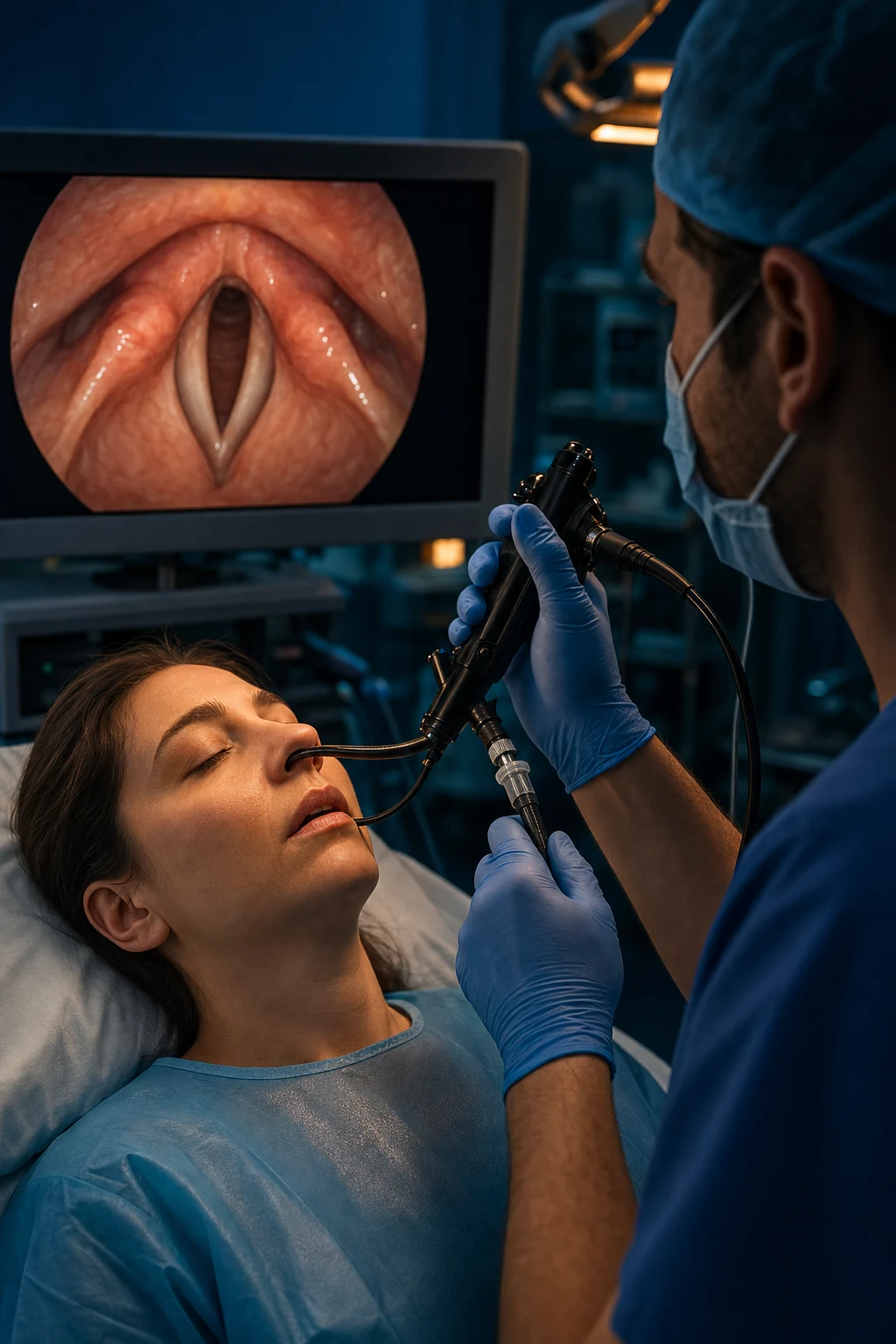
The third step is sedation, and the principle that governs it is absolute: the patient must remain cooperative and breathing. The role of the sedative is to reduce the anxiety and the discomfort of the procedure while preserving the airway reflexes and the respiratory drive that make the awake technique safe. Over-sedation is not a deeper version of the technique — it is its failure, because it converts a safe awake intubation into a cannot-intubate, cannot-ventilate situation with an unsecured airway[2][5].
The agent that best fits this profile, and the one the viva expects named as the ideal, is dexmedetomidine. It is a highly selective alpha-2 agonist that produces a unique cooperative, arousable sedation — the patient is calm and tolerant but can be woken to command — and, critically, it produces no respiratory depression: the respiratory rate and the drive are preserved, the airway reflexes are maintained, and the patient continues to breathe throughout. It also provides opioid-sparing analgesia and is sympatholytic, which moderates the hypertension and tachycardia of the airway instrumentation. These properties make it the agent of choice for AFOI, where the preservation of breathing is the entire point of the technique[5].
The alternatives each have a place but a clear disadvantage. Remifentanil provides excellent analgesia and suppresses the cough reflex powerfully, but it causes nausea, muscular rigidity at higher doses, and a narrow therapeutic margin — the patient who is adequately analgased is often close to apnoeic, which contradicts the principle. Propofol is titratable and familiar, but it depresses the respiratory drive and the airway reflexes, and it must be titrated with great care to avoid the loss of cooperation. The traditional midazolam with fentanyl combination is acceptable but it too depresses respiration, and the synergistic respiratory depression of the benzodiazepine-opioid pair is the classical pitfall. The key teaching is that the agent matters less than the principle: the patient must remain cooperative and breathing, and the sedative is titrated to that endpoint, not to sleep[2][5].
The sedation step is also the point at which the haemodynamic and cerebral oxygenation monitoring matters most. The sympatholysis of dexmedetomidine can produce bradycardia and hypotension, the airway stimulation can produce hypertension and tachycardia, and the critically ill patient may have little reserve; near-infrared spectroscopy monitoring of the cerebral oxygenation has been described as a sensitive early marker of the perfusion compromise during induction-phase haemodynamic change, and it illustrates the principle that the sedated-awake patient still warrants the haemodynamic vigilance that any induction warrants[4].
Steps 4 to 6: scope insertion, railroading, confirmation
The fourth, fifth, and sixth steps are the mechanical core of the technique, performed once the airway is topicalised and the patient is calmly sedated[2][5].
In the fourth step — insertion of the scope — the bronchoscope, preloaded with the lubricated endotracheal tube, is advanced through the nose (or the mouth, through a bite block) into the pharynx. The operator identifies the epiglottis first, then the vocal cords, and advances the scope between the cords into the trachea, confirming the position by the sight of the tracheal rings anteriorly and the carina ahead. The spray-as-you-go lidocaine is delivered onto the cords just before the scope passes through them, to abolish the residual cough. The view must be maintained throughout — losing the view means withdrawing to a recognised structure and re-advancing, never pushing blindly[2][5].
In the fifth step — railroading the tube — the endotracheal tube is advanced over the scope into the trachea. The scope acts as a guide wire, and the tube is railroaded over it. The common failure here is that the tube hangs up on the arytenoids or the cords as it is advanced; the fix is to withdraw the tube a centimetre, rotate it a quarter turn (so the bevel faces the obstruction), and re-advance. The scope is held still throughout the railroading so that its tip remains in the trachea[2][5].
In the sixth step — confirmation and induction — the scope position is confirmed by the tracheal rings and the carina, the endotracheal tube position is confirmed by capnography (the continuous waveform capnography is the gold standard for confirmation), and only then is general anaesthesia induced. The discipline of confirming the tube before inducing is the whole safety margin of the awake technique: the patient is intubated, the position is verified, and only then is the tone and the consciousness lost[2][5].
Oxygenation and safety during the awake technique
The awake technique is safe only if the patient is oxygenated throughout, because the procedure takes longer than an apnoeic intubation and the patient is at risk of desaturation if the airway is partially obstructed by the scope or the secretions[1][6].
Preoxygenation before the procedure begins is the first safeguard, and the modern teaching is that the quality of the preoxygenation should be measured rather than assumed. End-tidal oxygen measurement has been described as a sensitive metric of the denitrogenation achieved before the intubation, and it reflects the principle that the goal of preoxygenation is to replace the alveolar nitrogen with oxygen and so extend the safe apnoea period — a principle that applies as much to the awake technique as to the rapid sequence, because the patient who desaturates during an AFOI is one who must be rescued, and the rescue is harder in a difficult airway[6].
Continuous oxygen delivery during the procedure is the second safeguard, delivered through nasal specs, a modified face mask, or the working channel of the scope itself (the transnasal humidified rapid-insufflation ventilatory exchange, or THRIVE, technique extends the safe apnoea time further and is increasingly used). The patient who desaturates is reassessed for partial obstruction, secretions, or over-sedation, and the scope is withdrawn if the saturation does not recover[1][6].
The third safeguard is the monitoring of the haemodynamics and the cerebral perfusion throughout. The sedated-awake patient is not exempt from the haemodynamic lability of airway instrumentation; the induction-phase behaviour of the cerebral oxygenation relative to the mean arterial pressure has been described as a sensitive marker of the perfusion compromise, and it illustrates the principle that the awake technique warrants the same monitoring vigilance as any anaesthetic, not less[4].
Alternatives to the awake fibreoptic intubation
AFOI is the gold standard, but it is no longer the only awake option, and the modern algorithm presents a sequence of alternatives that the candidate is expected to know and to choose between on the merits of the case[5].
The leading alternative is the awake video laryngoscopy. A video laryngoscope is used in the awake, topically anaesthetised, lightly sedated patient, and the blade is introduced under vision while the patient breathes. It has the advantages of speed (the technique is faster than AFOI), of a familiar skill set (most anaesthetists are more practised with a video laryngoscope than a bronchoscope), and of a wide-angle view that the bronchoscope cannot provide. It requires less topicalisation than AFOI and it is increasingly preferred where the difficulty is anatomical — the anterior larynx, the limited mouth opening, the fixed cervical spine — rather than obstructing. Its weakness is that it cannot navigate a distorted or obstructed airway the way a flexible scope can, and it requires a mouth opening wide enough to admit the blade[5].
The awake intubating laryngeal mask airway is a second alternative: an intubating LMA is placed awake under topical anaesthesia, the airway is vented through it, and a tube is passed through the LMA into the trachea, either blindly or guided by a bronchoscope passed through the LMA. It is useful when the difficulty is at the level of the larynx and the supraglottic airway is achievable. [1]
The retrograde intubation is a third, rarely used alternative, in which a wire is passed through the cricothyroid membrane and out through the mouth, and the endotracheal tube is railroaded over the wire into the trachea. It is a technique of last resort, preserved for the case in which the fibreoptic and the video-laryngoscopic routes have failed or are unavailable[5].
The pragmatic teaching is that the choice between AFOI and the awake video laryngoscope is the choice most often examined, and the principle is anatomical: the flexible scope for the distorted, obstructed, or inaccessible airway; the video laryngoscope for the anatomically difficult but accessible airway[5].
Avoiding the airway entirely: the regional alternative
In a subset of the anticipated difficult airways, the safest airway is the one that is never instrumented — when the surgery is amenable to a regional anaesthetic technique that avoids general anaesthesia altogether. For the awake patient with a difficult airway presenting for limb, abdominal wall, or superficial surgery, a regional block performed under light sedation removes the airway from the operative plan entirely, and the difficult airway becomes a contingency rather than the primary event[3][5].
The erector spinae plane block and the transversus abdominis plane block, for example, provide abdominal wall analgesia for a range of surgical and trauma indications, and they illustrate the principle that a well-conducted regional technique can obviate the need for general anaesthesia and airway instrumentation in the selected patient. The decision is always hedged, however, by the requirement that the airway plan remains prepared — a regional block that fails or a surgery that converts to general anaesthesia must not leave the team without an awake airway strategy, and so the anticipated difficult airway is managed with the regional technique plus a fully prepared awake airway back-up, not with the regional technique alone[3].
Contraindications to the awake technique
The awake technique is the safest option for the anticipated difficult airway in most cases, but it has contraindications that the viva expects named, and each is a reason to convert to a different strategy rather than to push on[2][5].
- Patient refusal. A patient who, after explanation, declines the awake technique cannot be forced to undergo it; the alternative — an asleep technique with the best available equipment and a senior operator — is planned and consented.
- Inability to cooperate. The confused, the agitated, the intoxicated, and the young child who cannot hold still are relative contraindications to AFOI, because the technique depends on a cooperative, still patient. Deeper sedation is not the answer — it converts the awake technique into an asleep technique without the airway secured — and the alternative is the asleep technique with full preparation.
- Severe coagulopathy (nasal route). The nasal mucosa bleeds copiously when instrumented, and a coagulopathic or heavily anticoagulated patient can suffer catastrophic epistaxis into a difficult airway. The nasal route is avoided in these patients; the oral route or an awake video laryngoscopy is used instead. The nasal route is also avoided in the basilar skull fracture, where the scope can be misdirected into the cranial vault[5].
- Obstructing airway mass where instrumentation is dangerous. A critical obstruction — a large supraglottic tumour, a peritonsillar abscess, a retropharyngeal haematoma — can be disturbed by the scope, and the instrumentation itself can convert a partial obstruction into a complete one. These cases warrant a senior airway operator, a fully prepared surgical airway, and often an institutional protocol for the critical airway; the awake technique is performed with extreme care or replaced by a surgical airway under local anaesthesia[5].
The physiological difficult airway and the special contexts
Two contexts reshape the awake technique and are examined as the discriminating detail that separates the pass from the distinction[1][2].
The physiological difficult airway, seen most often in the emergency department and the intensive care unit, is the airway in which the anatomy may be favourable but the physiology is hostile: hypoxaemia shortens the safe apnoea period, hypotension and shock remove the cardiovascular reserve, and the patient may be agitated or unable to cooperate. The awake technique has a particular role here — it preserves the spontaneous ventilation and the cardiovascular tone that an apnoeic induction would abolish — but it is also harder to perform in the agitated, hypoxaemic patient, and the decision balances the time it takes against the stability it preserves. The recognition of the physiological difficult airway as a distinct entity is the major conceptual advance of the recent emergency and critical-care airway literature, and the awake technique is one of the strategies that it reshapes[1].
The syndromic and neuromuscular difficult airway — exemplified by spinal muscular atrophy and the congenital craniofacial syndromes — combines anatomical distortion with physiological fragility, and it carries a high incidence of difficult intubation that combines a difficult laryngoscopy with a difficult mask ventilation and a precarious cardiovascular and respiratory reserve. These patients are the classical indication for an awake, planned, senior-led technique, and the contemporary series document the anaesthetic care, the complications, and the airway management that these patients require across their lifespan[2].
Clinical
- Standard approach
- Evidence-based
Alternative
- Modified technique
- Risk-benefit
Red flags
[1] [1] [1] [1] [1]References
- [1]Ghaffar S, et al. Physiological difficult airway management in the emergency department J Pak Med Assoc, 2026.PMID 42363338
- [2]Black KM, et al. Anesthesia Care, Complications, and Airway Management for Patients With Spinal Muscular Atrophy: A Retrospective Chart Review From a Quaternary Children's Hospital Anesth Analg, 2026.PMID 42363899
- [3]Merchant N, et al. Comparing the analgesic utility & safety of erector spinae plane block versus thoracic epidural for multiple rib fracture trauma: a retrospective cohort analysis Injury, 2026.PMID 42361789
- [4]Ozgol I, et al. Induction-Phase rSO(2)-MAP Behaviour and Cross-Clamp Desaturation in NIRS-Guided Selective Carotid Endarterectomy: A Retrospective Cohort Study J Clin Med, 2026.PMID 42355788
- [5]Freund Y, et al. Improving the safety of emergency tracheal intubation Curr Opin Crit Care, 2026.PMID 42170830
- [6]Caputo ND, et al. End Tidal O(2): A Promising New Metric for Optimizing Preoxygenation and RSI Safety in the Emergency Department Acad Emerg Med, 2026.PMID 42340046