Anaes · Airway management
Direct laryngoscopy & orotracheal intubation — blade types, Cormack-Lehane grade, BURP, ETT selection and confirmation
Also known as Direct laryngoscopy · Orotracheal intubation · Cormack-Lehane grade · Macintosh blade · Miller blade · BURP manoeuvre · Endotracheal tube · Murphy eye · Sniffing position
Direct laryngoscopy is the technique by which the larynx and vocal cords are brought into a single line of sight so that a tracheal tube can be passed under direct vision, and it remains the index skill of airway management against which every alternative is measured. The framework rests on six ideas: the geometry of the sniffing position aligns the oral, pharyngeal and laryngeal axes; the curved Macintosh blade (placed in the vallecula) and the straight Miller blade (placed behind the epiglottis) lift the tongue and epiglottis by different mechanisms; the Cormack-Lehane grade records the view obtained and a grade of three or four defines the difficult intubation; the BURP manoeuvre and optimal external laryngeal manipulation improve a poor view and are distinct from cricoid pressure; the endotracheal tube is chosen by cuff, size and design, with the Murphy eye protecting the right upper lobe; and intubation is confirmed by sustained waveform capnography, because an unrecognised oesophageal intubation is fatal. Anchored to contemporary evidence on the optimisation of intubating conditions, the physiological difficult airway, double-lumen tube practice, and the airway management of neuromuscular disease.
On this page & tools
Your progress
Saved locally on this device.
8 MCQs with explanations
Target exams
Red flags
Overview & definition
Direct laryngoscopy is the technique by which a rigid laryngoscope is used to displace the tongue and soft tissues of the pharynx so that the larynx, the epiglottis and the vocal cords are brought into a single, direct line of sight, allowing a tracheal tube to be passed through the cords under vision. It is the index skill of airway management: the technique against which every alternative — video laryngoscopy, fibreoptic intubation, supraglottic rescue — is measured, and the procedure whose execution the airway viva and short-answer question dissect in the most detail[3].
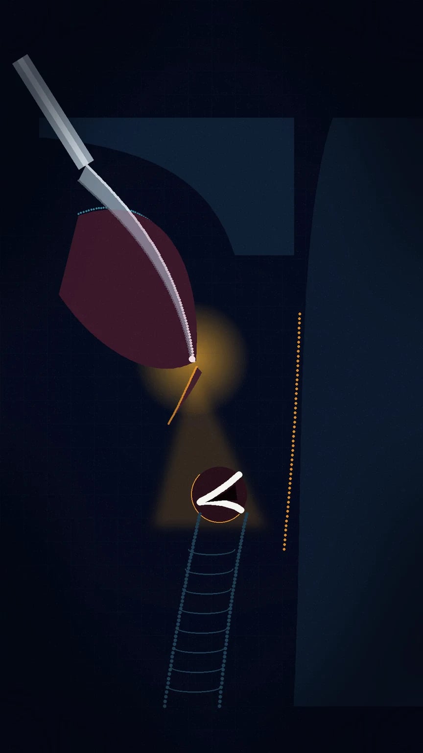
The exercise turns on a single geometric fact: the oral cavity, the pharynx and the larynx do not lie in a straight line. Three axes — the axis of the mouth, the axis of the pharynx, and the axis of the larynx — must be aligned before the glottis can be seen from the operator's eye. Alignment is achieved by positioning the head, by choosing a blade that displaces the tongue in the right direction, and by lifting (not levering) along the axis of the handle. When the axes are aligned and the tongue is swept left, the glottis comes into view; when they are not, only the epiglottis, or only the soft palate, is seen, and the intubation is graded as difficult[1][3].
The examined body of knowledge around direct laryngoscopy has six components, each developed below: the blade types and how each lifts the epiglottis; the sniffing position that aligns the three axes; the step-by-step technique; the Cormack-Lehane grade that records the view and defines difficulty; the BURP manoeuvre that improves a poor view; the endotracheal tube, its types and sizing; and the confirmation of tracheal placement, of which sustained waveform capnography is the definitive test. Each component is examined, and each carries a complication when it is done badly[1][2].
The laryngoscope: blade types
The modern laryngoscope is a handle (containing the light source and batteries) and a detachable blade (the introducer and the light carrier). Two blade designs dominate practice and are examined together: the Macintosh (curved) blade and the Miller (straight) blade. Their geometry differs, and with it the mechanism by which each exposes the glottis[3].
- The Macintosh blade is the curved blade and the most widely used in adult practice. It is placed in the vallecula — the recess between the base of the tongue and the epiglottis — and, on lifting, indirectly elevates the epiglottis by traction on the hypoepiglottic ligament. Because the epiglottis is not directly engaged, the blade traumaties it less, and the curved blade naturally sweeps the tongue leftward. Adult sizes are three and four, with size four reserved for the long airway.
- The Miller blade is the straight blade. It is placed posterior to the epiglottis and directly lifts it, exposing the glottis. The direct lift is advantageous when the larynx is anterior and high, when the epiglottis is long and floppy, and in infants, in whom the epiglottis is short, soft and angled, and the larynx sits high and anterior. Adult sizes are two and three. [1]
The examined distinction is therefore anatomical: the Macintosh blade lifts the epiglottis indirectly through the vallecula, the Miller blade lifts it directly. The Macintosh is the default in adult anaesthesia; the Miller is preferred for the anterior larynx, the infant, and the difficult floppy epiglottis. The competent laryngoscopist is fluent with both, because the blade that gives the best view is determined by the anatomy in front of them, not by habit[3][1].
The sniffing position and the line of sight
The position of the head is the single most powerful determinant of the laryngoscopic view, and the classical position is the sniffing the morning air position, described by Magill and refined by the radiographic studies that confirmed its geometric basis. The position has two components: flexion of the lower cervical spine (at around the C7 to T1 level), achieved by elevating the occiput on a pillow of about seven to ten centimetres; and extension of the atlanto-occipital joint, achieved by reclining the head at the skull base[3].
The aim is to align the three axes — oral, pharyngeal and laryngeal — into a single line of sight from the incisors to the glottis. Flexion of the neck brings the pharyngeal and laryngeal axes closer together; extension at the atlanto-occipital joint brings the oral axis into line with them. When all three are aligned, the glance from the operator's eye travels in a straight line over the tongue, around the curve of the pharynx, onto the glottic opening. When they are not aligned, the line of sight falls short, and the glottis is hidden behind the tongue or the epiglottis. In the obese patient, elevation must be greater — the ramped position, in which the external auditory meatus is level with the sternal notch, achieves the same alignment in the patient whose torso is thick[2].
The position is not negotiable. A patient induced flat on the bed, with the head level and the neck un-flexed, presents a difficult laryngoscopy even when the anatomy is favourable, because the axes cannot align. The pillow is therefore part of the intubating equipment, and achieving the sniffing position is the first action after induction, before the blade is inserted[3].
Technique of direct laryngoscopy
The technique is a sequence, and the examined candidate is expected to recite it in order, with the rationale for each step. The operator stands at the head of the bed, the patient in the sniffing position, the suction within reach and switched on, the tube, the stylet, the bougie and the supraglottic rescue device prepared[3].
- Position. Confirm the sniffing position, with the occiput elevated on a pillow and the head extended at the atlanto-occipital joint. The face is at the operator's working height.
- Hold the laryngoscope in the left hand. The laryngoscope is always held in the non-dominant (left) hand, regardless of the operator's handedness, so that the right hand is free to manipulate the tube. The grip is low on the handle, with the thumb pushing forward along the axis of the blade.
- Open the mouth. The right hand opens the mouth, often with a scissors action of the thumb and index finger pushing the lower lip away from the teeth.
- Insert the blade on the right side of the mouth. The blade is introduced from the right commissure of the mouth, with the tongue identified and kept to the left of the blade as the blade advances. Sweeping the tongue left is the central manoeuvre: a tongue that spills over the right side of the blade obstructs the view.
- Advance to the epiglottis. The blade is advanced over the curve of the tongue until the epiglottis is identified. For the Macintosh blade, the tip is placed in the vallecula; for the Miller blade, the tip is placed behind the epiglottis.
- Lift along the axis of the handle. The blade is lifted upward and forward at roughly 45 degrees along the long axis of the handle — a lift, never a lever or a pry. Levering against the upper incisors fractures teeth and does not expose the glottis; the force is a straight lift that draws the tongue and epiglottis up and out of the line of sight.
- Identify the landmarks. With the epiglottis elevated, identify the arytenoid cartilages first (the posterior cartilages are the last landmark to disappear and the first to find), then the posterior commissure, then the vocal cords. The glottic opening is a triangular gap between the cords.
- Pass the tube through the cords. The right hand advances the tube through the cords under direct vision from the right corner of the mouth. The tube is held in the right hand and introduced from the right; the cuff is passed just beyond the cords, so the cuff lies in the upper trachea.
- Remove the stylet, inflate the cuff, and confirm. The stylet is withdrawn, the cuff is inflated to a seal pressure of about 20 to 30 cm of water, and the tube is confirmed by sustained waveform capnography before the laryngoscope is withdrawn[3].
The common error is levering. The laryngoscope is not a bottle opener; it is a lever whose fulcrum is the base of the tongue, not the teeth, and whose force is applied by lifting the handle, not by rotating it against the incisors. A second common error is allowing the tongue to spill over the right edge of the blade, which obscures the view; the tongue must be gathered onto the left as the blade advances[1].
The Cormack-Lehane grade
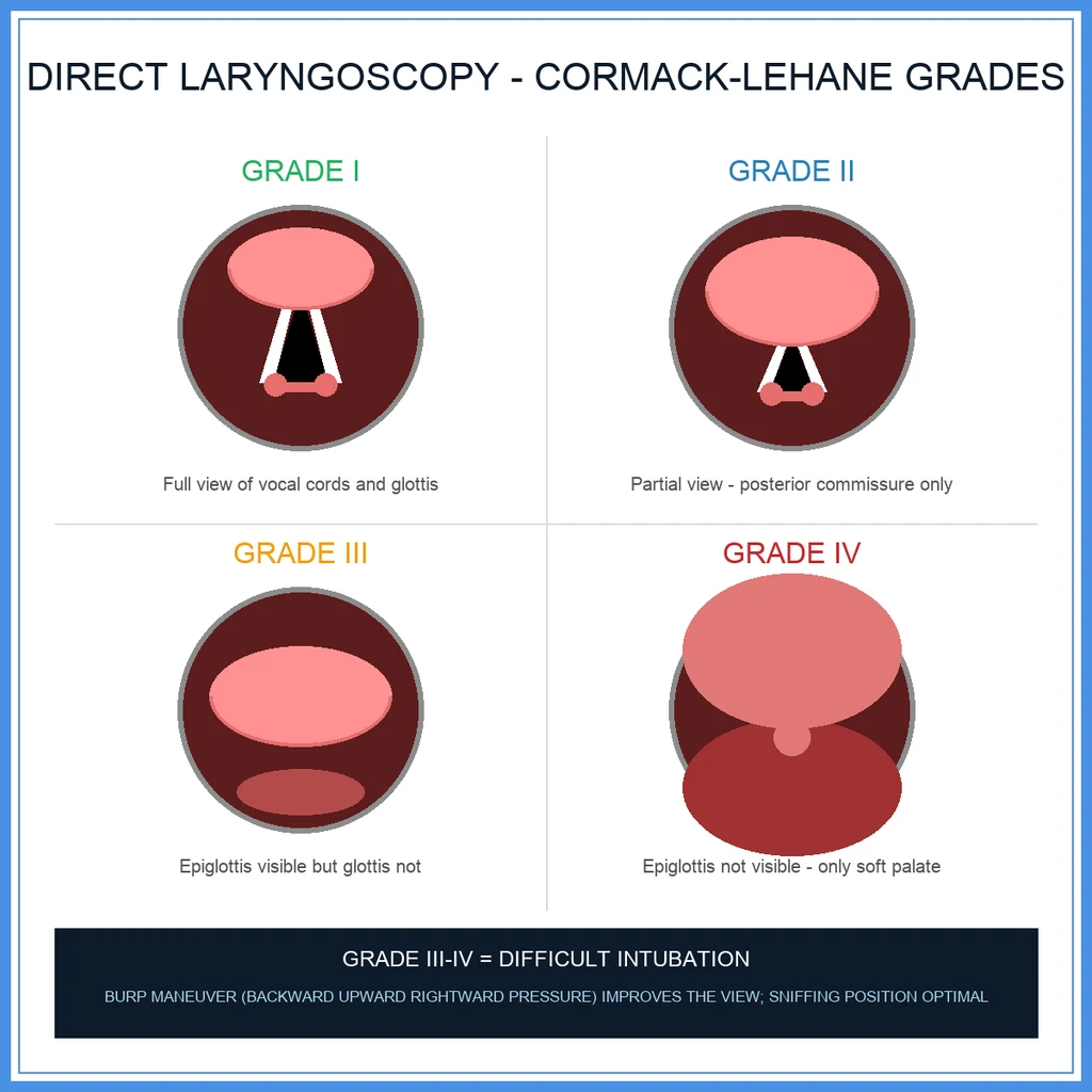
The view obtained at direct laryngoscopy is recorded by the Cormack-Lehane classification, the four-point scale described by Cormack and Lehane in 1984 and the universal language of the difficult airway. The grade is assigned by what the operator sees with the blade correctly placed and optimal external laryngeal manipulation applied[3].
- Grade I — most of the glottis is visible.
- Grade II — the posterior portion of the glottis, including the arytenoid cartilages and the posterior commissure, is visible; the anterior glottis is not.
- Grade III — only the epiglottis is visible; no part of the glottis can be seen.
- Grade IV — neither the glottis nor the epiglottis is visible; only the soft palate. [1]
A grade of three or four is the operational definition of the difficult direct laryngoscopy. Grade one and two are "easy", grade three and four are "difficult", and the boundary is the line an examiner draws. The grade predicts the difficulty of passing the tube, the number of attempts, and the risk of hypoxaemia and oesophageal intubation on repeated attempts. A modified three-point scale (the percentage-of-glottic-opening, or POGO, score) refines the grade two to three boundary, but the four-point Cormack-Lehane remains the universal record[3].
The grade is also the outcome that the bedside airway assessment (the LEMON tool, the Mallampati score) tries to predict, and the link is direct: a Mallampati class of three or four predicts a Cormack-Lehane grade of three or four. When a poor grade is encountered unanticipated, the disciplined response is to optimise the position, apply the BURP manoeuvre, and move to a video laryngoscope or a supraglottic rescue, rather than to repeat a failing attempt and incur hypoxaemia[1][2].
Optimising the view: BURP and external laryngeal manipulation
A poor Cormack-Lehane grade is not fixed; it is the starting point of a sequence of optimising manoeuvres, the most examined of which is the BURP manoeuvre — backward, upward, rightward pressure on the thyroid cartilage, applied by an assistant[3].
- Backward — pressure toward the vertebral column.
- Upward — pressure toward the ceiling (cephalad).
- Rightward — pressure that displaces the larynx slightly to the patient's right. [1]
The combined vector moves the larynx into the direct line of sight, improving the Cormack-Lehane grade by one or sometimes two levels. The operator identifies the optimal direction of pressure by manipulating the larynx with their own right hand (the optimal external laryngeal manipulation, or OELM), then instructs the assistant to reproduce it, because the assistant cannot see what the operator sees. BURP is best applied early, before the tube is passed, and is the single most effective bedside manoeuvre for the poor view[3].
BURP is distinct from cricoid pressure (the Sellick manoeuvre). Cricoid pressure is downward pressure on the cricoid cartilage, applied during a rapid sequence induction to occlude the oesophagus and prevent regurgitation. It is not intended to improve the laryngoscopic view, and in fact it often worsens it; BURP and cricoid pressure are applied to different cartilages (thyroid and cricoid respectively), for different purposes, and must not be conflated[2].
Endotracheal tube types and the Murphy eye
The endotracheal tube (ETT) is the device through which the airway is secured after the cords are visualised, and its design carries several examined features. The standard tube is made of polyvinyl chloride (PVC), marked with depth markings, a radio-opaque line for radiographic visualisation, and the internal diameter printed at the proximal end[4].
- Cuffed versus uncuffed. The modern adult tube is cuffed — a low-pressure, high-volume inflatable cuff near the tip that seals the trachea and protects the lower airway from aspiration. In the past, uncuffed tubes were standard in children under about eight to ten years, because the paediatric cricoid cartilage is the narrowest point of the airway (in adults the narrowest point is the vocal cords), and a correctly sized uncuffed tube was thought to seal there. The contemporary practice has shifted toward cuffed tubes in infants and children as well, with careful cuff pressure control, because a cuff reduces leak and the risk of repeat intubation; the uncuffed tube remains the default in the neonate and the small infant.
- Reinforced (armoured) tubes. A wire spiral embedded in the wall resists kinking and compression, and the reinforced tube is used for head, neck and maxillofacial surgery, where the tube is bent sharply or compressed by drapes or the surgical team. The wire spiral means the tube cannot be cut to length, and it cannot be used through a rigid stylet that distorts the spiral.
- Double-lumen tubes. A double-lumen tube (DLT) separates the right and left main bronchi, allowing one-lung ventilation for thoracic surgery and isolation of a lung. It is the classical device for lung isolation, though tubeless and bronchial-blocker strategies provide alternatives, and contemporary comparisons examine the conditions each provides against the airway trauma each imposes[4].
- The Murphy eye. The standard tube has a bevelled tip and a single side hole near the bevel — the Murphy eye — which provides an alternative gas pathway should the distal tip occlude against the tracheal wall, and which in particular protects the right upper lobe bronchus from obstruction if the tube is advanced too far or rotated[4].
The examined point is that the tube is chosen for the patient (size, cuffed or uncuffed) and for the operation (standard, reinforced, or double-lumen), and that the Murphy eye and the cuff together protect against the two common mechanical failures — occlusion and endobronchial intubation[4].
Tube sizing, depth and the paediatric airway
The choice of tube is governed by the patient, and the size is expressed as the internal diameter in millimetres. The adult female is typically intubated with a size 7.0 to 7.5 tube, and the adult male with a size 8.0 to 9.0, the smaller sizes reducing airway resistance and airway trauma. The paediatric size is estimated by formula (internal diameter equals four plus the age in years divided by four), and the uncuffed tube is used a half size smaller than the cuffed[4].
The depth of insertion is set so the cuff lies in the upper trachea, well above the carina and well below the cords. The rule of thumb is three times the internal diameter at the lips — about 21 centimetres for an adult woman and 23 centimetres for an adult man. A tube advanced too far enters the right main bronchus (because the right bronchus is wider, shorter and more vertical than the left), causing unilateral right-sided ventilation, contralateral lung collapse, and the risk of barotrauma — the endobronchial intubation that bilateral air entry and capnography alone may not detect. The depth is confirmed by bilateral, equal air entry on auscultation, and the tube is withdrawn if the right side alone is ventilated[4].
The paediatric airway carries two examined distinctions. First, as above, the narrowest point is the cricoid, not the cords — the anatomical rationale for the uncuffed tube in the small child. Second, the infant larynx is high and anterior (at around the C3 to C4 level, descending to C5 to C6 in the adult), which is why the straight Miller blade and the direct epiglottic lift suit the infant airway better than the curved Macintosh. These two facts together explain why paediatric intubation is a distinct skill[4][1].
Confirmation of intubation
An unrecognised oesophageal intubation is rapidly fatal, and the confirmation of tracheal placement is therefore the most consequential step of the whole procedure. Confirmation is layered, from the gold standard at placement to the definitive test over the following breaths[2][5].
- Direct visualisation — seeing the tube pass between the vocal cords at the moment of intubation is the gold standard during placement. Even when seen, it is supplemented by the confirmatory tests below, because a tube can be displaced after placement.
- Bilateral chest rise and auscultation — observing symmetrical chest movement and auscultating bilateral air entry over the axillae, and over the stomach to exclude gastric insufflation, is supportive but not definitive; both can be falsely reassuring in the oesophageal intubation.
- Sustained waveform capnography — the definitive confirmatory test. Carbon dioxide is exhaled from the lungs and not from the stomach, so a sustained square-wave trace of expired carbon dioxide over several breaths (classically six waveforms over twenty seconds) confirms tracheal placement. A trace that fades over the first few breaths suggests an oesophageal intubation after mask ventilation or the residual carbon dioxide of a recent drink. Capnography is mandatory wherever a tracheal tube or supraglottic airway is in place.
- Oesophageal detector device — a bulb or syringe that, applied to the tube, fills rapidly from the low-resistance trachea but not from the collapsible oesophagus; a useful adjunct where capnography is unavailable, as in the pre-hospital setting. [1]
The teaching, reinforced by the national airway audits, is that capnography is the single most reliable confirmation, and that auscultation, chest rise and tube misting are not. A tube is not confirmed until a sustained waveform is seen[2][5].
Complications of direct laryngoscopy and intubation
The complications of direct laryngoscopy and intubation are mechanical, physiological and airway-related, and the examined candidate is expected to group them and to name the preventable cause of each[1][2].
- Mechanical trauma. Dental trauma (chipped or avulsed incisors, most often the left upper central incisor) from levering the blade; lip and tongue laceration; pharyngeal and oesophageal perforation from the rigid stylet or the tube; laryngeal and vocal cord injury from the tube or an over-inflated cuff; and the long-term sequelae of prolonged intubation — subglottic stenosis, vocal cord granulomata, and tracheomalacia.
- Oesophageal intubation. The tube passed into the oesophagus rather than the trachea. It is rapidly fatal if unrecognised, causing gastric insufflation, hypoventilation and hypoxaemia. It is prevented by direct visualisation and confirmed by sustained waveform capnography; the disciplined operator declares it early and re-intubates rather than ventilating an unconfirmed tube.
- Endobronchial intubation. The tube advanced too far, entering the right main bronchus. It causes unilateral ventilation, contralateral collapse and ipsilateral barotrauma. It is detected by asymmetrical chest movement and auscultation, and corrected by withdrawing the tube to bilateral air entry.
- Aspiration. Regurgitation of gastric contents around the tube or during the intubation, prevented by pre-oxygenation, rapid sequence induction when indicated, and cricoid pressure. The cuff protects once the tube is in place, but does not protect during placement.
- Hypoxia from prolonged attempts. Each attempt consumes the apnoeic reserve; repeated failed attempts lead to desaturation, bradycardia and cardiac arrest. The disciplined limit is to abandon a failing attempt and re-oxygenate by mask before a further attempt.
- Cardiovascular response. Laryngoscopy and intubation are noxious stimuli that provoke tachycardia, hypertension, arrhythmia and a rise in intracranial and intra-ocular pressure, blunted by adequate depth of anaesthesia, opioid and, where indicated, a vasodilator or beta-blocker. In the patient with limited cardiovascular reserve, the response itself precipitates collapse[2].
The preventable causes run through this list: levering the blade (dental trauma), failing to confirm (oesophageal intubation), advancing too far (endobronchial), repeated attempts (hypoxia), and inadequate depth (the cardiovascular response). Each complication is examined against the manoeuvre that would have prevented it[1][2].
Optimising intubating conditions: depth, paralysis and pharmacology
The conditions for direct laryngoscopy — the stillness of the patient, the relaxation of the vocal cords, the absence of coughing and bucking — are themselves a determinant of success and of the Cormack-Lehane grade obtained. The optimisation of conditions is therefore part of the technique, and it rests on adequate depth of anaesthesia and full neuromuscular blockade[3][6].
The classical combination is an intravenous induction agent and a rapid-onset neuromuscular blocking drug. Propofol remains the default induction agent; remimazolam, an ultra-short-acting benzodiazepine, has been compared with propofol for the conditions it provides, with the advantage of haemodynamic stability and reversal with flumazenil, and is an examined contemporary alternative in the patient with limited cardiovascular reserve[6]. Rocuronium, at a dose of one milligram per kilogram, provides intubating conditions comparable to suxamethonium within sixty seconds and has become the default relaxant for the rapid sequence induction, with sugammadex available to reverse it. The optimisation of these conditions — full paralysis, adequate depth, optimal position — has been shown to improve even the intubating conditions for video laryngoscopy, and the same principle applies to direct laryngoscopy[3].
The examined point is that a poor view is sometimes a pharmacological failure rather than an anatomical one. A patient who is under-depth, lightly paralysed, or coughing on the blade presents a difficult laryngoscopy that resolves with depth and blockade, not with a larger blade or a repeated attempt. The disciplined laryngoscopist checks the conditions before blaming the anatomy[3][6].
Special contexts: video laryngoscopy, rescue and the physiological difficult airway
Direct laryngoscopy is not the only technique, and the competent operator knows when to move away from it. Video laryngoscopy — the angulated blade with a camera at the tip — provides a view of the glottis around the curve of the tongue, and it has become the first-line device for the anticipated difficult direct laryngoscopy, the predicted difficult airway, and the cervical-spine-injured patient who cannot be positioned. The optimisation of intubating conditions applies equally to video laryngoscopy, and the evidence supports full paralysis and optimal depth for both[3].
When direct laryngoscopy fails and video laryngoscopy is unavailable or also fails, the rescue is a supraglottic airway device — the second-generation device with an oesophageal drain tube is preferred — which maintains oxygenation while a definitive airway is secured. The contemporary supraglottic devices include non-inflatable, visually-directed masks that combine an airway channel with a viewing window, and they are examined as the bridge between the failed intubation and the surgical airway[5].
The physiological difficult airway, seen most often in the emergency department and the intensive care unit, reshapes the whole technique. The anatomy may be favourable, but the physiology is hostile: hypoxaemia shortens the safe apnoea period, hypotension and shock remove the cardiovascular reserve that induction erodes, and the patient may be agitated or unable to cooperate. The same direct laryngoscopy that is technically easy in the elective patient may be impossible in the physiologically fragile one, and the management must interleave resuscitation with the intubation — pre-oxygenation, vasopressor readiness, and an unhurried best attempt. The recognition of the physiological difficult airway as a distinct entity is the major conceptual advance of the recent emergency and critical-care airway literature, and it is the element most often missing from the candidate who recites the technique mechanically[2].
In the syndromic and neuromuscular contexts — exemplified by spinal muscular atrophy and the congenital craniofacial syndromes — the difficulty combines anatomical distortion with physiological fragility, and the airway plan is built around anticipation, senior presence, and a prepared rescue. These patients carry a high incidence of difficult intubation that the standard technique must adapt to, and they are the patients in whom an under-prepared induction is most likely to end in harm[1].
Clinical
- Standard approach
- Evidence-based
Alternative
- Modified technique
- Risk-benefit
Red flags
[1] [1] [1] [1] [1]References
- [1]Black KM, et al. Anesthesia Care, Complications, and Airway Management for Patients With Spinal Muscular Atrophy: A Retrospective Chart Review From a Quaternary Children's Hospital Anesth Analg, 2026.PMID 42363899
- [2]Ghaffar S, et al. Physiological difficult airway management in the emergency department J Pak Med Assoc, 2026.PMID 42363338
- [3]Ipsen EO, et al. Remifentanil Versus Rocuronium for Optimising Video Laryngoscopy Assisted Tracheal Intubation-The ROCVIDEO Trial Protocol Acta Anaesthesiol Scand, 2026.PMID 42304626
- [4]Jiang Y, et al. Effects of electroacupuncture-assisted tubeless anesthesia versus double-lumen endotracheal intubation anesthesia on the quality of postoperative recovery in thoracoscopic surgery: A propensity score-matched retrospective study Medicine (Baltimore), 2026.PMID 42363554
- [5]Migliorelli S, et al. Clinical Evaluation of a Non-inflatable Visual Laryngeal Mask Airway: A Prospective Service Assessment in Elective and Difficult Airway Management Cureus, 2026.PMID 42291977
- [6]Shionoya M, et al. Comparison of the Effects of Remimazolam and Propofol, With Epinephrine-containing Lidocaine, on Rocuronium-induced Muscle Relaxation Anesth Prog, 2026.PMID 42307548