Anaes · Neuromuscular blockade & reversal
Neuromuscular monitoring and the train-of-four
Also known as Train-of-four (TOF) monitoring · Peripheral nerve stimulation · Quantitative neuromuscular monitoring · Acceleromyography · Post-tetanic count
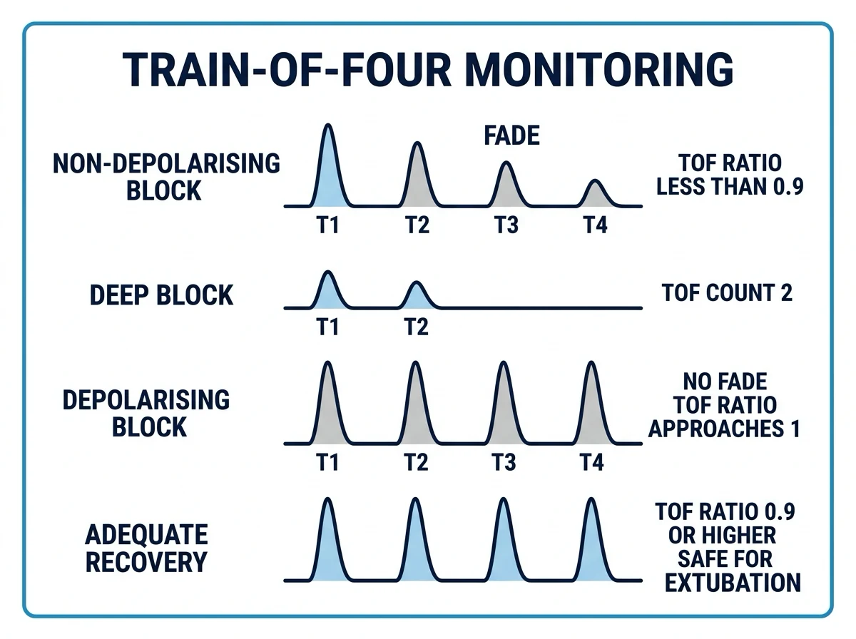
Neuromuscular monitoring is the objective assessment of the depth and recovery of a neuromuscular block by delivering a supramaximal electrical stimulus to a peripheral motor nerve and observing the evoked muscle response. The train-of-four — four stimuli at 2 Hz, 0.5 seconds apart — is the workhorse pattern: it yields a TOF count (the number of detectable twitches, 0 to 4) and a TOF ratio (T4 divided by T1). A non-depolarising block shows fade (ratio below 1.0, with sequential loss of T4 then T3 then T2 then T1) whereas a depolarising phase I block shows no fade (all four twitches reduced proportionally, ratio near 1.0). Residual neuromuscular blockade, defined as a TOF ratio below 0.9 at extubation, is the principal preventable cause of postoperative airway obstruction, aspiration and hypoxia; the 2023 ASA practice guideline mandates quantitative monitoring whenever a neuromuscular blocker is administered, and a confirmed TOF ratio of at least 0.9 before extubation.
On this page & tools
Your progress
Saved locally on this device.
10 MCQs with explanations
Target exams
Red flags
Overview — why neuromuscular monitoring matters
Neuromuscular monitoring is the objective assessment of the depth and recovery of a neuromuscular block by stimulating a peripheral motor nerve and measuring the evoked response of the muscle it supplies. It exists for a single, important reason: without it, the anaesthetist cannot reliably tell how deep a block is, how well it is recovering, or whether it is safe to extubate. The clinical and pharmacological signs that were once used as surrogates for recovery — tidal volume, grip strength, the ability to lift the head sustained for five seconds — are insensitive, and a patient can satisfy all of them and still have a train-of-four ratio of 0.5. The only reliable indicator of recovery is the evoked response itself, measured quantitatively.[1][3]
The clinical stakes are high. Incomplete recovery at the end of an anaesthetic — residual neuromuscular blockade — is a leading and largely preventable cause of airway obstruction, aspiration, hypoxia, reintubation and prolonged recovery-room stay. It remains common wherever monitoring is absent or purely subjective: in the POPULAR multicentre European observational study, postoperative pulmonary complications were significantly more frequent in patients who received a muscle relaxant and were not reversed with a confirmed adequate recovery, and the figure has changed little over the past two decades whenever quantitative monitoring is not routine.[9][8]
The modern consensus, formalised in the 2023 American Society of Anesthesiologists (ASA) practice guideline and reinforced by current reviews, is that quantitative monitoring — measuring the actual train-of-four ratio with a device such as an acceleromyograph — should be used whenever a neuromuscular blocker is administered, and that a train-of-four ratio of at least 0.9 should be demonstrated before extubation.[1][3] The older threshold of 0.7 is now known to be inadequate, because fade and clinical weakness persist up to a ratio of 0.9 and can be detected only by quantitative methods. The entire apparatus of monitoring — the patterns, the modalities and the thresholds — exists to close the gap between the assumption of recovery and the demonstration of it.
A short history and the conceptual basis
The principle that an evoked response could be used to monitor a block dates to the 1950s, but the modern apparatus crystallised in the work of Savarese, Ali and colleagues in the early 1970s, who introduced the train-of-four specifically as a stimulus pattern that did not require a control (baseline) twitch height — a property that made it usable in routine anaesthesia, where a baseline is rarely available.[10] The double-burst stimulation was added by Engbaek, Viby-Mogensen and colleagues in the late 1980s as a tactile refinement, and the post-tetanic count by Viby-Mogensen for the deep-block range.[16][11]
The train-of-four was named and validated at the Massachusetts General Hospital and the Harvard anaesthesia programme; it displaced the older single-twitch and tetanic patterns because it carried its own internal control (T1) within the stimulus, so a baseline was unnecessary. The term "train-of-four ratio" (T4/T1) entered the anaesthesia vocabulary from this work and is now the universal unit of recovery.
The conceptual basis is straightforward. A neuromuscular blocker reduces the safety margin of neuromuscular transmission — the gap between the end-plate potential and the threshold for generating a muscle action potential. The greater the block, the smaller the safety margin, and the smaller the evoked response. Fade — the progressive reduction of successive responses within a short burst of stimuli — is a separate phenomenon, arising because a competitive (non-depolarising) block depletes the immediately releasable pool of acetylcholine faster than it can be replenished, so each successive stimulus in a train releases less transmitter and the postsynaptic response is progressively more affected. Fade is therefore the electrophysiological fingerprint of a non-depolarising block; its absence (with globally reduced twitches) is the fingerprint of a depolarising block. Every monitoring pattern in clinical use exploits these two phenomena — the amplitude of the response (which tracks block depth) and the relationship between successive responses (which reveals the type of block and, quantitatively, the degree of recovery). [1]
The peripheral nerve stimulator
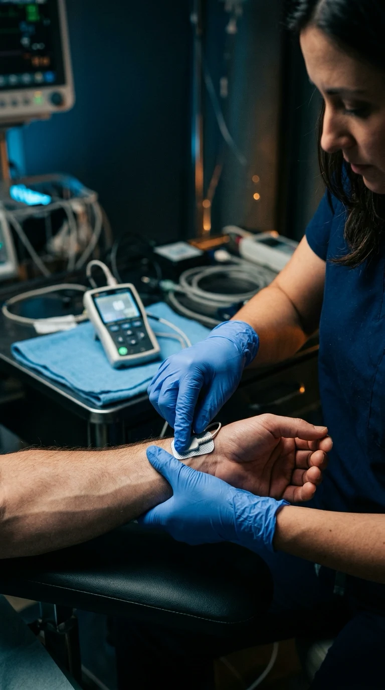
A peripheral nerve stimulator (PNS) is a constant-current device that delivers a brief electrical pulse to a superficial peripheral motor nerve, depolarises the motor axons, and produces a synchronous muscle contraction whose amplitude is then observed or measured. Three parameters define every stimulus the device delivers. [1]
[1]Supramaximal stimulation is the single most important concept in the mechanics of monitoring. The current is increased from threshold until the evoked response no longer grows — every motor axon in the nerve has been recruited — and then a 10 to 25 percent margin is added so that any subsequent change in the response can be attributed to changes at the neuromuscular junction rather than to changes in the stimulus strength. If the stimulus were submaximal, a small drift in current (from a drying electrode, a change in skin impedance, or a displaced probe) would be indistinguishable from a change in block depth, and the monitoring would be meaningless. The routine clinical supramaximal current at the ulnar nerve is 40 to 60 mA with surface electrodes.[3]
The electrodes are usually surface ECG-style electrodes, which are non-invasive and adequate for routine use. Needle electrodes (subcutaneous, insulated to the tip) are reserved for situations of poor access or high skin impedance (oedema, dressings, obesity) but add no sensitivity when the surface electrodes are well placed. The negative electrode is placed over the nerve and the positive electrode along its course or transversely across the wrist; for the ulnar nerve the negative electrode goes on the volar wrist just radial to the flexor carpi ulnaris tendon, with the positive a few centimetres proximal or across the forearm. Polarity matters because the cathode (negative) is the depolarising electrode: the motor axons are activated under the cathode, so placing the cathode over the nerve gives the cleanest response.[3]
Stimulation sites and the muscle group that responds
The site of stimulation and the muscle whose response is monitored are inseparable: a peripheral motor nerve is stimulated, and a specific muscle supplied by that nerve is observed. Four classical sites are used in routine anaesthetic practice, and the choice of site carries a real clinical consequence because different muscles differ in their sensitivity to a non-depolarising block. [1]
The ulnar nerve at the wrist, with the response assessed at the adductor pollicis (thumb adduction), is the classical and most reliable site, and it is the reference muscle on which all the recovery thresholds — including the TOF ratio of 0.9 — were validated. The adductor pollicis is a small peripheral muscle and is highly sensitive to non-depolarising block, so it is the last muscle to recover; if it has recovered, the airway muscles can be assumed to have recovered as well. For quantitative (acceleromyography) monitoring the thumb must be free to move, which means the arm cannot be tucked tightly at the side; the contralateral arm is often used when the surgical field occupies one side.[3]
The facial nerve is stimulated at the stylomastoid foramen or just in front of the tragus, with the response assessed at the orbicularis oculi (eyelid twitch) or corrugator supercilii. It is convenient and visible, which is why it is widely used to judge the onset of block for intubation, but the facial muscles are less sensitive to non-depolarising block than the adductor pollicis and recover earlier. A face that looks fully relaxed may coexist with a hand that still has only two twitches of four. Relying on the facial nerve to judge recovery therefore overestimates recovery and is a recognised source of failed extubation. The safe rule is to use the facial nerve for onset and the adductor pollicis for recovery.[3]
The posterior tibial nerve at the ankle (toe flexion, especially of the hallux) and the common peroneal nerve at the fibular head (extensor digitorum longus, foot dorsiflexion) are used when the arms are inaccessible (prone positioning, abdominal surgery with both arms tucked). They are valid sites but are less well validated for the quantitative 0.9 threshold and should not be the sole basis for an extubation decision in a high-risk patient. [1]
The reason site choice matters so much is the uneven sensitivity of muscles to non-depolarising block. The diaphragm, the laryngeal adductors and the abdominal muscles are the most resistant to non-depolarisers — they require roughly 1.5 to 2 times the dose of the adductor pollicis to achieve the same depth, and they recover first. The adductor pollicis, the strap muscles of the neck, the pharyngeal muscles and the masseter are the most sensitive — they are blocked first and recover last. This is the central-to-peripheral gradient of block: the muscles an anaesthetist most needs to be relaxed for abdominal surgery (the diaphragm and abdominal wall) need more drug and recover sooner, while the muscles most important for airway protection (the pharynx, larynx and strap muscles) need less drug and recover later. The implication for monitoring is direct: the adductor pollicis is a sentinel — if the sentinel muscle is recovered, the muscles that matter for airway safety are recovered too.[3]
The train-of-four (TOF) — stimulus pattern, count and ratio
The train-of-four is the workhorse stimulus pattern of neuromuscular monitoring. Four identical supramaximal stimuli are delivered in quick succession at 2 Hz — that is, one stimulus every 0.5 seconds, the four twitches spanning 1.5 seconds from the first stimulus to the fourth (a 2 Hz train delivers four stimuli at t = 0, 0.5, 1.0 and 1.5 s, totalling 1.5 s). Two quantities are derived from the trace.[3][10]
The TOF count is the number of detectable twitches out of four — an integer from 0 to 4 that grades the depth of block in the surgical phase. It is the readout used at the bedside during the case to titrate block depth to surgical need. The TOF ratio is the height (or, for quantitative methods, the measured amplitude) of the fourth twitch divided by that of the first — T4 divided by T1 — a continuous measure of fade and therefore of recovery. The TOF ratio is the readout that matters for safe extubation: a ratio of at least 0.9 is the target, because clinical airway protection can be impaired up to that level.[1][8]
The TOF count is read by eye (qualitatively) or by device (quantitatively) and grades the surgical block as follows. A count of 4 with no fade is either no block or very light recovery. A count of 4 with fade (a felt or measured ratio below 0.6 to 0.7) marks the onset of recovery and is the trigger to consider reversal. A count of 3 marks a light block, suitable for most superficial surgery but not for abdominal wall relaxation. A count of 2 marks a moderate surgical block. A count of 1 marks a deep block, sufficient for most abdominal and thoracic work. A count of 0 marks a profound block — but the train cannot then discriminate further depth, which is why the post-tetanic count exists.[3]
During recovery, the twitches reappear in the reverse order of their disappearance — T1 reappears first, then T2, then T3, then T4 — and as they reappear the ratio climbs back toward 1.0. A rule of thumb is that the TOF ratio is reasonably quantifiable once four twitches are present: below a count of 4 the ratio is by definition undefined (T4 is absent), and the count is the only useful readout. [1]
Non-depolarising block — fade and the sequential loss of twitches
A non-depolarising neuromuscular blocker (for example rocuronium, vecuronium, atracurium, cisatracurium or pancuronium) is a competitive antagonist at the postsynaptic muscle-type nicotinic receptor. It binds the receptor without activating it, so there is no depolarisation and no fasciculation, and the result is flaccid paralysis. Its electrophysiological signature on the train-of-four is fade — successive twitches diminish in amplitude, so the fourth twitch is smaller than the first and the TOF ratio falls below 1.0.[3]
Fade arises for two complementary reasons. First, a competitive block reduces the safety margin of neuromuscular transmission: at any moment a fraction of the postsynaptic receptors are occupied by the blocker and unavailable to acetylcholine, so the end-plate potential is closer to threshold. Second, repeated stimulation of the motor nerve within a short train depletes the immediately releasable pool of acetylcholine (the vesicles closest to the active zone) faster than mobilisation can replenish it, so each successive stimulus releases less acetylcholine. At a fully functioning junction the safety margin absorbs this depletion without effect; at a partially blocked junction the safety margin is eroded, the later twitches fail to reach threshold in some fibres, and the response fades. The deeper the block, the more pronounced the fade, until the later twitches disappear altogether.[3]
As a non-depolarising block deepens, the twitches disappear in a characteristic sequence: T4 disappears first, then T3, then T2, and finally T1. This orderly disappearance is the basis of the TOF count as a depth scale. A TOF count of 3 means T4 has gone but T1 to T3 remain; a count of 1 means only T1 remains; a count of 0 means the block is too deep for any twitch to be elicited by the train. The asymmetry — last in, first out — is because the later responses are the most affected by fade, so they are the first to disappear as the safety margin narrows. Conversely, during recovery the twitches reappear in the reverse order (T1 first), and the ratio climbs back toward 1.0. [1]
This pattern has a clinical corollary that examiners like to probe. The relationship between the TOF count and the approximate TOF ratio during recovery is as follows: when the count is 1, the ratio is unmeasurable and the block is deep; when the count is 2, the ratio is roughly 0.3 to 0.4 and recovery is not safe; when the count is 3, the ratio is roughly 0.5 to 0.7 and recovery is still not safe; when the count is 4, the ratio may be anywhere from 0.4 to over 0.9 — and this is the crux, because a felt TOF count of 4 is widely and wrongly taken as evidence of recovery, when in fact the ratio can still be in the dangerous 0.4 to 0.7 range. A count of 4 is necessary but never sufficient for safe extubation. [1]
Depolarising (phase I) block — no fade
A depolarising block — produced by suxamethonium (succinylcholine) in its early phase I form — has a fundamentally different appearance on the train-of-four, and this difference is a classic exam discriminator. Because suxamethonium is a nicotinic agonist, it depolarises the motor end plate persistently and in effect behaves like a sustained excess of acetylcholine at every receptor. All four twitches are reduced proportionally and in parallel, with no fade: the fourth twitch is reduced to the same extent as the first, so the TOF ratio remains near 1.0 even though the absolute twitch height is low.[3]
The practical point follows directly: in a phase I depolarising block the TOF ratio stays close to 1.0 and the train does not show fade, whereas in a non-depolarising block the ratio falls and the fourth twitch fades away first. Absence of fade with globally reduced twitches is therefore characteristic of depolarisation; fade with sequential loss of T4 to T1 is characteristic of competitive (non-depolarising) block. Examiners expect this distinction verbatim.[3]
A phase II (dual) block develops after repeated or large doses of suxamethonium (typically greater than 4 to 6 mg/kg, or a continuous infusion). The membrane, having been persistently depolarised, begins to behave like a non-depolarising junction: fade appears, the TOF ratio falls, tachyphylaxis develops, and the block becomes partly reversible by acetylcholinesterase inhibitors. Phase II is uncommon in modern practice but is tested, and its recognition — fade appearing during what should be a depolarising block — should prompt a change of relaxant rather than escalation of the dose. [1]
Tetanic stimulation
A tetanic stimulus is a sustained burst of high-frequency stimulation — classically 50 Hz for 5 seconds, sometimes 100 Hz for shorter durations. Because a voluntary contraction is a fused tetanus at roughly 40 to 50 Hz, a tetanic stimulus is the monitoring pattern that most closely mimics a sustained voluntary effort, and the ability to sustain a tetanic contraction without fade is the most demanding bedside test of full recovery.[14]
In a fully recovered junction, a 5-second 50 Hz tetanus produces a sustained contraction of unchanged amplitude — there is no fade. In a partially blocked junction, the tetanic contraction fades progressively over the 5 seconds (tetanic fade), revealing residual block that a single train-of-four might miss. The ability to sustain a tetanus without fade correlates with a TOF ratio above roughly 0.7 to 0.8, but tetanus is painful in an awake or lightly anaesthetised patient and is largely avoided as a routine monitoring tool. Its principal modern role is as the first component of the post-tetanic count, where it is used to provoke post-tetanic facilitation in a deeply blocked muscle, and as a research tool to elicit subtle fade. [1]
A clinically important and frequently tested phenomenon follows any tetanus. After a tetanic stimulus there is a brief period — minutes — of post-tetanic facilitation (transiently improved transmission as mobilisation replenishes the releasable pool), followed by post-tetanic exhaustion (a transient depression of subsequent responses). For this reason a tetanic stimulus should never be applied immediately before a quantitative measurement of the TOF ratio: the ratio after a tetanus will not reflect the true steady-state block and may over-read (from facilitation) or under-read (from exhaustion). The standard guidance is to wait at least 1 to 2 minutes after any tetanus, or any PTC, before measuring a TOF ratio for an extubation decision.[18]
The post-tetanic count (PTC) for deep and profound block
When a non-depolarising block is deep enough that the TOF count is zero, the train-of-four can no longer discriminate between degrees of block — all the clinician knows is that the block is at least deep enough to abolish T1. To grade a block of this depth, the post-tetanic count (PTC) is used.[11][3]
The PTC stimulus consists of two parts delivered in sequence. First, a 5-second, 50 Hz tetanus is delivered to the nerve. Then, after a brief pause of exactly 3 seconds, a series of single 1 Hz stimuli (15 to 20 of them) are delivered and the number of palpable or measured twitches is counted. The tetanus transiently improves neuromuscular transmission by mobilising acetylcholine from the reserve pool into the immediately releasable pool (post-tetanic facilitation), so a deeply blocked muscle that showed no twitch to the train-of-four may produce a few twitches after the tetanus. The number of these post-tetanic twitches — the PTC — is proportional to the depth of block: a higher count means a shallower block, a lower count means a deeper block.[11]
[1]The PTC is clinically important because it allows profound block to be titrated and graded. A PTC of 0 indicates an intense ("profound") block in which even post-tetanic facilitation cannot elicit a response — this is the depth required for the deep-block surgical conditions of, for example, microsurgery, off-pump cardiac surgery or transoesophageal echo without bucking, and it is the depth at which the higher sugammadex dose of 4 mg per kg is appropriate for reversal of an aminosteroid block. A PTC of 1 to 2 indicates a deep block — sufficient for abdominal wall relaxation during closure — and corresponds to the depth at which the diaphragm may begin to move. A PTC of 5 to 6 predicts that the TOF count is about to reappear (the PTC is leading the recovery by a few minutes). The PTC therefore links block depth to depth-based dosing of reversal, which is the central insight of the modern monitoring-guided approach.[3]
Two practical cautions apply. First, the PTC itself delivers a tetanus, so it induces post-tetanic facilitation/exhaustion and invalidates a TOF ratio measured immediately afterwards — wait 1 to 2 minutes before re-measuring. Second, a PTC is uncomfortable and should not be used in an awake patient. The PTC is a deep-block tool, not a recovery tool. [1]
Double-burst stimulation (DBS)
Double-burst stimulation (DBS) is a pattern devised by Engbaek and Viby-Mogensen specifically to make fade easier to detect by feel than the train-of-four.[16] Two short bursts of stimuli are delivered a short interval apart, and the clinician compares the strength of the second burst with the first by tactile assessment. The classical pattern, DBS 3,3, delivers two bursts of three stimuli at 50 Hz, the bursts separated by 750 ms; a variant, DBS 3,2, delivers three stimuli then two. Because each burst is a brief tetanus, the response to each burst is a brief sustained contraction, and the difference in strength between the two bursts is more palpable than the subtle difference between individual twitches of a train-of-four.[16][14]
[1]The important limitation is that DBS remains a subjective method: the clinician feels the two bursts and judges whether the second is weaker, and this judgement cannot be made reliably once the ratio is above about 0.3 to 0.4. DBS therefore detects more residual blockade than a felt train-of-four (which loses sensitivity above 0.4), but it cannot quantify the ratio and cannot be relied on to confirm safe extubation. The classic studies of Brull, Ehrenwerth and Silverman showed that the tactile detection threshold for both TOF fade and DBS fade is around 0.3 to 0.4, meaning that the felt assessment misses the dangerous residual range of 0.4 to 0.9 entirely.[14][15]
For that, a quantitative method is required. The role of DBS in modern practice is therefore as a better subjective test than the felt TOF — useful in units without a quantitative monitor, or as an adjunct — but never as a substitute for a measured ratio. [1]
Subjective (qualitative) monitoring and its limits
Subjective (qualitative) monitoring means assessing the evoked response by eye or by feel — watching or palpating the thumb for fade across a train-of-four or a double-burst. It has been the historical default and is still common in practice, but it is now known to be insufficiently sensitive for safe practice.[2][14]
The core problem is the human sensory threshold for fade. The classical work of Brull, Silverman and colleagues in the 1990s established that the eye and finger cannot reliably detect fade once the train-of-four ratio is above about 0.4 — above this threshold the difference between T4 and T1 is too small to perceive, and a felt "no fade" judgement is returned even when the true ratio is 0.5, 0.6, 0.7 or 0.8.[14][15] A patient with a ratio of 0.6 or 0.7 — still clearly residual blockade and still clinically unsafe — will therefore show no perceptible fade and will appear "recovered" to subjective assessment.
The consequence is direct: subjective monitoring systematically under-detects residual neuromuscular blockade, and centres that rely on it report high rates of residual blockade at extubation. The felt TOF count of 4, in particular, is widely misinterpreted as proof of recovery when in fact the underlying ratio may be anywhere in the dangerous 0.4 to 0.9 range. Surveys of current practice consistently show that quantitative monitoring is underused, with many practitioners still relying on subjective assessment, which is precisely the gap that the 2023 ASA guideline and current reviews set out to close.[2][1]
[1]Objective (quantitative) monitoring — the modalities
Objective (quantitative) monitoring measures the evoked muscle response with a device and returns an actual TOF ratio rather than a subjective impression. It is the recommended standard whenever a neuromuscular blocker is used, and the 2023 ASA guideline is unambiguous that it should be the default rather than the exception.[1] Five modalities are in use, each measuring a different physical correlate of the muscle contraction. They differ in what they measure, their accuracy and their clinical availability.
Acceleromyography (AMG) is the most widely deployed clinical method. A miniature piezoelectric transducer is fixed to the thumb (the thenar eminence or the nail), and the thumb is allowed to move freely in response to ulnar nerve stimulation; Newton's second law (force equals mass times acceleration) means that, for a constant thumb mass, the acceleration of the twitch is proportional to the evoked force, and the transducer converts the acceleration into an electrical signal whose peak is the measured twitch amplitude. AMG is simple, portable, well validated and cheap, and is the modality most often meant by "quantitative monitoring" in clinical practice. Its limitations are that it requires a free-moving thumb (which is not always available in tucked arms or lateral positioning), that it is sensitive to thumb fixation and preload (the baseline tension in the thumb), and that AMG-derived TOF ratios tend to read slightly higher than MMG-derived ratios — the so-called "AMG offset" — which is why a TOF ratio of 1.0 by AMG is sometimes taken as the practical safe target rather than 0.9.[3][18]
Mechanomyography (MMG) measures the isometric force of the twitch directly with a force transducer. The hand is fixed in a jig so that the thumb pulls against a preload of 100 to 300 g and the evoked force is recorded. MMG is the research standard against which other methods are validated, because it measures the quantity of physiological interest (force) directly and precisely. It is impractical for routine clinical use because the apparatus is bulky and requires rigid fixation of the hand, and it cannot be moved once set up. The TOF ratio thresholds (including 0.9) were originally defined by MMG.[18]
Electromyography (EMG) measures the compound muscle action potential (the electrical response of the muscle fibres) directly, with surface or needle electrodes over the muscle belly, rather than the mechanical contraction. EMG is attractive because it does not require a free-moving thumb — the electrodes can be placed on any accessible muscle — and because it measures the neural response, which is closer to the pharmacological site of action of the blocker. Modern EMG monitors (such as the TOF-Cuff, which combines a blood-pressure cuff with EMG electrodes over the underlying muscles) have made EMG a practical clinical option, with good agreement with MMG. EMG is sensitive to electrical interference (diathermy, mains) and to direct muscle stimulation if the electrodes are misplaced.[3]
Kinemyography (KMG) uses a bend sensor (a piezoresistive strip) taped between the thumb and the index finger; the bending of the strip during thumb adduction is converted into a signal proportional to the strength of contraction. KMG is portable and does not require rigid fixation; it is used in some integrated monitors but is less widely deployed than AMG. [1]
Phonomyography (PMG) measures the low-frequency acoustic signal (muscle sound, the evoked thump) generated by a contracting muscle with a microphone. PMG is theoretically attractive because the microphone can be placed on any superficial muscle (including the diaphragm at the chest wall), but it is sensitive to ambient noise and remains largely a research tool. [1]
The decisive advantage shared by all quantitative methods is that they measure the ratio rather than estimate it, and so they reveal residual blockade — a ratio between roughly 0.4 and 0.9 — that subjective methods cannot detect. Integrated advanced monitoring combining neuromuscular monitoring with depth-of-anaesthesia and target-controlled infusion is increasingly described, including in paediatric practice, and reflects the direction of travel toward fully quantitative, monitored anaesthesia.[3]
Residual neuromuscular blockade — definition, threshold and consequences
Residual neuromuscular blockade (rNMB) is incomplete recovery of neuromuscular function at the end of anaesthesia, defined quantitatively as a train-of-four ratio below 0.9 at extubation.[1][8] The 0.9 threshold superseded the older 0.7 threshold once quantitative monitoring showed that clinically important fade and weakness persist between 0.7 and 0.9: at a ratio of 0.7 the upper airway muscles are still weak, the ventilatory response to hypoxia is blunted, and the risk of airway obstruction and aspiration is increased. Only at 0.9 and above can airway protective reflexes and respiratory mechanics be considered reliably recovered.[8][12]
The pathophysiological basis for the 0.9 threshold is well established by a series of classic studies. Eriksson and colleagues showed that even partial paralysis (a TOF ratio of 0.6 to 0.8) depresses the hypoxic ventilatory response by impairing the carotid body chemoreflex, an effect that persists up to a ratio of 0.9 — vecuronium, in particular, impairs the chemoreflex directly, not merely through respiratory muscle weakness.[12] Sundman, Eriksson and colleagues used videoradiography to show that partial paralysis disrupts pharyngeal and upper oesophageal function, with a striking incidence of misdirected swallows (contrast entering the airway) and pharyngeal pooling at TOF ratios between 0.6 and 0.9 — explaining the aspiration risk that is invisible to bedside inspection.[13] These two effects together — a blunted response to hypoxia and a weakened swallow — are why the upper airway is the organ most endangered by rNMB, and why a patient can look clinically well and still be unsafe to extubate.[12][13]
The clinical consequences of rNMB are the reason the topic carries such weight. A patient extubated with a TOF ratio below 0.9 may have: [1]
Even after sugammadex reversal, recurrence of blockade (recurarisation) in the recovery room has been described, particularly with insufficient dosing or with redistribution, underscoring that recovery must be confirmed quantitatively rather than assumed from the choice of reversal agent.[4][7] The lesion in rNMB is fundamentally a failure of measurement: the patient has not recovered, but no one has measured the ratio to find out.
The incidence of residual blockade — and how to reduce it
The incidence of residual neuromuscular blockade at extubation, in studies from the 1980s to the present, has stubbornly remained 30 to 40 percent when reversal is with neostigmine and monitoring is qualitative or absent.[8][9] This figure is the single most powerful argument for the modern approach. The introduction of sugammadex for aminosteroid reversal, with depth-guided dosing, has reduced the incidence of a TOF ratio below 0.9 to under 5 percent in well-conducted studies and meta-analyses, and in the strongest recent trial, sugammadex-based reversal was associated with fewer postoperative pulmonary complications than neostigmine-based reversal.[7][8]
The mechanisms behind these figures are pharmacological and clinical. Neostigmine is an acetylcholinesterase inhibitor that raises synaptic acetylcholine to outcompete the blocker; its efficacy is ceiling-limited (it cannot raise acetylcholine above the maximum the nerve can release, so a profound block cannot be fully reversed by neostigmine), and it has a slow onset (5 to 10 minutes) and a fixed dose-response that frequently under- or over-shoots. Sugammadex encapsulates the aminosteroid relaxant (rocuronium or vecuronium) in plasma, removing it from the junction by chemical chelation; its efficacy is not ceiling-limited for these agents, its onset is rapid (1 to 3 minutes), and its dose is titrated to depth. The pharmacological asymmetry explains the clinical asymmetry.[7][17]
[1]The 2023 ASA guideline and the consensus
The 2023 American Society of Anesthesiologists practice guideline on neuromuscular blockade monitoring is the most influential recent statement of the standard of care.[1] Its central recommendation is unambiguous: quantitative neuromuscular monitoring should be used whenever a neuromuscular blocker is administered. The guideline further recommends that reversal be guided by the monitored depth of block, and that extubation be deferred until a train-of-four ratio of at least 0.9 is documented — ideally together with clinical signs of recovery such as a sustained 5-second head lift and a strong hand grip.[1]
The guideline's impact has been to shift the default from subjective to quantitative monitoring and to retire the 0.7 ratio as a safe endpoint. Reviews published since reinforce the same points: quantitative monitoring, depth-appropriate reversal, and a confirmed ratio of 0.9 or higher together reduce the incidence of residual blockade and its pulmonary complications.[3][1] The 2023 update of the Good Clinical Research Practice consensus for pharmacodynamic studies of neuromuscular agents further formalised the standardisation of monitoring methods, recording and reporting — including the explicit recognition that AMG, MMG and EMG read slightly differently and that the modality should always be stated.[18]
The 0.95 threshold has also been explored. An exploratory analysis of pooled randomised controlled trials (Blobner and colleagues) found that extubating at a TOF ratio of 0.95 rather than 0.9 produced a measurable but small additional reduction in residual signs, and the authors speculated that 0.95 might become a future target as quantitative monitoring becomes universal.[8] For now, 0.9 is the standard and 0.95 is the stretch target.
Monitoring-guided reversal — sugammadex versus neostigmine
Reversal of a non-depolarising block is now guided by the monitored depth of block, and the choice and dose of agent depend on it. This depth-based dosing is the practical payoff of the entire monitoring apparatus.[3]
[1]For the aminosteroids rocuronium and vecuronium, sugammadex — a modified gamma-cyclodextrin that encapsulates the relaxant in the plasma — reverses the block rapidly and completely, and its dose is titrated to depth: 2 mg per kg for a block where the TOF count has returned to 1 to 2 (shallow to moderate recovery, equivalent to the reappearance of T2), 4 mg per kg for a deep block where the PTC is detectable but the TOF count is 0, and 16 mg per kg for immediate reversal of an intubating dose of rocuronium 1.2 mg per kg (the "chemical reversal" of a rapid-sequence intubation that has gone wrong, the modern alternative to the older suxamethonium-then-wait approach). Sugammadex-based reversal, by achieving a rapid and reliable return to a TOF ratio of 0.9 or above, reduces the incidence of residual blockade to below 5 percent in well-monitored practice.[3][7]
For non-aminosteroid blocks (atracurium, cisatracurium, mivacurium) and for cost-conscious aminosteroid practice, neostigmine — an acetylcholinesterase inhibitor that raises synaptic acetylcholine to outcompete the blocker — remains the reversal agent, given with glycopyrrolate (or atropine in bradycardic patients) to block its muscarinic effects. Neostigmine must be administered only once spontaneous recovery is under way (a TOF count of 4 with fade, or at least 1 to 2 twitches), because giving it against a profound block cannot achieve full reversal (the ceiling effect) and may paradoxically deepen it by augmenting a desensitising block at high dose. The usual dose is 0.04 to 0.07 mg per kg with glycopyrrolate 0.01 to 0.02 mg per kg. The need to wait for recovery, and the ceiling on its efficacy at deeper blocks, is the principal reason neostigmine is associated with more residual blockade than sugammadex.[6][7] Hybrid approaches — for example half-dose neostigmine combined with a low dose of sugammadex — have been explored as cost-saving strategies and show non-inferiority for shallow-to-moderate aminosteroid blocks, but they do not displace the principle that the dose is chosen by the monitored depth.[6]
A final point on neostigmine that examiners like. Excess neostigmine itself can cause weakness. Acetylcholinesterase inhibition raises synaptic acetylcholine, and at high concentrations acetylcholine depolarises the postsynaptic membrane in a manner analogous to suxamethonium, producing a depolarising (phase II) block. Giving neostigmine above approximately 0.07 mg per kg therefore risks worsening the block it was meant to reverse — a paradoxical "neostigmine-induced weakness" that can itself cause residual blockade and postoperative hypoxia. This is one reason the modern dose has drifted downward from the older 0.07 to 0.08 mg per kg toward 0.04 to 0.05 mg per kg when quantitative monitoring is used to confirm the endpoint.[3]
Practical clinical workflow
The practical workflow that the modern evidence supports is straightforward and is the frame an examiner will expect.[1][3]
Whenever a neuromuscular blocker is given, apply a quantitative monitor (an acceleromyograph at the adductor pollicis is the standard), set the current to supramaximal, and use the train-of-four to track block depth through the case. If a profound block is needed, use the post-tetanic count to grade it. Plan reversal according to the monitored depth: sugammadex dosed to depth for aminosteroid blocks, neostigmine titrated to a recovered train-of-four for the others. Finally, before extubation, confirm a TOF ratio of 0.9 or higher on the quantitative monitor and check clinical criteria such as a sustained head lift and grip strength. Wait at least 1 to 2 minutes after any PTC or tetanus before measuring the extubation ratio, to avoid post-tetanic distortion. [1]
The current place in practice, however, still lags the guideline. Surveys show that quantitative monitoring is underused in many settings and that subjective assessment — which cannot reliably confirm recovery — remains common, and this gap between evidence and practice is itself a recurrent exam theme.[2] The implication for the individual anaesthetist is to adopt quantitative monitoring as a default, to use it to guide depth-based reversal, and never to extubate on the basis of a felt train-of-four alone.
Special situations
Paediatrics. The same principles apply in children, but the technical execution is harder: small hands, small electrodes, and a restless waking child who dislodges the transducer. AMG at the adductor pollicis is validated in children over infants, and integrated quantitative monitoring including neuromuscular, depth and TCI is increasingly described in paediatric anaesthesia.[3] The 0.9 threshold applies at all ages. The choice of relaxant and reversal follows the same logic as in adults, with weight-based dosing.
The intensive care unit. Prolonged infusions of non-depolarisers (most often cisatracurium or vecuronium) in ICU patients with ARDS or severe shivering in therapeutic hypothermia are a special case. Prolonged blockade risks critical illness polyneuromyopathy, and the depth of block that is required for ARDS (a PTC of 0 to 2, to allow lung-protective ventilation) is much deeper than is needed for surgery. Monitoring in ICU is essential to avoid over-blocking, and daily sedation holds with re-titration are standard. Reversal in ICU is rarely with sugammadex (because the goal is sustained block) but is occasionally needed at the end of an infusion. [1]
Obesity. Dosing of relaxants and reversal in obesity should use ideal body weight for the relaxant (to avoid overdose) and ideal or adjusted body weight for sugammadex (the dose is titrated to depth, not weight), because sugammadex distributes poorly into fat and weight-based dosing on total body weight risks overdose. Neostigmine is dosed on ideal body weight. [1]
Myasthenia gravis. Myasthenic patients are sensitive to non-depolarisers (resistant to suxamethonium) and need roughly 30 to 50 percent of the usual relaxant dose; quantitative monitoring is essential to avoid over-block, and reversal is often unnecessary if small doses have been used. [1]
Renal and hepatic disease. Atracurium and cisatracurium are organ-independent (Hofmann elimination and ester hydrolysis) and are unaffected; rocuronium and vecuronium have prolonged duration in renal failure and need careful monitoring; sugammadex is renally excreted and is contraindicated in severe renal impairment (eGFR below 30); neostigmine is renally excreted and prolonged in renal failure. [1]
Pitfalls and sources of error
The seven classic errors in neuromuscular monitoring
The single most classic crisis
The classic exam crisis of this topic is the patient who reaches the end of an anaesthetic with a felt TOF count of 4 and no perceptible fade, who is extubated on that basis, and who within minutes develops upper airway obstruction, stridor, hypoxia and the need for reintubation in the recovery room. The true TOF ratio on a quantitative monitor would have been 0.6 to 0.8, in the invisible range of residual blockade. The teaching point is that a felt TOF count of 4 is necessary but never sufficient for extubation, and that a quantitative ratio is the only way to detect the dangerous middle range. The prevention is universal quantitative monitoring and the 0.9 threshold.[1][8]
The complementary crisis is the cannot-intubate-cannot-oxygenate scenario after rocuronium-induced arrest of the diaphragm, in which the immediate administration of sugammadex 16 mg per kg chemically reverses the intubating dose of rocuronium 1.2 mg per kg within minutes and restores spontaneous ventilation — the modern alternative to a surgical airway. This is the practical payoff of depth-based reversal dosing, and it depends entirely on the monitoring concept that an aminosteroid block of any depth is sugammadex-reversible. [1]
Summary
Neuromuscular monitoring is the objective assessment of block depth and recovery through evoked responses, and it is the only reliable safeguard against residual neuromuscular blockade. The train-of-four — four stimuli at 2 Hz — gives a count (depth) and a ratio (recovery); fade is the non-depolarising signature, and its absence with globally reduced twitches is the depolarising signature. The post-tetanic count grades the deep-block range where the TOF count is zero, and the double-burst stimulation improves the tactile detection of fade but does not quantify it. Quantitative (acceleromyography) monitoring is the recommended standard whenever a relaxant is given, the safe-extubation threshold is a TOF ratio of at least 0.9 (not the older 0.7), and reversal is dosed by the monitored depth — sugammadex for aminosteroids, neostigmine for all non-depolarisers with a recovered train-of-four. Residual blockade, defined as a TOF ratio below 0.9, causes upper airway obstruction, impaired swallow and aspiration, a blunted hypoxic ventilatory response, hypoxaemia and reintubation; it occurs in 30 to 40 percent of patients reversed with neostigmine and in under 5 percent reversed with depth-guided sugammadex. The lesson, taught by every modern guideline, is to monitor quantitatively, to reverse by depth, and never to extubate on a felt train-of-four alone.[1][3][8]
Red flags
[1]References
- [1]Renew JR, et al. The Impact of the 2023 American Society of Anesthesiologists Practice Guidelines for Monitoring and Antagonism of Neuromuscular Blockade on Quantitative Neuromuscular Monitoring Two Years Later Anesth Analg, 2026.PMID 42361319
- [2]Tao JC, et al. Current status of neuromuscular reversal and neuromuscular monitoring in China: a cross-sectional survey BMC Anesthesiol, 2026.PMID 42185770
- [3]Krönauer T, et al. [Muscle relaxation and neuromuscular monitoring : Current findings and recommendations for the clinical practice] Anaesthesiologie, 2026.PMID 42334564
- [4]Felix F, et al. Recurarization in the Post-anesthesia Care Unit One Hour After Sugammadex Administration: A Case Report Cureus, 2026.PMID 42306367
- [5]Jia X, et al. Sugammadex Vs. Neostigmine for Cerebral Perfusion During Emergence From General Anesthesia in Patients Undergoing Carotid Endarterectomy: A Double-Blind Randomized Controlled Trial CNS Neurosci Ther, 2026.PMID 42144995
- [6]Chen Z, et al. Half-dose neostigmine and low dose sugammadex as a reversal agent for vecuronium: a pilot randomized trial Anaesth Crit Care Pain Med, 2026.PMID 42217574
- [7]Leslie K, et al. Sugammadex versus neostigmine for reversal of neuromuscular blockade and postoperative pulmonary complications (SNaPP): an international, randomised, controlled, phase 4 trial Lancet Respir Med, 2026.PMID 42263720
- [8]Blobner M, et al. Use of a train-of-four ratio of 0.95 versus 0.9 for tracheal extubation: an exploratory analysis of POPULAR data Br J Anaesth, 2020.PMID 31607388
- [9]Kirmeier E, et al. Post-anaesthesia pulmonary complications after use of muscle relaxants (POPULAR): a multicentre, prospective observational study Lancet Respir Med, 2019.PMID 30224322
- [10]Ali HH, Utting JE, Gray TC, Savarese JJ Train-of-four nerve stimulation in the management of prolonged neuromuscular blockade following succinylcholine Anesthesiology, 1975.PMID 122883
- [11]Eriksson LI, Lennmarken C, Staun P, Viby-Mogensen J Reduced hypoxic chemosensitivity in partially paralysed man. A new property of muscle relaxants? Acta Anaesthesiol Scand, 1996.PMID 8792879
- [12]Wyon N, Joensen H, Yamamoto Y, Lindahl SG, Eriksson LI Carotid body chemoreceptor function is impaired by vecuronium during hypoxia Anesthesiology, 1998.PMID 9856722
- [13]Sundman E, Witt H, Olsson R, Ekberg O, Kuylenstierna R, Eriksson LI The incidence and mechanisms of pharyngeal and upper esophageal dysfunction in partially paralyzed humans: pharyngeal videoradiography and simultaneous manometry after atracurium Anesthesiology, 2000.PMID 10754616
- [14]Brull SJ, Ehrenwerth J, Silverman DG Visual and tactile assessment of neuromuscular fade Anesth Analg, 1993.PMID 8394051
- [15]Brull SJ, Ehrenwerth J, Connelly NR, Silverman DG Visual assessment of train-of-four and double burst-induced fade at submaximal stimulating currents Anesth Analg, 1991.PMID 1952146
- [16]Drenck NE, Ueda N, Olsen NV, Engbaek J, Jensen E, Skovgaard LT, Viby-Mogensen J Manual evaluation of residual curarization using double burst stimulation: a comparison with train-of-four Anesthesiology, 1989.PMID 2929994
- [17]Cammu G, de Baerdemaeker L, den Blauwen N, de Mey JC, Struys M Diaphragmatic and intercostal electromyographic activity during neostigmine, sugammadex and neostigmine-sugammadex-enhanced recovery after neuromuscular blockade: A randomised controlled volunteer study Eur J Anaesthesiol, 2017.PMID 27902641
- [18]Fuchs-Buder T, et al. Good clinical research practice (GCRP) in pharmacodynamic studies of neuromuscular blocking agents III: The 2023 Geneva revision Acta Anaesthesiol Scand, 2023.PMID 37345870