Anaes · Airway management
Rapid sequence induction (RSI) and modified RSI — preoxygenation, the cricoid pressure debate, and the modern approach
Also known as Rapid sequence induction · RSI · Rapid sequence intubation · Modified RSI · Sellick manoeuvre · Cricoid pressure · THRIVE · Apnoeic oxygenation · Preoxygenation
Rapid sequence induction (RSI) is the technique devised to secure the airway of a patient at risk of pulmonary aspiration, the single most feared complication of anaesthesia, by minimising the interval between the loss of consciousness and the placement of a cuffed tracheal tube. The framework rests on four ideas: preoxygenation denitrogenates the functional residual capacity and buys a finite, measurable safe apnoea period whose lower limit is now trackable by end-tidal oxygen; an induction agent followed by a rapid-onset neuromuscular blocker produces unconsciousness and a relaxed jaw within 45 to 60 seconds; cricoid pressure (the Sellick manoeuvre) was the classical safeguard against regurgitation but is now an examined controversy — anatomically unreliable, capable of worsening the laryngoscopic view, and increasingly applied selectively or released if it interferes; and the modified RSI, the modern default, layers gentle mask ventilation, apnoeic oxygenation with high-flow nasal cannula (THRIVE), video laryngoscopy, and rocuronium reversible by sugammadex onto the original sequence, extending safe apnoea and rescuing the cannot-intubate-cannot-oxygenate situation. Anchored to the contemporary evidence on end-tidal oxygen optimisation, cervical spine movement under cricoid pressure, succinylcholine versus rocuronium outcomes, the safety of emergency tracheal intubation, prehospital intermittent-bolus maintenance, and physiological difficult-airway management in the emergency department.
On this page & tools
Your progress
Saved locally on this device.
8 MCQs with explanations
Target exams
Red flags

Overview & definition
Rapid sequence induction (RSI) is the technique devised to secure the airway of a patient at risk of pulmonary aspiration, the single most feared complication of anaesthesia, by minimising the interval between the loss of consciousness and the placement of a cuffed tracheal tube. The principle is simple and unchanged since its description: give an induction agent to abolish consciousness, give a rapid-onset neuromuscular blocker to relax the jaw, and place the tube before the airway is left unprotected and before regurgitated gastric contents can reach the lungs[4].
The motivation is anatomical and chemical. The vocal cords and the cuff of the tracheal tube are the only mechanical barrier between the stomach and the lungs. In the patient with a full stomach, a bowel obstruction, an obstructed labour, or delayed gastric emptying from opioids or autonomic neuropathy, the lower oesophageal sphincter is not a reliable seal, and any period of unconsciousness with an unprotected airway invites regurgitation and aspiration. Mendelson showed in 1946 that the aspiration of acidic gastric contents produces a sterile chemical pneumonitis whose severity tracks the volume and the acidity of the aspirate; the cuffed tube, placed swiftly, is the prophylaxis[4].
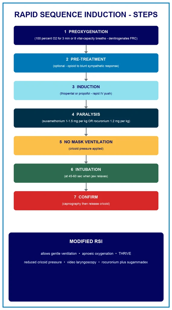
The classical sequence was a fixed, seven-step ritual that excluded any face-mask ventilation between induction and intubation, on the reasoning that positive-pressure ventilation could insufflate the stomach, increase the regurgitation pressure, and so defeat the purpose of the technique. The modern, examined position is that this classical rule is neither sacred nor safe in every patient: in the obese, the pregnant, the child, and the critically ill, the functional residual capacity is small and the oxygen consumption is high, so the safe apnoea period collapses to under two minutes, and the rigid refusal to ventilate produces a hypoxic patient before the tube is placed. The modified RSI — the contemporary default in emergency and critical-care practice — preserves the principle of a swift, planned, single-tube placement while layering on gentle mask ventilation, apnoeic oxygenation with high-flow nasal cannula, video laryngoscopy, and a rocuronium-sugammadex pair that gives the operator a pharmacological rescue from the cannot-intubate-cannot-oxygenate situation[4][6].
The topic is examined heavily because it sits at the intersection of pharmacology, physiology, and airway technique, and because the modern literature has refined — and in places overturned — several of the classical tenets: cricoid pressure, the no-ventilation rule, and the primacy of suxamethonium[2][3][6].
Indications for RSI
The indication for an RSI, rather than a standard inhalational or intravenous induction with mask ventilation, is any situation in which the stomach is presumed full and the risk of regurgitation and aspiration is therefore higher than the baseline of the fasted elective patient[4].
- The full stomach. A patient who has eaten within the fasting window (typically six hours for solids, two for clear fluids), or who cannot give a reliable fasting history (the confused, the traumatised, the intoxicated).
- Pregnancy, second and third trimester. The progesterone-relaxed lower oesophageal sphincter, the mechanically displaced and compressed stomach, and the increased intra-gastric pressure together raise the aspiration risk; pregnancy is the textbook indication for RSI in every exam syllabus.
- Bowel obstruction and peritonitis. The obstructed or inflamed gut holds air and fluid under pressure, and the patient is often actively vomiting.
- Active vomiting or haematemesis. An immediate aspiration threat.
- Trauma and the emergency patient. Pain, opioids, shock, and delayed gastric emptying combine to invalidate any fasting assumption; every trauma intubation is an RSI.
- Delayed gastric emptying. Opioid use, diabetes mellitus (autonomic neuropathy), renal failure, and the critically ill all slow gastric emptying and raise the residual volume.
- Haemodynamic instability requiring immediate anaesthesia. The patient who needs anaesthesia now, for a procedure or for control of the airway, in whom there is no time to fast.
- The emergency and the critical-care intubation. The default technique for nearly every intubation in the emergency department and the intensive care unit, because these patients cannot be assumed to be fasted and are often physiologically fragile[6].
The contraindication to a classical RSI is the anticipated difficult airway in a patient who can still cooperate — the syndromic, the post-radiation, the obstructed, or the known cervical-spine-injured patient with stridor at rest — in whom an awake fibreoptic intubation secures the airway before the loss of tone. The RSI assumes that the airway can be managed once the patient is paralysed; where that assumption is unsafe, the technique changes[6].
The classical 7-step sequence
The classical RSI is a fixed, rehearsed sequence of seven steps, taught because it forces the operator to preoxygenate, to paralyse, and to intubate without pausing to ventilate. The order is inviolable, and the rationale of each step is the examined point[4].
- Preoxygenation. One hundred per cent oxygen for three minutes of tidal breathing, or eight vital-capacity breaths, to denitrogenate the functional residual capacity and build the alveolar oxygen reservoir that funds the apnoeic period (detailed below)[1].
- Pre-treatment (optional). An opioid — classically fentanyl — to blunt the sympathetic and the vagal responses to laryngoscopy and intubation, given three minutes before induction. This step is optional and is omitted in the rapidly deteriorating patient.
- Induction. The induction agent given as a rapid intravenous push: thiopental or propofol in the cardiovascularly stable patient, ketamine or etomidate in the unstable. The dose is reduced in shock (detailed below).
- Paralysis. The neuromuscular blocker given immediately after the induction agent, in a paralytic dose: suxamethonium 1 to 1.5 mg per kg, or rocuronium 1.2 mg per kg. Onset of full paralysis is at 45 to 60 seconds[3].
- No face-mask ventilation. Cricoid pressure (the Sellick manoeuvre) is applied BEFORE the loss of consciousness, and no positive-pressure mask ventilation is given between induction and intubation, to avoid insufflating the stomach.
- Intubation. Performed as soon as the jaw relaxes — at 45 to 60 seconds after the paralytic — with a direct or video laryngoscope, and the cuff inflated as soon as the tube is through the cords.
- Confirmation and release. Tracheal placement is confirmed by waveform capnography over six breaths, and ONLY THEN is the cricoid pressure released.
The discipline of the sequence is its strength: it is a single, unhurried, rehearsed act, and the operator who has run it in simulation performs it cleanly in crisis. The examined weakness, returned to throughout, is that several of its fixed rules — the no-ventilation step, the obligatory cricoid pressure, the primacy of suxamethonium — have been revised by the modern evidence[2][4].
Preoxygenation: denitrogenation and the safe apnoea period
Preoxygenation is the single most important preparatory step, because it is the step that funds the apnoeic period that follows. Its purpose is to replace the nitrogen in the functional residual capacity (the reservoir gas in the lungs at end-expiration) with oxygen, so that when the patient stops breathing, the alveoli contain a reservoir of oxygen that diffuses into the blood for as long as the alveolar-to-venous partial pressure gradient is maintained[1].
The standard technique is one hundred per cent oxygen for three minutes of tidal breathing, or eight vital-capacity breaths. The two are approximately equivalent in the healthy lung: three minutes of tidal breathing washes out the nitrogen at the breathing rate, while eight vital-capacity breaths achieves the same denitrogenation faster by exchanging a larger volume per breath. The end-point is a fraction of expired oxygen (end-tidal oxygen) approaching 90 per cent, which is the objective evidence that the functional residual capacity has been denitrogenated. The end-tidal oxygen is the metric that confirms a successful preoxygenation, and it is increasingly advocated as the bedside target that should replace the clock[1].
The reward is the safe apnoea period — the time from the cessation of breathing to the fall of the arterial saturation to below 90 per cent. In a healthy, fasted, normal-weight adult, fully preoxygenated, the safe apnoea period is approximately 3 to 8 minutes. This is the window in which the tube must be placed. The figure is not a constant: it is shortened by anything that reduces the functional residual capacity (obesity, pregnancy, the supine position, splinting) or increases the oxygen consumption (sepsis, the child, the agitated patient), and it is lengthened by apnoeic oxygenation (THRIVE, detailed below)[1][4].
The teaching point, examined repeatedly, is that the safe apnoea period in the patient who actually needs an RSI is often much shorter than the textbook 3 to 8 minutes. The obese patient may desaturate inside two minutes, the pregnant patient similarly, and the child inside one minute. These patients are the primary indication for the modified RSI with apnoeic oxygenation, because the classical sequence that refuses to ventilate will produce a hypoxic patient before the tube is placed[4][6].
Induction agents and the haemodynamically unstable patient
The induction agent is given as a rapid intravenous push immediately before the neuromuscular blocker, and its purpose is to abolish consciousness so that the laryngoscopy and the intubation are not experienced, and so that the sympathetic response to laryngoscopy is blunted. The choice of agent and the dose are governed by the cardiovascular reserve, and the most common error — and the most common cause of peri-intubation harm — is the failure to adjust for shock[4][6].
- Thiopental (3 to 5 mg per kg) — the classical induction agent for RSI; a rapid-onset barbiturate that produces unconsciousness in one arm-brain circulation time. It is a myocardial depressant and a venodilator, and the standard dose will cause profound hypotension in the hypovolaemic or the shocked patient.
- Propofol (1.5 to 3 mg per kg) — the modern standard; rapid and smooth, but a more potent vasodilator than thiopental, and equally hazardous in shock at the standard dose.
- Ketamine (1 to 2 mg per kg) — the agent of choice for the haemodynamically unstable patient; a dissociative anaesthetic that preserves the sympathetic tone and the blood pressure, and a bronchodilator that suits the asthmatic. Its traditional disadvantages (a rise in intracranial pressure, emergence phenomena) have been tempered by re-evaluation, and its haemodynamic stability makes it the default in the emergency and the critical-care RSI.
- Etomidate (0.2 to 0.3 mg per kg) — a haemodynamically neutral imidazole that is the alternative for the unstable patient; the recurring controversy is its transient suppression of adrenal steroidogenesis, which has been linked to worse outcomes in sepsis, and which is the reason some practitioners prefer ketamine. [1]
The cardinal rule, examined and bed-side alike, is that the patient in shock will cardiovascularly collapse with a standard induction dose. The mechanisms are that the induction agent removes the sympathetic drive that has been holding the vascular tone, and that the positive-pressure ventilation that follows the intubation reduces the venous return. The practical responses are three: reduce the dose (a half or a quarter of the standard dose is common in shock), choose a haemodynamically stable agent (ketamine or etomidate), and have a vasopressor drawn up and ready (metaraminol, phenylephrine, or an adrenaline infusion) to treat the post-induction hypotension immediately[4][6].
The national audits of emergency intubation consistently find that cardiovascular instability is the most common major peri-intubation adverse event, exceeding the rate of difficult intubation, and that the induction agent given at an unadjusted dose to the shocked patient is the most preventable cause. The high-scoring candidate frames the induction agent not as a recipe but as a haemodynamic decision[4].
Neuromuscular blocking agents: suxamethonium versus rocuronium
The neuromuscular blocker produces the jaw relaxation and the abolition of the gag and the vocal-cord reflex that make the intubation possible, and in the RSI it must act fast — within 45 to 60 seconds — so that the airway is secured before the safe apnoea period expires. The two agents that achieve this are suxamethonium (succinylcholine) and rocuronium in a high dose, and the choice between them is a perennial exam question and a live clinical debate[3].
Suxamethonium (1 to 1.5 mg per kg) is a depolarising neuromuscular blocker that produces fasciculations followed by paralysis within 45 to 60 seconds, and which is metabolised by plasma cholinesterase with a recovery to spontaneous ventilation in around 5 to 10 minutes. It has the fastest, most reliable onset of any agent, and it has been the traditional first choice for RSI on that ground alone. Its disadvantages are well-rehearsed and heavily examined: [1]
- Hyperkalaemia — a small rise in serum potassium occurs in every patient, but a life-threatening rise occurs in the denervated, the burns (after 24 to 48 hours), the crush, the renal failure, and the neuromuscular disease patient, where up-regulation of the acetylcholine receptors produces a dangerous potassium efflux.
- Anaphylaxis — suxamethonium remains one of the most common triggers of anaesthetic anaphylaxis.
- Malignant hyperthermia — the suxamethonium-halothane susceptibility is a contraindication in the known susceptible patient.
- Bradycardia — repeated doses produce a muscarinic bradycardia, requiring atropine.
- Prolonged apnoea — the patient with a genetic plasma-cholinesterase deficiency (the dibucaine-number variants) cannot metabolise the drug, and the paralysis lasts for hours. [1]
Rocuronium at 1.2 mg per kg is a non-depolarising aminosteroid that achieves intubating conditions at 45 to 60 seconds, comparable to suxamethonium, and which is reversible by sugammadex 16 mg per kg — the modified gamma-cyclodextrin that encapsulates the rocuronium molecule and restores neuromuscular function within minutes. The availability of sugammadex has been the decisive change: rocuronium now offers the rapid onset of suxamethonium WITHOUT the hyperkalaemia, the anaphylaxis risk of suxamethonium, the malignant hyperthermia, or the prolonged apnoea, and WITH a pharmacological rescue from the cannot-intubate-cannot-oxygenate situation that suxamethonium cannot match[3].
The contemporary outcome literature finds that the two agents achieve equivalent first-pass intubation success and equivalent rates of complications, and that the choice increasingly favours rocuronium in the emergency and critical-care setting precisely because its side-effect profile is cleaner and its reversibility is a safety net[3]. The traditional contraindication to rocuronium — that its longer duration made the cannot-intubate-cannot-oxygenate situation unsurvivable — has been removed by sugammadex, and this is the modern teaching point[3][4].
Cricoid pressure: the Sellick manoeuvre and the controversy
Cricoid pressure — the Sellick manoeuvre — is the backward pressure applied to the cricoid cartilage to compress the oesophagus against the body of the sixth cervical vertebra, occluding the oesophageal lumen and preventing the passive regurgitation of gastric contents into the pharynx during the induction. It is applied before the loss of consciousness (so that the semi-conscious patient does not gag and vomit), and it is held until the tracheal tube is confirmed by capnography[2].
The technique is a single-handed or a bimanual manoeuvre: the operator places the thumb and the index finger on the cricoid cartilage and applies a force of 10 newtons to the awake patient, rising to 30 newtons with the loss of consciousness. The cricoid is the only complete tracheal ring, which is why the pressure occludes the oesophagus behind it without narrowing the airway — in the ideal anatomy[2].
The controversy is that the ideal anatomy is not always present, and the manoeuvre is not always beneficial. The examined objections are four: [1]
- Anatomical unreliability. Magnetic resonance imaging studies have shown that the oesophagus is frequently displaced laterally to the cricoid, and that the cricoid pressure compresses the oesophagus only in the minority of patients in whom the anatomy is midline. The mechanical premise of the manoeuvre is often not met.
- Impaired laryngoscopic view. Cricoid pressure displaces the larynx posteriorly and laterally, and it is a recognised cause of a worse Cormack-Lehane grade. The very manoeuvre intended to protect the airway can make the intubation harder.
- Airway obstruction. Excessive force, or pressure applied to the thyroid rather than the cricoid, can obstruct the airway and make mask ventilation impossible — the opposite of the intended effect.
- Cervical spine movement. The bimanual technique, and the single-handed technique in the suspected cervical-spine-injured patient, produce measurable cervical spine movement, which is a concern in the trauma patient whose spine is not yet cleared[2].
The modern, evidence-based position is that cricoid pressure is optional, not obligatory. It may still be used in the patient with a clear aspiration risk, but it should be applied lightly, and — critically — it should be reduced or released immediately if it interferes with the laryngoscopic view, with mask ventilation, or with the intubation. The mantra is that the cricoid pressure should never be the obstacle to a successful intubation; a tube in the trachea is the best protection against aspiration, and a cricoid pressure that prevents the tube is self-defeating[2][4].
The candidate who recites cricoid pressure as a fixed, mandatory step of the RSI without acknowledging the controversy marks themselves as the uncritical rote learner; the high-scoring candidate presents the technique, the rationale, the objections, and the modern selective-or-release approach[2].
The modified RSI: the modern default
The modified RSI is the contemporary evolution of the classical sequence, and it is the default technique in modern emergency and critical-care airway practice. It preserves the core principle — a swift, planned, single-tube placement after induction and paralysis — but it revises the fixed rules that the evidence has shown to be unsafe in the patient who actually needs an RSI. The modifications are four[4][6].
- Gentle mask ventilation during the apnoeic period. The classical no-ventilation rule is abandoned in the patient at risk of rapid desaturation. Gentle positive-pressure mask ventilation (the lowest pressure that moves the chest, to minimise gastric insufflation) maintains the arterial oxygenation during the interval between induction and intubation. This is the single most important modification: it acknowledges that the hypoxaemia that the classical rule risks is a greater harm than the theoretical aspiration it prevents, especially in the obese, the pregnant, the child, and the critically ill[6].
- Reduced or omitted cricoid pressure. As detailed above, cricoid pressure is applied selectively and released if it interferes. The modified RSI does not depend on it.
- Apnoeic oxygenation (THRIVE). Transnasal humidified rapid-insufflation ventilatory exchange — high-flow nasal cannula oxygen at 70 L per min, with the mouth closed — delivers oxygen continuously into the nasopharynx during the apnoeic period. Because the alveolar-to-venous oxygen gradient continues to draw gas into the lungs even during apnoea, the continuous nasal flow replenishes the alveolar reservoir and dramatically extends the safe apnoea period, in some studies to over 30 minutes in the favourable patient. THRIVE is now a standard adjunct of the modified RSI[4].
- Video laryngoscopy as first-line. The video laryngoscope (the hyperangulated blade with the camera on the blade) provides a view of the glottis that the direct laryngoscope cannot, and it is increasingly the first-line device in the emergency and the difficult airway, improving the first-pass success and reducing the trauma of repeated attempts.
- Rocuronium instead of suxamethonium. The rocuronium-sugammadex pair offers the rapid onset of suxamethonium without its side-effect profile and with a pharmacological rescue, as detailed above. The modified RSI defaults to rocuronium 1.2 mg per kg[3].
The modified RSI is not a different technique from the classical RSI; it is the same technique adapted to the patient in front of the operator. The candidate who can name the classical sequence AND the modifications, and explain WHY each modification was made, has grasped the examined point: the RSI is a principle (swift, single-tube placement), and the rules are the servants of the principle, not its masters[4][6].
Apnoeic oxygenation and THRIVE
Apnoeic oxygenation is the physiological principle that underpins the most consequential of the modified-RSI adjuncts, and it deserves its own treatment because it is the answer to the central problem of the RSI — the finite safe apnoea period[4].
The principle is that during apnoea, the alveolar-to-venous oxygen partial-pressure gradient continues to drive oxygen from the alveoli into the blood. The blood continues to take up oxygen from the alveolar gas, the alveolar gas volume falls, and if a continuous supply of oxygen is provided to the upper airway, the oxygen is drawn into the alveoli to replace the volume absorbed. The result is that the arterial oxygenation is maintained — for as long as the upper-airway oxygen supply continues and for as long as the alveoli do not collapse — even though the patient is not breathing. [1]
The classical preoxygenation funds this gradient for a few minutes by pre-filling the functional residual capacity with oxygen. THRIVE extends it dramatically by delivering a continuous, high-flow (70 L per min), humidified nasal oxygen throughout the apnoeic period, replenishing the alveolar reservoir as fast as it is absorbed. In the favourable patient, THRIVE has extended the safe apnoea period to over 30 minutes, and it has transformed the practice of the difficult airway and the morbidly obese airway, where the classical safe apnoea period was measured in one or two minutes[4].
The caveats are two. First, THRIVE does not ventilate — the carbon dioxide continues to rise, at roughly 1 to 2 kPa in the first hour, and the respiratory acidosis eventually becomes a problem in the prolonged apnoea. Second, THRIVE works best where the upper airway is patent; the high nasal flow provides some continuous positive airway pressure that splints the upper airway, but an obstructed airway (the grossly obese, the obstructing tumour) limits the benefit. The technique is an adjunct that extends the window, not a substitute for the tube[4].
Confirmation of tracheal placement and post-intubation care
The seventh step of the classical sequence — the confirmation of the tracheal tube and the release of the cricoid pressure — is the step that converts an attempted intubation into a secured airway, and the failure to confirm is a recurring cause of the catastrophes that the audits record[4].
Waveform capnography is the mandatory standard for the confirmation. The detection of carbon dioxide in the exhaled gas over six breaths confirms that the tube is in the trachea or a main bronchus, not in the oesophagus. The waveform must be a square-wave trace that persists across the breaths; a single trace that then flattens is the signature of an oesophageal intubation that has cleared the small reservoir of carbon dioxide from the stomach. The colour-change capnometry (the chemical detector) is a fallback where waveform capnography is unavailable, but it is not the standard. The clinical signs — the chest movement, the auscultation, the condensation in the tube — are supplementary and unreliable, and they do not replace the capnography[4].
Cricoid pressure is released only after the capnographic confirmation. Releasing it before the confirmation, in the patient whose tube is in fact in the oesophagus, allows the regurgitation that the manoeuvre was preventing, and it is the error the sequence is designed to prevent[2].
After the confirmation, the tube is secured, the patient is ventilated with a tidal volume and a rate that achieve a normocapnia (or a permissive hypercapnia in the asthmatic and the raised intracranial pressure patient), and the haemodynamics are stabilised — the post-intubation hypotension treated with fluid, vasopressor, and the reduction of the anaesthetic depth. The maintenance of anaesthesia is then transitioned to an inhalational agent or a total intravenous technique, and where the RSI was performed in the prehospital or the retrieval context, an intermittent bolus regime (ketamine and a relaxant) is a recognised maintenance strategy that balances the depth and the haemodynamic stability[5].
Special contexts: obesity, pregnancy, the child, and the critical illness
Four contexts reshape the RSI because they alter the safe apnoea period, the aspiration risk, or the haemodynamic reserve, and they are the contexts most often examined in the applied scenario[6].
Obesity reduces the functional residual capacity (the weight of the chest wall and the abdominal contents splint the diaphragm), increases the oxygen consumption, and raises both the aspiration risk and the difficult-airway probability. The obese patient is the textbook indication for the modified RSI: a ramped position (the head and the shoulders elevated so that the external auditory meatus is level with the sternal notch), full preoxygenation to an end-tidal oxygen above 90 per cent, THRIVE throughout, gentle mask ventilation, and a senior operator with a video laryngoscope from the outset. The classical refusal to ventilate will produce a hypoxic patient inside two minutes[6].
Pregnancy is the classical indication for RSI: the aspiration risk (progesterone-relaxed lower oesophageal sphincter, the compressed stomach, the increased intra-gastric pressure) and the difficult-airway risk (the laryngeal and the pharyngeal oedema, the displaced anatomy, the breast enlargement) are both elevated, and the safe apnoea period is shortened by the increased oxygen consumption and the reduced functional residual capacity. The technique demands a left lateral tilt to relieve the aortocaval compression, a reduced induction dose (the volatile requirement and the cardiac output are altered), a smaller endotracheal tube (the oedematous larynx), and a senior obstetric anaesthetist from the outset[4].
The child desaturates very rapidly (a small functional residual capacity and a high metabolic rate, so the safe apnoea period may be under one minute), and the RSI must be modified accordingly: full preoxygenation, gentle mask ventilation, and a paralysing dose that is weight-calculated. The child is also the context where the suxamethonium hyperkalaemia risk is highest in the undiagnosed neuromuscular disease, which is a further reason for the rocuronium preference[3].
The critical illness and the emergency department patient is the context that has driven most of the modern modifications. These patients combine the aspiration risk (not fasted) with the physiological difficult airway (hypoxaemic, hypotensive, acidotic, often shocked), and the RSI is the technique that must both secure the airway and protect the haemodynamics. The physiological difficult airway is the concept that the anatomically easy airway becomes impossible when the patient desaturates or arrests on induction, and it demands resuscitation before, during, and after the intubation — fluid, vasopressors, and an unhurried, well-prepared single best attempt. The recognition of the physiological difficult airway as a distinct entity is the major conceptual advance of the recent emergency and critical-care airway literature, and it is the element most often missing from the candidate who recites the classical sequence mechanically[6].
Clinical
- Standard approach
- Evidence-based
Alternative
- Modified technique
- Risk-benefit
Red flags
[1] [1] [1] [1] [1] [1]References
- [1]Caputo ND, et al. End Tidal O(2): A Promising New Metric for Optimizing Preoxygenation and RSI Safety in the Emergency Department Acad Emerg Med, 2026.PMID 42340046
- [2]Kumar J, et al. Cervical Spine Movements With Single-Handed Versus Bimanual Cricoid Pressure Under Simulated Cervical Immobilization: A Randomized Controlled Trial J Neurosurg Anesthesiol, 2026.PMID 42333579
- [3]O'Connell DH, et al. Outcomes of Succinylcholine and Rocuronium for Rapid Sequence Intubation in the Emergency Department West J Emerg Med, 2026.PMID 42258841
- [4]Freund Y, et al. Improving the safety of emergency tracheal intubation Curr Opin Crit Care, 2026.PMID 42170830
- [5]Sheridan B, et al. Maintenance of prehospital anaesthesia using an intermittent bolus regime in blunt trauma patients with a high GCS and hemodynamic reserve: a retrospective cohort study Scand J Trauma Resusc Emerg Med, 2026.PMID 42351216
- [6]Ghaffar S, et al. Physiological difficult airway management in the emergency department J Pak Med Assoc, 2026.PMID 42363338