Anaes · Perioperative medicine
Anaesthesia in remote sites and the MRI environment
Also known as MRI anaesthesia · Remote site anaesthesia standards · MRI zones safety · Ferromagnetic projectile risk · Non-operating room anaesthesia MRI
Exam-exhaustive remote-site and MRI anaesthesia: organisational standards equal to the OR, MRI Zones I–IV, ferromagnetic projectile risk, implant labelling, MRI-conditional monitoring, quench response, and practical GA/sedation logistics for ANZCA Final and equivalents.
On this page & tools
Your progress
Saved locally on this device.
Target exams
Red flags
Why this is examined / the one-line answer
Remote-site anaesthesia (MRI, CT, endoscopy, IR) is a classic fellowship safety viva: same patient, worse environment. MRI multiplies risk with an always-on magnet, limited access to the airway, noisy monitoring, and specialised equipment rules. Examiners want organisational standards plus MRI physics applied to real decisions — not a physics monologue.[1][2]
One-liner: I insist on OR-equivalent staffing and monitoring, complete ferromagnetic and implant screening, MRI-conditional equipment, a clear airway/induction plan relative to Zone IV, continuous visible monitoring, and rehearsed projectile/quench responses. [1]
AAGBI/NACCS 2019 MRI anaesthesia guidelines and ACR MR Safe Practices are the named anchors.[1][2][3]
Preoperative assessment and risk stratification
Remote-site standards (apply to all NORA, including MRI)
Before accepting any remote case, confirm: [1]
| Requirement | Exam language |
|---|---|
| Trained anaesthesia help available | Not “radiographer will shout if needed” alone |
| Oxygen, suction, ventilation, resuscitation drugs | Immediately available and checked |
| Standard monitoring capability | SpO2, NIBP, ECG, capnography for GA/deep sedation |
| Recovery pathway | Staffed recovery or PACU-equivalent — not a corridor |
| Communication | Phone/alarm to main theatre/arrest team that works in that suite |
| Lighting, space, scavenging as applicable | Especially for GA in MRI |
| Case appropriateness | Can this wait for a better environment? |
Rule: If you cannot provide equivalent safety to the operating theatre, relocate or defer non-urgent imaging under anaesthesia.[1]
MRI-specific history
- Prior MRI problems, claustrophobia, pain on lying flat
- Implants: pacemaker/ICD, neurostimulators, cochlear implants, aneurysm clips, stents, tissue expanders, metal fragments (eye/orbit history, sheet-metal work), bullets, medication patches with foil, drug delivery pumps
- Pregnancy (risk–benefit for MRI; avoid gadolinium unless essential)
- Renal function if gadolinium considered (NSF risk in severe CKD — radiology protocols)
- Airway predictors — because the head is often in the bore and access is delayed [1]
Screening culture
Everyone (patient, anaesthetist, ODP, radiographer, parent if allowed) completes MRI screening. Pocket check: pens, scissors, phones, bleeps, stethoscopes, gas cylinders, ordinary laryngoscope batteries, steel oxygen cylinders — none enter Zone IV unless labelled safe/conditional for that field strength.[2][3]
Applied physics that changes the plan (keep it clinical)
Three fields
- Static field (B0) — typically 1.5 T or 3 T: projectile effect, implant torque/translation, always on.
- Gradient fields — switching: noise, peripheral nerve stimulation, ECG artefact.
- Radiofrequency — tissue heating (SAR), burns from loops of monitoring wires or skin-to-skin contact. [1]
Implant labels (must say cleanly)
| Label | Meaning | Action |
|---|---|---|
| MR Safe | No known hazards in MRI | Proceed with normal screening |
| MR Conditional | Safe only under specified conditions (field strength, spatial gradient, SAR, coil) | Follow exact conditions; document |
| MR Unsafe | Hazard | Do not scan (or specialised research pathways only) |
Unknown implant = do not scan until identified.[2][3]
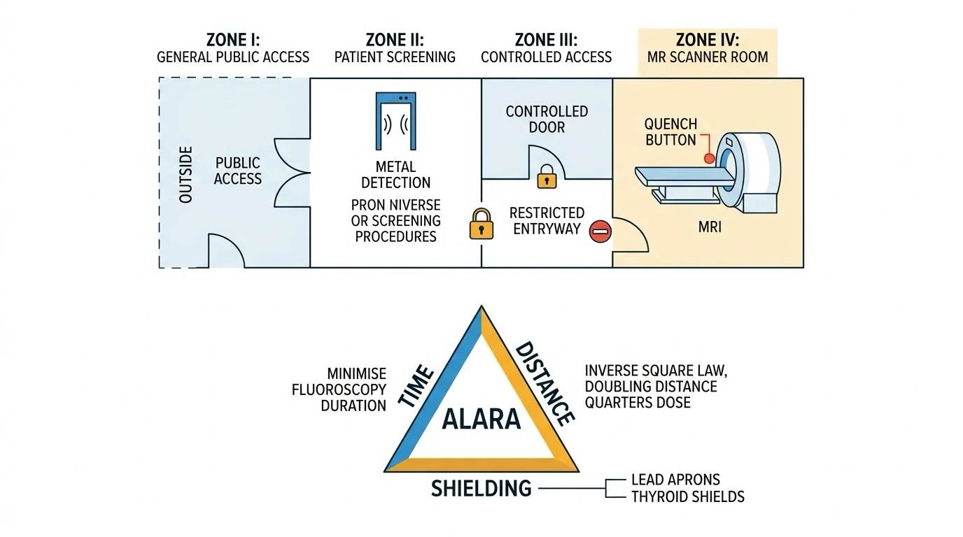
Zones I–IV (ACR)
| Zone | What it is | Anaesthetist action |
|---|---|---|
| I | General public access | Normal |
| II | Interface/screening | History, consent, change into safe attire |
| III | Restricted — serious hazard if unscreened entry | Controlled access; only screened people/equipment |
| IV | Magnet room | Conditional equipment only; quench button awareness |
Zone III/IV access control is a core safety design, not bureaucracy.[2]
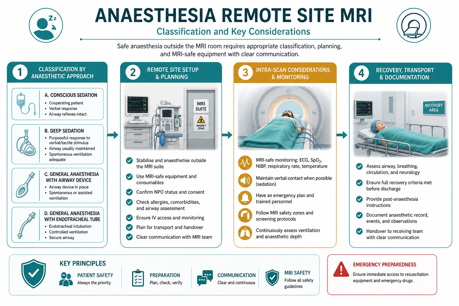
Anaesthetic goals
- Safety culture first — screening and equipment discipline.
- Secure airway and ventilation before the head becomes inaccessible when needed.
- Reliable monitoring with MRI-conditional systems and artefact awareness.
- Immobility adequate for image quality with minimal residual sedation risk.
- Exit strategy — how to get the patient out and resuscitated in seconds to minutes.
- Same standards as main theatre for drugs, checks, and recovery.[1]
Technique options and decision matrix
Where to induce
Paediatric MRI is commonly GA with ETT or SGA per local practice; parental presence only if screened and local policy allows.[1]
Monitoring in MRI
- SpO2: MRI-conditional probe; watch for artefact
- Capnography: mandatory for GA/deep sedation — long sampling lines increase delay
- NIBP: conditional cuff; cycle times may lag
- ECG: special electrodes/leads; ST analysis unreliable; used mainly for rate/rhythm
- Temperature: consider for long GA (RF heating vs environmental cold)
- Visual patient viewing + audible alarms routed to the control room [1]
Wire rules: avoid coiled leads (induction heating/burns); place insulating padding; prevent skin-to-skin contact loops.[1][3]
Equipment checklist (say in viva)
MRI-conditional: anaesthesia machine/ventilator or long Mapleson/circle adaptations per suite design, infusion pumps (or keep pumps in Zone III with long lines if validated), laryngoscope (fibreoptic/plastic battery designs as approved), airway trolley plan for remove-from-magnet-first resuscitation, non-ferromagnetic oxygen source strategy, defibrillator kept outside Zone IV with drill to evacuate patient to it. [1]
Steel oxygen cylinders are classic projectile hazards — never take a standard cylinder into the magnet room. [1]
Intraoperative management
Team brief: field strength, zones, implant status, induction location, airway plan, who watches monitors in control room, emergency “stop scan / remove patient” command. [1]
Airway: preoxygenate; plan VL/FOI as indicated before entering bore. Once scanning, airway access may require table withdrawal — build that into the crisis time estimate. [1]
Maintenance: TIVA (propofol ± remifentanil) popular to reduce machine complexity; volatiles if MRI-conditional vapouriser/machine available. Avoid unnecessary deep paralysis without airway security. [1]
Hearing protection for patients (gradient noise). [1]
Contrast: gadolinium by radiology protocol; anaphylaxis plan identical but execute outside magnet if possible. [1]
Crisis pivots
Projectile event
Do not chase the object into the magnet unplanned. Protect patient, stop scan, call for help, follow local MRI emergency protocol; only ferromagnetic object removal by trained teams with field considerations. [1]
Cardiac arrest in Zone IV
Priority teaching: start CPR as able, remove patient from magnet room to a designated resuscitation area where full ALS equipment (defibrillator) is available — do not bring a standard defibrillator into the bore. Assign roles in advance.[1]
Airway obstruction under sedation in the bore
Stop scan, withdraw table, jaw thrust/airway adjuncts, deepen or convert to secured airway in safe zone, call for help. [1]
Quench
Sudden helium boil-off: white fog, asphyxiation, cold injury, pressure risks. Evacuate Zone IV, avoid entering fog, account for staff/patient, oxygen outside, engineering/MRI physicist notify. Quench is rare and reserved for life-threatening emergencies (e.g. person trapped by ferromagnetic object) — not for routine arrests if patient can be removed.[2]
Thermal burn under ECG electrodes
Stop scan, inspect skin, redesign lead routing, padding, replace electrodes with approved types. [1]
Postoperative / recovery
- Recover in a staffed area with full monitoring until return of airway reflexes and baseline consciousness
- Do not recover alone in the scanner control room
- Handover implant/contrast issues
- Day-case MRI GA: standard discharge criteria, escort, written advice [1]
Special populations
ICU patients: full transfer checklist; MRI-conditional ventilator or manual ventilation plan; infusions reformulated to conditional pumps; many ICU devices are unsafe — strip and replace systematically. [1]
CIED patients: only if MR-conditional device and cardiology protocol (programming, monitoring); otherwise alternative imaging. [1]
Pregnancy: MRI without gadolinium preferred when imaging essential; anaesthesia rare — multidisciplinary. [1]
Claustrophobia without GA: psychology/sedation alternatives; do not force unsafe under-sedation in bore. [1]
SAQ answer scaffold
You are asked to provide GA for a 4-year-old for MRI brain in a 3 T scanner. Outline your management. [1]
- Remote standards (3): staffing, monitoring, recovery, emergency help, equipment check.[1]
- MRI safety (3): zones, screening of child and staff, remove ferromagnetic items, conditional equipment only.[2]
- Anaesthetic technique (4): induction location, airway choice, TIVA vs volatile as available, ear protection, temperature.
- Crisis plan (3): stop scan, withdraw table, resus outside Zone IV, quench awareness.
- Recovery (2): PACU-level care until safe.
Viva stem bank and model phrases
Stem 1: “Is remote site anaesthesia lower risk because no surgery?”
Model: “No — standards equal the operating theatre; the environment is often higher risk because help and equipment are farther away.”[1]
Stem 2: “What are the MRI zones?”
Model: “I to IV — public, screening interface, restricted controlled access, and the magnet room itself.”[2]
Stem 3: “Why is the oxygen cylinder dangerous?”
Model: “A ferromagnetic cylinder can become a high-speed projectile in the static field, which is always on.” [1]
Stem 4: “MR Conditional means?”
Model: “Safe only under specified conditions of field strength and scanning parameters — not blanket permission for any MRI.” [1]
Stem 5: “Cardiac arrest in the scanner?”
Model: “Call for help, start CPR, remove the patient from Zone IV to the designated resus area with a defibrillator — I do not take a standard defibrillator to the magnet.”[1]
Stem 6: “Why do ECG leads burn?”
Model: “RF energy can induce currents, especially in loops; we use approved leads, avoid coils, and pad the skin.” [1]
Stem 7: “When do you refuse the case?”
Model: “Unsafe implant status, inadequate equipment/staffing, or when imaging can wait until a safe pathway exists.” [1]
Common traps
- “It’s only a scan” complacency
- Taking a bleep/phone/scissors into Zone IV
- Standard steel cylinder in magnet room
- Deep sedation without capnography
- No plan to extricate for ALS
- Assuming all modern implants are safe
- Recovering unmonitored in a dark control room [1]
MRI anaesthesia — ZONE
Examiner mental map
- Remote-site standards = OR.
- Zones + screening.
- Safe/Conditional/Unsafe.
- Conditional monitoring/airway plan.
- Arrest = extricate.
- Quench = evacuate.
That is the MRI anaesthesia pass package. [1]
References
- [1]Wilson SR, Shinde S, Appleby I, et al. Guidelines for the safe provision of anaesthesia in magnetic resonance units 2019: Guidelines from the Association of Anaesthetists and the Neuro Anaesthesia and Critical Care Society of Great Britain and Ireland Anaesthesia, 2019.PMID 30714123
- [2]Kanal E, Barkovich AJ, Bell C, et al. ACR guidance document on MR safe practices: 2013 J Magn Reson Imaging, 2013.PMID 23345200
- [3]Greenberg TD, Hoff MN, Gilk TB, et al. ACR guidance document on MR safe practices: Updates and critical information 2019 J Magn Reson Imaging, 2020.PMID 31355502