Anaes · Measurement & monitoring physics
Humidity and heat
Also known as Humidity · Absolute humidity · Relative humidity · Dew point · Latent heat of vaporisation · Heat and moisture exchanger · Inadvertent perioperative hypothermia
Humidity and heat are the physics behind two questions the anaesthetist answers every day — is the gas I am delivering kind to the airway, and is my patient's temperature safe? The framework rests on six exam-critical ideas. First, HUMIDITY is described three ways: ABSOLUTE humidity (the mass of water vapour per unit volume of gas, in mg/L), RELATIVE humidity (the percentage of water vapour actually present relative to the maximum the gas can hold at that temperature), and the DEW POINT (the temperature at which the gas becomes fully saturated and water begins to condense). Second, HEAT and TEMPERATURE are not synonyms: heat is the THERMAL ENERGY content of a body, measured in joules, while temperature is the DEGREE of hotness on a scale, measured in degrees. Third, heat is transferred by four mechanisms — CONDUCTION (direct molecular contact), CONVECTION (movement of a warmed fluid or gas), RADIATION (infrared transfer between surfaces, accounting for about 60 per cent of perioperative loss), and EVAPORATION (from the surgical wound and the respiratory tract). Fourth, the LATENT HEAT OF VAPORISATION of water is about 2.4 MJ/L — the energy needed to convert liquid water to vapour — which is why evaporation is such a powerful coolant and why a dry gas draws heat and moisture from the airway. Fifth, medical gases from a cylinder or machine are essentially dry and cold, so they must be HUMIDIFIED to prevent desiccation of the mucosa, ciliary dysfunction, retained secretions and heat loss; the two methods are the PASSIVE heat and moisture exchanger (HME — a hygroscopic membrane that retains exhaled heat and water, single-use, low resistance) and the ACTIVE heated water-bath humidifier (more effective, used for long-term ventilation). Sixth, under anaesthesia the core temperature falls by 1 to 1.5 degrees C in the first hour from REDISTRIBUTION hypothermia (vasodilatation moves core heat to the periphery), then falls more slowly by ongoing loss; prevention rests on forced-air warming, fluid and blood warming, radiant warming and an adequate ambient temperature, because hypothermia causes wound infection, coagulopathy and increased blood loss, shivering with cardiac stress, and prolonged drug action. Built on the hygrometric-properties-of-passive-humidifiers study (Lellouche 2026), the ventilatory-failure-from-retained-HMEF report (Zou 2026), the circuit-humidification-during-mechanical-ventilation review (Tsai 2026), the intraoperative-thermal-injury-from-warming-device report (Yeh 2026), the airway-warming-devices systematic review (Lee 2026), the blood-warming-device-performance study (Williams 2026), the risk-factors-for-postoperative-hypothermia review (Ji 2026), and the double-wall-warmer randomised trial (Kabra 2026).
On this page & tools
Your progress
Saved locally on this device.
8 MCQs with explanations
Target exams
Red flags
Why this matters to the anaesthetist
Two things make an anaesthetist reach for the physics of humidity and heat. The first is the airway: medical gases from a cylinder or machine are cold and essentially dry, and delivering them straight into a trachea for hours would desiccate the mucosa, stop the cilia beating, and leave the patient with retained secretions and infection. The second is temperature: anaesthesia abolishes thermoregulation, the patient cools, and a core a degree or two below normal measurably raises wound infection, blood loss, shivering and prolonged recovery. Understanding humidity, heat transfer, the latent heat of vaporisation, and the devices that humidify and warm lets the anaesthetist protect both the airway and the core [1][7].
Humidity — absolute, relative and the dew point
Humidity describes the amount of water vapour held in a gas, and it is expressed in three distinct ways that an examiner expects you to keep separate: [1]
- Absolute humidity — the mass of water vapour per unit volume of gas, expressed in mg/L. This is the quantity the airway actually experiences.
- Relative humidity — the ratio, as a percentage, of the water vapour actually present to the maximum the gas can hold at that temperature. Fully saturated gas has a relative humidity of 100 per cent.
- Dew point — the temperature at which the gas becomes fully saturated (relative humidity 100 per cent) and water begins to condense. Cooling a gas below its dew point forces condensation, which is how a cold breathing-circuit limb "rains out" water. [1]
A key relationship: the maximum water vapour a gas can hold RISES with temperature. So warming a sample of gas at constant water content LOWERS its relative humidity (it can now hold more), while cooling it RAISES the relative humidity until, at the dew point, it reaches 100 per cent. Alveolar gas at 37 degrees C is fully saturated with an absolute humidity of about 44 mg/L; the nose and upper airway normally warm and humidify inspired room air (which may hold only 10 mg/L) up to this alveolar condition [1].
Heat versus temperature
Heat and temperature are not the same thing, and candidates lose marks for treating them as interchangeable: [1]
- Heat is the thermal energy content of a body, measured in joules (J). It depends on how much matter there is and at what temperature.
- Temperature is the degree of hotness of a body, measured on a scale — degrees Celsius, Kelvin, or Fahrenheit. It is the property that determines the direction of heat flow (heat flows from a higher to a lower temperature).
- Specific heat capacity links them: it is the energy needed to raise the temperature of a unit mass of a substance by one degree, in J/kg/K. Water has a high specific heat capacity (about 4.2 kJ/kg/K), which is why the body — mostly water — resists rapid temperature change. [1]
The classic illustration: a hot spark is at a very high temperature but contains little heat (tiny mass), while a bath of warm water holds a great deal of heat at a modest temperature. Heat is what is lost and gained in the perioperative period; temperature is what the oesophageal probe reads [7].
Heat transfer mechanisms
Heat moves from one place to another by four mechanisms, all of which operate around an anaesthetised patient: [1]
- Conduction — heat flow through direct molecular contact between two bodies in touch, from the hotter to the cooler. A patient lying on a cold operating table loses heat by conduction into the mattress; it is usually a small fraction of total loss because the contact area and the conductivity of foam are low.
- Convection — heat carried away by the movement of a warmed fluid or gas. Warm air next to the skin rises and is replaced by cooler air (natural convection), or is blown away (forced convection in the laminar airflow of an operating theatre). Convection is a significant route of loss in theatre.
- Radiation — infrared electromagnetic energy transferred between surfaces that are not in contact, in proportion to the fourth power of their temperature difference. The patient radiates infrared to the cooler walls, ceiling and equipment. Radiation is the single largest route of perioperative heat loss, accounting for about 60 per cent of the total.
- Evaporation — the heat carried away when a liquid turns to vapour (see latent heat below). It operates from the exposed surgical wound (especially a large open abdomen) and from the respiratory tract when dry gases are breathed. [1]
Latent heat of vaporisation
When a liquid evaporates it takes energy to break the intermolecular bonds and turn molecules from liquid into vapour; that energy is drawn from the surroundings as heat. The latent heat of vaporisation is the energy required to convert a unit mass (or, in clinical usage, a litre) of liquid to vapour at constant temperature, without any change in the temperature reading. [1]
For water, the latent heat of vaporisation is approximately 2.4 MJ/L — a very large number. This is why evaporation is such a powerful coolant: every litre of water that evaporates from a wound or respiratory tract removes 2.4 MJ of energy from the patient. It is also why sweating cools the skin so effectively, and why a dry medical gas (which the airway must humidify itself) is a source of both water and heat loss [7].
Why humidify medical gases
Gas from a cylinder or an anaesthetic machine is delivered essentially dry (close to 0 mg/L water content) and cool. The upper airway normally warms, humidifies and filters inspired gas so that gas reaching the alveolus is at 37 degrees C and fully saturated (about 44 mg/L). When the natural airway is bypassed by a tracheal tube, or when high flows of dry gas are delivered for any length of time, that conditioning is lost and the airway mucosa pays the price. The reasons to humidify are: [1]
- mucosal desiccation — the mucosa dries and becomes damaged;
- ciliary dysfunction — the hydrated mucus layer thins and the cilia slow and then stop beating, because cilia need a fluid sol layer to move;
- secretion retention — mucus becomes viscous and tenacious, plugging small airways;
- atelectasis and infection — retained, infected secretions cause lobular collapse and pneumonia;
- heat and water loss — the patient must spend energy (latent heat) evaporating water to humidify the gas itself, contributing to perioperative cooling [1].
For short surgical cases with a natural airway and low flows, the nose does the work. For an intubated, ventilated patient, an artificial humidifier is needed [3].
Humidification methods — passive HME and active humidifiers
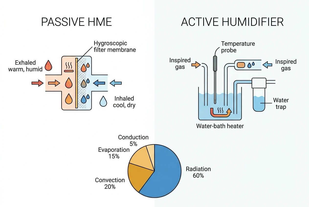
There are two broad approaches to humidifying inspired gas: [1]
- Passive — the heat and moisture exchanger (HME). A small housing containing a hygroscopic or hydrophobic membrane (paper, foam, ceramic or a salt-impregnated fleece) is placed between the tracheal tube and the breathing circuit. On EXPIRATION the warm, saturated exhaled gas meets the cool membrane; water condenses onto it and the membrane is warmed. On the next INSPIRATION the dry, cool inspired gas picks up that stored heat and water. The HME therefore recycles the patient's OWN heat and moisture — it is a passive device needing no power or water. It is single-use, low resistance, simple and cheap, and typically achieves an absolute humidity of about 25 to 32 mg/L at the carina. Its limitations are a lower efficiency than an active humidifier, an increase in dead-space and resistance (which matters in small children and during weaning), and the rare but serious risk of a waterlogged or obstructed filter causing ventilatory failure [1][2].
- Active — the heated water-bath humidifier. Inspired gas is bubbled through, or passed over, a water reservoir that is heated by an electrical element; the gas leaves the chamber warmed and fully saturated. A heated wire in the inspiratory limb prevents rain-out as the gas travels to the patient, and temperature probes at the airway guard against overheating. An active humidifier can deliver gas at near-physiological humidity (44 mg/L at 37 degrees C) regardless of the patient's own output, so it is more effective than an HME and is preferred for long-term ventilation in ICU and for very small children. Its costs are complexity, a power and water requirement, the risk of infection from a colonised water bath, and a thermal-injury risk if a probe or heater fails [3][4].
Perioperative heat loss — redistribution hypothermia
The temperature course of a general-anaesthetised patient has a characteristic shape with three phases: [1]
- Phase 1 — redistribution (the first hour). The core temperature falls by 1 to 1.5 degrees C in the first hour after induction. This is NOT principally from net heat loss to the environment; it is from REDISTRIBUTION. Anaesthesia abolishes the normal tonic thermoregulatory vasoconstriction that keeps the warm core blood central and the periphery cool. Vasodilatation lets the warm core blood flow into the periphery, and the core reading drops even though total body heat has barely changed. Warming applied DURING this phase cannot fully prevent the fall, because the heat is being moved internally, not just lost — but it limits the depth of the drop.
- Phase 2 — linear, slow fall (the next 2 to 3 hours). Once the core and periphery have equilibrated, the patient cools more slowly as ongoing heat LOSS (radiation, convection, evaporation) exceeds metabolic heat production, which anaesthesia has also reduced.
- Phase 3 — plateau. When the core temperature falls low enough to trigger vasoconstriction again (if it is permitted — regional anaesthesia and vasodilators block it), heat loss falls and the temperature plateaus. [1]
Risk factors for a greater drop include low body mass index, very young or old age, major surgery, a large open wound, cool theatre temperatures, and regional or combined anaesthesia [7].
The four routes of perioperative heat loss
During the linear phase the anaesthetised patient loses heat to the environment by four routes. The approximate proportions, for a typical adult in a standard theatre, are: [1]
- Radiation — about 60 per cent. Infrared loss to the cooler walls, ceiling, equipment and windows. This is the dominant route and the reason raising the ambient temperature and using a reflective or warm-air barrier helps.
- Convection — a large minority (around a quarter to a third). Heat carried away by air currents, enhanced by the laminar airflow in modern theatres.
- Evaporation — variable, often 8 to 10 per cent, more with a large wound. From the exposed surgical wound (especially an open abdomen or thorax, or a burns excision) and from the respiratory tract when dry, unhumidified gas is breathed. Because each litre evaporated removes 2.4 MJ, a large open wound can make evaporation the dominant loss.
- Conduction — small, a few per cent. Into the operating table and any cold fluid in contact with the skin. Underwater or warmed mattresses reduce this. [1]
These figures shift with the surgical exposure and the warming strategy, but the principle — radiation first, convection next, then evaporation, then conduction — is a reliable exam answer [7].
Patient warming strategies
Prevention of inadvertent perioperative hypothermia is now a recognised quality indicator. The strategies, in rough order of impact, are: [1]
- Forced-air warming (a Bair Hugger-type blanket). Temperature-controlled air is blown into a perforated blanket draped over the patient, creating a warm boundary layer against the skin that suppresses convective loss and establishes a positive heat gradient back into the body. It is the single most effective cutaneous warming method and the standard of care for most surgery [5].
- Fluid and blood warming. Each unit of cold stored blood or each litre of room-temperature crystalloid cools the patient; a rapid-infusion or in-line fluid warmer brings these to near body temperature before they enter the circulation, and is essential for major resuscitation and massive transfusion [6].
- Radiant warming. An overhead radiant heater, mainly for neonates and in the pre-induction period, delivers infrared heat directly.
- Increasing ambient temperature. Raising the theatre temperature to around 23 degrees C or above reduces the radiation and convection gradients; there is a trade-off with staff comfort.
- Wrapping and insulation. A plastic wrap, a reflective blanket, a head cover and a warmed mattress reduce exposed surface area and conductive loss, and are particularly important before induction and during transfer.
- Airway warming and humidification. An HME or active humidifier returns the heat and moisture of exhaled gas, reducing respiratory heat and water loss [1].
Active warming must be applied safely: a malfunctioning blanket or a mis-sited probe can cause a thermal burn [4], and in the neonate a dedicated double-wall incubator-style warmer outperforms simple overhead radiant heat [8].
Perioperative hypothermia — consequences
Even mild hypothermia — a core 1 to 2 degrees C below normal (around 35 to 36 degrees C) — has measurable, evidence-based harms, which is why maintaining normothermia is a quality standard: [1]
- Wound infection. Hypothermia triggers vasoconstriction, which reduces wound oxygen tension and impairs neutrophil bacterial killing, raising surgical-site infection rates.
- Coagulopathy and increased blood loss. Hypothermia impairs platelet function and the coagulation enzyme cascade, increasing blood loss and the transfusion requirement.
- Shivering and cardiac stress. Postoperative shivering can raise oxygen consumption several-fold, increasing myocardial oxygen demand and provoking ischaemia in the vulnerable patient.
- Prolonged drug action. The metabolism of anaesthetic agents and neuromuscular blockers is slowed, prolonging recovery and the effect of muscle relaxants.
- Patient discomfort. Cold and shivering are among the complaints patients remember most. [1]
These consequences, taken together, lengthen hospital stay and increase morbidity [7].
Measurement of humidity
Humidity is measured by several physical principles, each suited to a different setting: [1]
- Wet and dry bulb hygrometer (psychrometer). Two thermometers sit side by side; one has its bulb wrapped in a wet wick. Evaporation from the wet bulb cools it, so it reads lower than the dry bulb. The temperature DIFFERENCE is inversely related to the humidity — a large difference means dry air, a small difference means humid air — and the relative humidity is read off standard psychrometric tables. This is the classic, accurate laboratory method.
- Capacitive sensor. A thin polymer or metal-oxide dielectric absorbs water in proportion to the humidity, changing the capacitance of the element, which is read electronically. Fast, small and accurate, it is the basis of most modern electronic hygrometers, including those used to test HME performance.
- Resistive sensor. A hygroscopic salt (such as lithium chloride) changes its electrical resistance as it absorbs water, giving a resistance proportional to the humidity. Simple and cheap but less accurate than the capacitive type and slower to respond.
- Dew-point (chilled-mirror) hygrometer. A mirror is cooled until dew just condenses on its surface, detected optically; the temperature at that moment is, by definition, the dew point. It is the reference standard for high-accuracy calibration. [1]
In clinical practice the capacitive sensor dominates; in the laboratory the wet and dry bulb and the chilled mirror remain the references against which HME and humidifier outputs are measured [1].
Clinical
- Standard approach
- Evidence-based
Alternative
- Modified technique
- Risk-benefit
Red flags
[1]References
- [1]Lellouche F, et al. Evaluation of the Hygrometric Properties and Resistances of Several Passive Humidifiers After Prolonged Use Respir Care, 2026.PMID 42296038
- [2]Zou H, et al. Ventilatory failure following active humidification of a retained HMEF in an intubated infant: a case report Front Surg, 2026.PMID 42211546
- [3]Tsai RB, et al. Optimizing dry powder delivery during invasive mechanical ventilation via circuit absolute humidity control Int J Pharm, 2026.PMID 42142673
- [4]Yeh CC, et al. Unrecognized intraoperative thermal injury to the foot following arthroscopic anterior cruciate ligament reconstruction: a case report Patient Saf Surg, 2026.PMID 42304488
- [5]Lee JJ, et al. Airway warming devices in the context of single and multimodal strategies for intraoperative hypothermia: a network meta-analysis Syst Rev, 2026.PMID 41904534
- [6]Williams S, et al. Perceived Performance Traits of Blood Warming Devices Among Special Warfare Medics J Spec Oper Med, 2026.PMID 41861467
- [7]Ji Y, et al. Risk Factors Contributing to Inadvertent Postoperative Hypothermia in Adult Patients Following Hepatobiliary Surgery J Perianesth Nurs, 2026.PMID 41071148
- [8]Kabra NS, et al. A randomized controlled trial to evaluate the efficacy and safety of a double-walled incubator compared to a radiant warmer in the care of extremely low-birth-weight infants J Trop Pediatr, 2026.PMID 42275631