Anaes · Applied cardiovascular & respiratory physiology
Acid-base: the Stewart approach
Also known as Stewart acid-base · Strong ion difference · SID · Total weak acid · Atot · Physicochemical acid-base
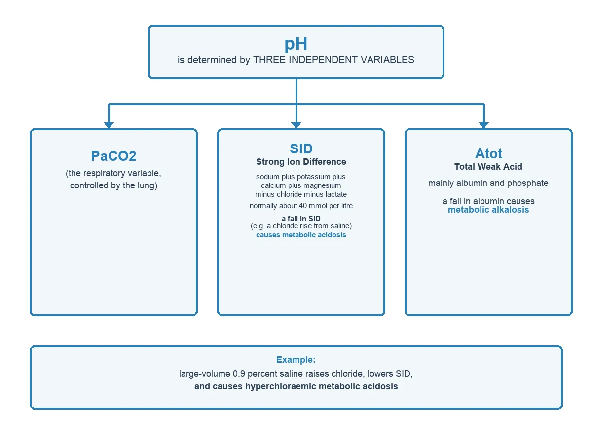
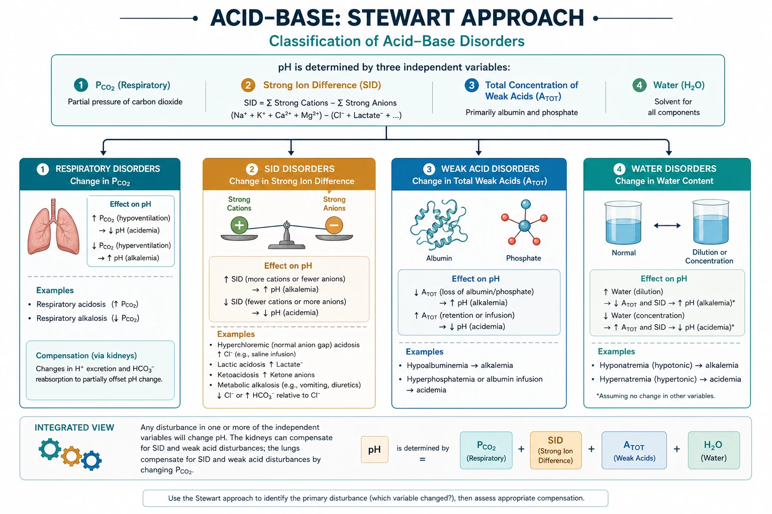
Peter Stewart's physicochemical approach reinterprets acid-base in terms of three independent variables that determine the dependent variable, pH, by mass-action: the carbon dioxide tension, the strong ion difference (the net charge of fully dissociated ions, dominated by sodium and chloride), and the total concentration of weak acid (chiefly albumin and phosphate). The framework rests on five exam-critical ideas: only three independent variables set the pH — PaCO2, the strong ion difference, and total weak acid; the strong ion difference (normally about 38 to 42 mmol per litre, dominated by sodium minus chloride) is the principal metabolic lever, and a fall in the SID (a chloride rise) causes metabolic acidosis while a rise causes alkalosis; total weak acid is mainly albumin, so hypoalbuminaemia causes a metabolic alkalosis that can mask a coexisting acidosis; bicarbonate is a DEPENDENT variable in Stewart's view (it changes to maintain electroneutrality), not a cause; and the Stewart approach explains the hyperchloraemic metabolic acidosis of large-volume normal saline, the hypoalbuminaemic alkalosis of critical illness, and unmeasured anions (the strong ion gap) that the bicarbonate method can miss. Built on the Stewart acid-base recovery study (Samara 2026), the dialysis acid-base comparative analysis (Kroustalakis 2025), the physicochemical COVID acid-base study (de Souza 2024), the balanced-crystalloids versus saline studies (Carrigan 2026, Sweety 2026), and the robotic-surgery (CO2 pneumoperitoneum) acid-base study (Pitimada 2026).
On this page & tools
Your progress
Saved locally on this device.
8 MCQs with explanations
Target exams
Red flags

Why this matters to the anaesthetist
Hyperchloraemic acidosis from 0.9% saline, hypoalbuminaemic alkalosis masking lactic acidosis, and the "normal anion gap" that is not normal once albumin is corrected are everyday ICU and theatre problems. Stewart's physicochemical approach does not replace Henderson–Hasselbalch; it explains why bicarbonate changes when chloride, free water, and albumin change. Primary candidates must define SID and Atot, list independent versus dependent variables, and apply Stewart to saline acidosis and unmeasured anions. [1]
Stewart's central principle
Peter Stewart's core claim: the hydrogen ion concentration of a solution is not an independent primary "set" quantity. In plasma, once electroneutrality, conservation of mass, and weak-acid dissociation equilibria are satisfied, [H+] is determined by three independent variables: [1]
- PaCO2 (respiratory)
- Strong ion difference (SID)
- Total weak acid concentration (Atot) — mainly albumin and inorganic phosphate [1]
Dependent variables include pH, [H+], [HCO3−], and the dissociated weak acid anions [A−]. Bicarbonate is not an independent control knob — it moves when the independents move. [1]
This is why "giving bicarbonate" can raise measured HCO3− yet leave the underlying SID problem unaddressed if the clinical issue is chloride or unmeasured anions. [1]
The strong ion difference (SID)
Strong ions are fully dissociated at physiologic pH: principally Na+, K+, Ca2+, Mg2+, Cl−, lactate−, and other fixed anions. [1]
Apparent SID (SIDa) ≈ [Na+] + [K+] + [Ca2+] + [Mg2+] − [Cl−] − [lactate−] (all in mEq/L) [1]
Normal SIDa ≈ 40 mEq/L (range often quoted 38–42). [1]
Effective SID (SIDe) is calculated from the charge contributions of HCO3−, albumin, and phosphate (the known weak-acid anions). [1]
Strong ion gap (SIG) = SIDa − SIDe [1]
A raised SIG implies unmeasured anions (ketoacids, toxins, many organic acids) — analogous in spirit to a corrected anion gap but grounded in Stewart bookkeeping. [1]
Qualitative rules: [1]
- ↓ SID → acidosis (e.g. hyperchloraemia, free-water excess diluting SID, loss of sodium relative to chloride)
- ↑ SID → alkalosis (e.g. vomiting with Cl− loss, diuretics, contraction alkalosis, massive citrate/acetate metabolism) [1]
Total weak acid (Atot)
Atot is dominated by albumin (and phosphate). Albumin is a weak non-volatile acid. [1]
- Hypoalbuminaemia lowers Atot → metabolic alkalosis tendency
- Hyperphosphataemia raises Atot → metabolic acidosis tendency [1]
Clinically, a critically ill patient with albumin 15 g/L has a built-in alkalinising force that can mask a concurrent lactic acidosis: pH and base excess look milder than the SIG/lactate suggest. Always correct the anion gap for albumin (roughly +2.5–3 mmol/L gap per 10 g/L albumin fall below normal — teaching approximation). [1]
Chloride — the pivotal strong anion
Because sodium is regulated tightly for tonicity and volume, chloride is often the strong ion that moves SID in iatrogenic acid–base disorders. [1]
0.9% sodium chloride has [Na+] = [Cl−] = 154 mmol/L. Relative to plasma SID, it delivers a chloride-rich, SID-zero fluid. Infusing large volumes narrows plasma SID (relative hyperchloraemia) → hyperchloraemic metabolic acidosis. Balanced crystalloids (e.g. Plasma-Lyte, Hartmann's/lactated Ringer's) have lower [Cl−] relative to [Na+] and organic anions that metabolise to bicarbonate-equivalent, preserving SID better. [1]
Stewart versus bicarbonate / base-excess approach
| Feature | Traditional (Boston/Copenhagen) | Stewart |
|---|---|---|
| Primary metrics | pH, PCO2, HCO3−, BE, AG | pH, PCO2, SID, Atot, SIG |
| Metabolic story | HCO3− gain/loss | SID and Atot changes |
| Saline acidosis | Hyperchloraemic / dilutional labels | ↓SID from relative ↑Cl− |
| Hypoalbuminaemia | Correct AG | ↓Atot alkalosis |
| Unmeasured anions | Anion gap | SIG |
| Clinical agreement | Four primary disorders | Same disorders, more mechanism |
You must still diagnose respiratory vs metabolic and anion-gap vs non-gap patterns fluently — Stewart adds mechanism, especially for chloride and albumin. [1]
Free-water disorders and SID
Hyponatraemia with free-water excess dilutes all strong ions; because SID is a difference, dilution reduces SID → acidosis (dilutional). Hypernatraemic contraction can raise SID → alkalosis. This links tonicity management to acid–base. [1]
Renal and GI SID physiology (exam layer)
- Diarrhoea: loss of HCO3− and organic anions; plasma Cl− often rises relatively → normal-gap acidosis (↓SID).
- Vomiting / NG suction: loss of HCl → hypochloraemic ↑SID alkalosis.
- Renal tubular acidosis: inability to excrete acid or reclaim HCO3− alters net SID handling via urine chemistry.
- Mineralocorticoid excess: Na+ retention and H+/K+ wasting → alkalosis. [1]
Hyperchloraemic metabolic acidosis from saline — clinical
Large-volume 0.9% NaCl perioperatively: rising Cl−, falling BE, falling HCO3−, possible hyperkalaemia trend from acidosis, and renal vasoconstriction concerns in some literature. Balanced fluids reduce this physicochemical hit. Note: lactate in Hartmann's is not "lactic acidosis" when metabolised normally; in severe liver failure metabolism may slow. [1]
Unmeasured anions and toxins
Raised SIG/AG: lactate, ketones, uraemic acids, salicylate, methanol (formate), ethylene glycol (glycolate), pyroglutamate, etc. Stewart does not name the anion — laboratory context does. Osmolar gap complements toxic alcohol workup. [1]
Anaesthetic and perioperative relevance
- Choose crystalloid with SID/chloride profile in mind for large infusions.
- Interpret ABG with albumin and Cl− visible, not pH alone.
- Post-hypercapnia: residual metabolic alkalosis if renal compensation occurred.
- Massive transfusion: citrate (metabolised to HCO3−) → alkalosis later; also volume and Cl− load from products vary.
- Hyperchloraemia may affect renal afferent tone — one argument in fluid debates. [1]
Clinical worked example
Patient after 5 L of 0.9% NaCl: Na 140, Cl 115, HCO3 18, pH 7.28, PaCO2 36, albumin normal, lactate 1.0. [1]
SIDa roughly (ignoring minor ions) Na+K − Cl − lactate ≈ 140+4 − 115 − 1 ≈ 28 (low). SIG not raised. Diagnosis: hyperchloraemic metabolic acidosis from ↓SID. Treatment: stop saline, use balanced fluid, treat cause of volume need; bicarbonate rarely first-line if perfusion intact and pH not extreme. [1]
Second example: albumin 18 g/L, pH 7.40, HCO3 24, lactate 6. The "normal" pH hides alkalinising hypoalbuminaemia plus acidifying lactate — compute corrected gap/SIG mentally before reassurance. [1]
↓ SID acidosis
- Hyperchloraemia (saline)
- Lactate/ketones (also raise SIG)
- Free-water excess dilution
- Some diarrhoea patterns
↑ SID alkalosis
- Vomiting (Cl loss)
- Diuretics / contraction
- Citrate load metabolism
- Mineralocorticoid excess
Equations and electroneutrality
Electroneutrality: sum of cations = sum of anions. [Na+] + [K+] + [Ca2+] + [Mg2+] + [H+] = [Cl−] + [HCO3−] + [A−] + [OH−] + [other anions] Ignoring tiny [H+] and [OH−] at physiologic pH, the charge space of weak acids and bicarbonate is fixed once SID and Atot and PCO2 are set. [1]
Viva traps
- Independent variables are PaCO2, SID, Atot — not HCO3−.
- Hypoalbuminaemia causes alkalosis, not acidosis.
- Lactate lowers SID and raises SIG — both true.
- Hartmann's lactate is not an acid load if metabolised.
- Stewart and HH must agree on measured pH — if not, measurement error. [1]
Link to base excess
Base excess quantifies metabolic component empirically; Stewart explains mechanism of that metabolic component. BE can be partitioned (e.g. Fencl–Stewart, Stewart–Figge) into sodium-chloride effects, free-water, albumin, phosphate, and unmeasured anions — useful in complex ICU gases. [1]
SAQ: independent variables
"In the Stewart approach the plasma hydrogen ion concentration is determined by three independent variables: the partial pressure of carbon dioxide, the strong ion difference, and the total concentration of weak acids. Bicarbonate and pH are dependent variables. A fall in strong ion difference, for example when chloride rises during saline infusion, causes a metabolic acidosis. A fall in albumin lowers total weak acid and causes a metabolic alkalosis that may mask concurrent lactic acidosis." [1]
Fencl–Stewart partitioning (overview)
Complex blood gases can be partitioned into: free-water effect (sodium), chloride effect (corrected chloride), albumin effect, phosphate effect, and residual SIG. You need not compute every term in a viva, but naming the partition shows mastery. [1]
Relationship to base excess
Base excess is the amount of acid or base needed to return pH to 7.40 at PCO2 40 mmHg at 37 °C fully saturated. It quantifies the metabolic component without explaining mechanism. Stewart explains whether that metabolic component is chloride, albumin, or unmeasured anions. [1]
Balanced crystalloid chemistry
Hartmann's: Na 131, Cl 111, lactate 29, K 5, Ca 2 (approx) — SID effective after lactate metabolism near 28–30 before full accounting. Plasma-Lyte: acetate/gluconate, lower Cl relative to Na. 0.9% NaCl: SID zero, Cl 154. The physicochemical after-effect on plasma SID differs accordingly. [1]
Teaching case: DKA in Stewart language
Ketone anions increase unmeasured strong anions → SIDa falls if Na and Cl unchanged → acidosis; SIG rises. Insulin therapy metabolises ketoanion → SID recovers → pH rises. Saline resuscitation can add hyperchloraemic acidosis as ketones clear — mixed picture common on the ward. [1]
Primary exam expansion
Worked numerical SID examples
Example A — saline acidosis: Na 140, K 4, Cl 118, lactate 1 → rough SIDa ≈ 140+4−118−1 = 25 mEq/L (low) → metabolic acidosis with normal SIG if SIDe matches. [1]
Example B — vomiting: Na 140, K 3, Cl 90, lactate 1 → SIDa ≈ 52 (high) → metabolic alkalosis. [1]
Example C — lactic acidosis: Na 140, K 4, Cl 100, lactate 10 → SIDa ≈ 34 (low) and SIG raised because lactate is sometimes handled as measured strong anion (if included in SIDa, SIG may not show it — be consistent with definition used in teaching). State your definition in the answer. [1]
Anion gap formula and albumin correction
AG = Na − (Cl + HCO3); sometimes include K. Normal ~8–12 mmol/L depending on analyser. Corrected AG ≈ observed AG + 0.25 × (normal albumin g/L − observed albumin g/L) with unit care (some use 2.5 per 10 g/L). Stewart's SIG is the physicochemical cousin. [1]
Hyperchloraemia and the kidney
High chloride delivery to macula densa may alter afferent tone (tubuloglomerular feedback), a proposed mechanism linking saline to reduced GFR compared with balanced fluids in some studies. Even if trials debate outcomes, the physiology story is coherent for exams. [1]
Citrate, acetate, lactate as SID actors
These organic anions are strong ions while present; when metabolised to CO2/H2O essentially they leave behind sodium (or other cations) effectively increasing SID → alkalinising. Failed metabolism (shock liver) leaves the anion present → persistent SID reduction or SIG elevation. [1]
Teaching: you still need Henderson–Hasselbalch
Stewart does not abolish pH = pKa + log([HCO3−]/(0.03 PCO2)). It explains why [HCO3−] has its value. In the exam, open with traditional disorder classification, then add Stewart mechanism for chloride and albumin questions. [1]
Massive transfusion acid–base timeline
Early: lactic acidosis from hypoperfusion, hyperchloraemia from products/fluids, citrate load. Later: citrate metabolised → rebound metabolic alkalosis; hypocalcaemia from citrate binding. Stewart language clarifies the phases. [1]
Extended viva dialogue
Examiner: What are the independent variables in Stewart's approach? [1]
Candidate: PaCO2, the strong ion difference, and the total weak acid concentration Atot, mainly albumin and phosphate. pH, hydrogen ion concentration and bicarbonate are dependent variables fixed once the independents and physical chemistry constraints are set. [1]
Examiner: Explain saline-induced metabolic acidosis using SID. [1]
Candidate: 0.9 percent sodium chloride contains 154 mmol/L of sodium and 154 mmol/L of chloride, a strong ion difference of zero. Plasma normal SID is about 40 mEq/L. Large saline infusions raise the chloride relative to sodium, reduce SID, and the equilibria force hydrogen ion up and bicarbonate down — hyperchloraemic metabolic acidosis. [1]
Examiner: How does hypoalbuminaemia affect acid–base? [1]
Candidate: Albumin is a weak acid. Lowering albumin lowers Atot and produces a metabolic alkalosis tendency. This can mask a concurrent high-SIG acidosis such as lactate. Always correct the anion gap for albumin or think in SIG terms before declaring the metabolic picture normal. [1]
Examiner: Does Stewart replace base excess and the anion gap? [1]
Candidate: No. Base excess quantifies the metabolic component; the anion gap screens for unmeasured anions. Stewart explains mechanisms — especially chloride and albumin — and formalises unmeasured anions as the strong ion gap. Use traditional classification first, then Stewart for mechanism. [1]
Clinical synthesis: Read pH, PCO2, bicarbonate, chloride, albumin and lactate together every time. Fluids are acid–base drugs. [1]
Worked SAQ model answers
SAQ: Explain the Stewart approach to acid–base and its clinical usefulness (10 marks)
Stewart's physicochemical approach states that plasma hydrogen ion concentration is determined by three independent variables: the partial pressure of carbon dioxide, the strong ion difference (SID), and the total concentration of non-volatile weak acids (Atot), chiefly albumin and phosphate. Bicarbonate and pH are dependent variables that take the values required to satisfy electroneutrality and dissociation equilibria. [1]
SID is the difference between fully dissociated strong cations and anions, normally about 40 mEq/L. A fall in SID produces metabolic acidosis; a rise produces metabolic alkalosis. The common iatrogenic example is large-volume 0.9% saline, which delivers equal sodium and chloride (SID zero) and reduces plasma SID by relative hyperchloraemia. Balanced crystalloids reduce this effect. Lactate and ketoanions also reduce SID and appear as unmeasured anions when quantified by the strong ion gap (SIDa minus SIDe). [1]
Atot changes matter in critical illness. Hypoalbuminaemia lowers Atot and causes a metabolic alkalosis that can mask lactic acidosis; the anion gap must be corrected for albumin. Hyperphosphataemia increases Atot and acidifies. [1]
Stewart does not replace Henderson–Hasselbalch or the traditional four primary acid–base disorders. It provides mechanism for hyperchloraemic acidosis, free-water effects, albumin effects and unmeasured anions, which is why it is useful in complex ICU blood gases and perioperative fluid choices. [1]
SAQ: Worked interpretation (5 marks)
A patient receives 6 L of 0.9% NaCl. Na 141, Cl 116, HCO3 17, pH 7.29, PaCO2 35, albumin normal, lactate 1.2. SIDa is reduced by hyperchloraemia; SIG is not raised. Diagnosis: hyperchloraemic metabolic acidosis from reduced SID. Management: stop saline, use balanced fluid, restore perfusion if needed; bicarbonate is not first-line if pH is only moderately reduced and ventilation is intact. [1]
Red flags
- Only three independent variables set pH: PaCO2, SID, Atot. Bicarbonate is dependent.
- Fall in SID (e.g. chloride rise from saline) causes hyperchloraemic metabolic acidosis.
- Hypoalbuminaemia lowers Atot and alkalinises — can mask acidosis.
- Strong ion gap detects unmeasured anions when albumin is low better than naive AG.
- Stewart agrees with traditional four disorders; adds chloride/albumin/SIG clarity. [1]
References
- [1]Samara EM. Stewart-based characterisation of blood acid-base recovery over 48 h following exercise under hot condition in camels (Camelus dromedarius) Anim Biosci, 2026.PMID 41856088
- [2]Kroustalakis N, et al. Dialysis and Acid-Base Balance: A Comparative Physiological Analysis of Boston and Stewart Models J Clin Med, 2025.PMID 41303241
- [3]de Souza SP, et al. Physico-chemical characterization of acid base disorders in patients with COVID-19: A cohort study World J Nephrol, 2024.PMID 38983762
- [4]Carrigan K, et al. Balanced fluid bolus: Should we prefer balanced crystalloids over normal saline? J Perinatol, 2026.PMID 41826663
- [5]Sweety S, et al. Balanced electrolyte solution vs isotonic saline in the resuscitation of children with diabetic ketoacidosis: A randomized controlled trial World J Clin Pediatr, 2026.PMID 41884024
- [6]Pitimada M, et al. Acid-base balance and respiratory mechanics during robotic-assisted surgery: an observational study J Robot Surg, 2026.PMID 42010144