Anaes · Applied cardiovascular & respiratory physiology
Carbon dioxide transport
Also known as CO2 transport · Carbon dioxide carriage · Haldane effect · Chloride shift · CO2 dissociation curve · Bicarbonate buffer
Carbon dioxide is the waste product of metabolism and the chemical driver of ventilation, and its transport has a subtlety oxygen transport lacks: the CO2 dissociation curve is steep and near-linear, and the Haldane effect lets oxygenation in the lung unload CO2. The framework rests on five exam-critical ideas: CO2 is carried in three forms — as bicarbonate (about 70 percent), as carbamino compounds on haemoglobin (about 20 percent) and dissolved (about 10 percent); bicarbonate is made in the red cell by carbonic anhydrase and exported in exchange for chloride (the chloride shift, via the band-3 anion exchanger); the CO2 dissociation curve is steep and near-linear (so CO2 content tracks PCO2 closely, unlike the sigmoid oxygen curve); the Haldane effect — deoxygenated haemoglobin carries more CO2 than oxygenated — is the dominant mechanism of CO2 uptake in the tissues and release in the lung; and CO2 is the acid load of the body, buffered by the bicarbonate system (Henderson-Hasselbalch), with large body stores that equilibrate slowly. Built on the mechanistic CO2-transport-in-blood model (O'Neill 2017), the CO2-derived-variables review (Mallat 2025), the venous-to-arterial CO2-content study (Ospina-Tascon 2025), the haemoglobin-electrolyte interaction study (Valsecchi 2025), the membrane-oxygenator gas-exchange model (Monsefi 2025), and the band-3 chloride-bicarbonate exchanger study (Fawaz 2012).
On this page & tools
Your progress
Saved locally on this device.
8 MCQs with explanations
Target exams
Red flags
Why this matters to the anaesthetist
CO2 is the primary waste gas the lung must eliminate, the dominant acute ventilatory stimulus, the main acute determinant of pH via the Henderson–Hasselbalch relationship, and a cerebral vasodilator. Capnography is continuous CO2 physiology at the airway. Primary candidates must state carriage fractions, explain the chloride shift, draw the CO2 dissociation curve, define the Haldane effect, and write Henderson–Hasselbalch. [1]
Overview of CO2 production and elimination
Whole-body V̇CO2 at rest ≈ 200 mL/min (RQ × VO2; RQ ≈ 0.8 if VO2 250). Steady state: elimination = production. PaCO2 is set by the balance PaCO2 ∝ V̇CO2 / VA. Hypercapnia means relative alveolar hypoventilation (or inspired CO2); hypocapnia means relative hyperventilation (or low production). [1]
The three forms of CO2 carriage
In arterial blood total CO2 content ≈ 48 mL/dL; mixed venous ≈ 52 mL/dL; a–v difference ≈ 4 mL/dL. [1]
Approximate distribution of the additional CO2 in venous blood (classic teaching percentages for total carriage vary slightly by source; learn one consistent set): [1]
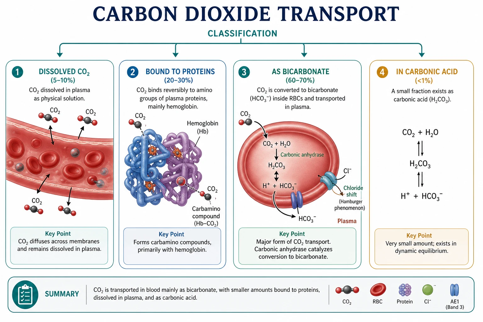
- Bicarbonate (~70% of transported CO2) — formed mainly in red cells
- Carbamino compounds (~20%) — CO2 bound to N-terminal amino groups of Hb (and plasma proteins)
- Dissolved (~10%) — Henry's law: dissolved CO2 ≈ 0.03 × PCO2 mmol/L (or ~0.07 mL/dL per mmHg depending on units) [1]
Examiners accept: bicarbonate majority, then carbamino, then dissolved — with Haldane effect amplifying carbamino and buffering differences between oxy and deoxy Hb. [1]
Bicarbonate formation and the chloride shift
In tissue capillaries: [1]
- Dissolved CO2 enters the red cell.
- Carbonic anhydrase catalyses CO2 + H2O ⇌ H2CO3 ⇌ H+ + HCO3− (orders of magnitude faster than uncatalysed).
- H+ is buffered by deoxyhaemoglobin (excellent buffer — Haldane linkage).
- HCO3− exits the red cell via band-3 anion exchanger (AE1, SLC4A1) in exchange for Cl− entering — the chloride (Hamburger) shift.
- Plasma [Cl−] is slightly lower in venous than arterial blood; red-cell chloride is higher in venous blood.
- In the lung, oxygenation reverses the process: H+ released as Hb binds O2, HCO3− re-enters, CO2 is regenerated and exhaled; Cl− shifts out. [1]
Without carbonic anhydrase (acetazolamide inhibition), CO2 transport and tissue/ventilatory equilibria are disturbed — used therapeutically for altitude and some types of metabolic alkalosis, with respiratory side effects. [1]
The CO2 dissociation curve
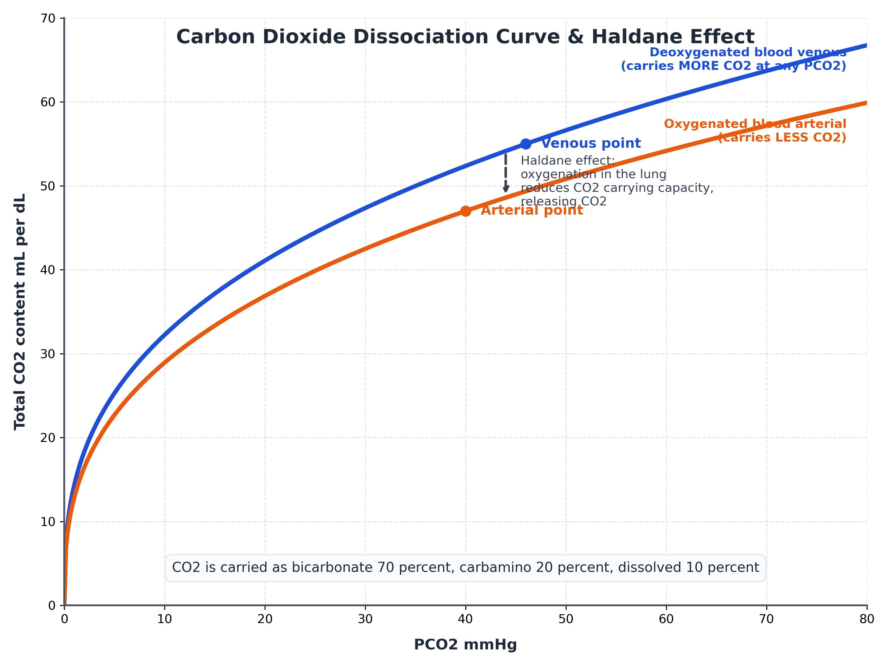
Plot CO2 content (y) against PCO2 (x). Compared with the ODC: [1]
- More linear over the physiological range
- Steeper — content changes more per mmHg
- Separate curves for oxygenated vs deoxygenated blood: deoxy curve higher (Haldane) [1]
Physiological a–v points: arterial PCO2 40 mmHg, venous ~46 mmHg, with content difference ~4 mL/dL. Because the curve is steep, small PCO2 changes move substantial CO2 stores. [1]
The Haldane effect — dominant for CO2 exchange
Deoxyhaemoglobin:
- Is a better H+ buffer (binds protons from carbonic acid)
- Forms carbamino compounds more readily [1]
Thus at any given PCO2, deoxygenated blood holds more CO2. In tissues, O2 unloading promotes CO2 loading; in lungs, O2 loading promotes CO2 unloading. Quantitatively the Haldane effect accounts for a large share of the a–v CO2 content difference — often taught as roughly half of CO2 exchange, comparable to or exceeding the simple dissolved difference. Do not confuse with Bohr (which is about O2 affinity changing with CO2/H+). [1]
Henderson–Hasselbalch and the bicarbonate buffer
pH = pKa + log10([HCO3−] / (0.03 × PCO2)) [1]
with pKa ≈ 6.1 for the carbonic acid system at body temperature, [HCO3−] in mmol/L, PCO2 in mmHg, solubility coefficient 0.03 mmol/L/mmHg. [1]
Normal: HCO3− 24 mmol/L, PCO2 40 → ratio 24/1.2 = 20 → log10(20) ≈ 1.3 → pH ≈ 7.4. [1]
Acute respiratory acidosis: raise PCO2, ratio falls, pH falls; acute buffering raises HCO3− slightly (~1 mmol/L per 10 mmHg acute); renal compensation over days raises HCO3− further (~3–4 mmol/L per 10 mmHg chronic). [1]
CO2 stores and compartments
CO2 is stored in blood, interstitial fluid, muscle, bone (as carbonate), and fat. Body stores are large (~120 L CO2 equivalent) compared with O2 stores — hence PaCO2 changes more slowly than PaO2 during apnoea if starting from normal, but during high production states (MH) EtCO2 can rocket. Different compartments equilibrate at different rates — explaining hysteresis between ventilation changes and arterial sampling. [1]
Capnography physiology
- EtCO2 approximates PACO2 when VD alv is small and sampling is good.
- a–ET gradient normally 2–5 mmHg; rises with alveolar dead space (PE, low CO, COPD).
- Sudden fall in EtCO2: embolism, low CO, disconnect, cardiac arrest.
- Rise in EtCO2: hypoventilation, rebreathing, hypermetabolism (MH, thyrotoxicosis, release of tourniquet).
- Shape: shark-fin obstructive pattern; cleft in spontaneous efforts; rebreathing elevates baseline. [1]
CO2-derived variables in critical care
- Pv-a CO2 gap: elevated with low flow / inadequate perfusion relative to CO2 production.
- Respiratory quotient on circuit: VCO2/VO2.
- Dead-space calculations from Enghoff/Bohr using CO2. [1]
Hypercapnia and hypocapnia — systemic effects
Hypercapnia: respiratory acidosis, sympathetic activation, cerebral vasodilatation (raised CBF/ICP), pulmonary vasoconstriction, right shift of ODC, narcosis at very high levels, arrhythmias, residual neuromuscular block potentiation. [1]
Hypocapnia: respiratory alkalosis, cerebral vasoconstriction (lower ICP temporarily), left shift ODC, coronary vasoconstriction risk, reduced respiratory drive after deliberate hyperventilation stops, arrhythmias, free Ca2+ changes. [1]
Anaesthetic relevance
- Minute ventilation is the immediate CO2 control knob under GA.
- Soda lime and circle systems manage inspired CO2; exhaustion → rebreathing.
- Laparoscopy: systemic CO2 absorption raises V̇CO2 load — increase VE.
- MH crisis: rising EtCO2 is an early flag.
- Permissive hypercapnia in ARDS trades pH for lung protection — know the physiology of tolerance. [1]
Bohr effect
- CO2/H+ alter O2 affinity
- Right shift in tissues
- Helps O2 unloading
- About the ODC
Haldane effect
- O2 binding alters CO2 carriage
- Deoxy-Hb holds more CO2
- Helps CO2 loading/unloading
- About the CO2 curve
Worked Henderson–Hasselbalch
Acute PaCO2 80 mmHg, HCO3− still 24: ratio = 24 / (0.03×80) = 24/2.4 = 10; log10(10)=1; pH≈7.1. After chronic compensation HCO3− might be ~36: ratio 36/2.4=15; log10(15)≈1.18; pH≈7.28 — improved but not normal. [1]
Viva traps
- Percentages of carriage — bicarbonate dominant.
- Carbonic anhydrase is intracellular (RBC), not primarily plasma.
- Bohr vs Haldane naming swap is an instant fail.
- Dissolved CO2 still matters for diffusion gradients despite being 10%.
- RQ links V̇CO2 to diet and to the alveolar gas equation. [1]
Extended tissue-to-lung narrative
At the tissues, PO2 falls and PCO2 rises. O2 leaves Hb (Bohr-facilitated). Deoxy-Hb buffers H+ and binds CO2 as carbamino. HCO3− generated via carbonic anhydrase leaves the cell (chloride shift). Blood arrives at the lung with higher total CO2 content. Alveolar gas has low PCO2 (~40 arterial equilibrium). CO2 diffuses out; oxygenation flips Hb to R-state; H+ is released; HCO3− re-enters; CO2 is exhaled. The cycle repeats every red-cell transit — about three-quarters of a second in pulmonary capillaries at rest. [1]
SAQ: describe CO2 carriage from tissue to lung
"Carbon dioxide produced in tissues dissolves in plasma and enters red cells. Inside red cells carbonic anhydrase forms carbonic acid that dissociates into hydrogen ion and bicarbonate. Hydrogen ion is buffered by deoxyhaemoglobin. Bicarbonate leaves the cell in exchange for chloride via band-3 — the chloride shift. Some carbon dioxide binds to haemoglobin as carbamino compounds. About seventy percent of transported carbon dioxide is as bicarbonate, twenty percent carbamino, and ten percent dissolved. In the lung oxygenation of haemoglobin reverses these processes — the Haldane effect — and carbon dioxide is exhaled." [1]
Capnography waveform phases
Phase I: dead-space gas, CO2 near zero. Phase II: mixture, steep upstroke. Phase III: alveolar plateau; slope increases with V/Q scatter. Phase 0: inspiration downstroke. End-tidal value is end of phase III. Alpha angle and beta angle jargon appears in some texts. Shark-fin: slow emptying of obstructed lung. Curare cleft: spontaneous effort during plateau. [1]
CO2 and cerebral blood flow equation style statement
CBF changes approximately 1–2 mL/100 g/min per mmHg PaCO2 change in the middle range (teaching approximations vary; some quote ~3% per mmHg). Below ~20 mmHg and above ~80 mmHg responses flatten. Use numbers cautiously but state the direction confidently. [1]
Laparoscopic hypercapnia differential
Rising EtCO2 during laparoscopy: CO2 insufflation absorption (expected), hypoventilation, apparatus rebreathing, subcutaneous emphysema, or rarely CO2 embolism (usually sudden fall then instability). Treat by increasing VE, checking system, communicating with surgeon about pressure/time. [1]
Primary exam expansion
Quantitative contents
Arterial total CO2 content ~48 mL/dL; venous ~52 mL/dL. Of the 4 mL/dL difference, classic breakdown attributes roughly 2.5–3 mL to bicarbonate pathway (with chloride shift), ~1 mL to carbamino (Haldane-linked), and ~0.3–0.5 mL to dissolved — exact splits vary by textbook; the order of magnitude is what matters. [1]
Carbonic anhydrase isoforms
CA II in red cells is the key high-activity isoform for CO2 transport. CA inhibitors (acetazolamide) cause CO2 retention in tissues and compensatory hyperventilation, renal HCO3− loss, and are used for altitude and glaucoma. In the eye and choroid plexus, CA supports fluid secretion — linking to CSF production reduction. [1]
CO2 stores washout and wash-in
Hyperventilating a patient blows off stores; EtCO2 falls quickly then more slowly as compartments empty. Stopping hyperventilation, CO2 reaccumulates. This kinetics explains why short hyperventilation before intubation only briefly keeps PaCO2 low, and why MH produces sustained high CO2 production that outpaces ventilation. [1]
Venous and tissue hypercapnia in low flow
When perfusion falls, CO2 accumulates in tissues and venous blood — raised PvCO2 and Pv−a CO2 gap — while EtCO2 may fall because less blood delivers CO2 to the lung. Interpreting both ends of the circuit is mandatory in shock. [1]
Respiratory acidosis management principles
Increase alveolar ventilation (rate or tidal volume carefully), treat obstruction, reduce apparatus dead space, treat high V̇CO2 causes, and only use bicarbonate in special circumstances (e.g. extreme acidaemia with limited ventilatory options) because CO2 generated from HCO3− + H+ must still be ventilated. [1]
Hypercapnia permission in ARDS
Lung-protective ventilation accepts higher PaCO2 to avoid VILI. Physiology trade-offs: pulmonary hypertension, right heart strain, intracranial considerations (avoid in raised ICP), and catecholamine surges. Contraindications to permissive hypercapnia include uncontrolled raised ICP and sometimes severe RV failure or pregnancy depending on context. [1]
Bohr–Haldane simultaneous action paragraph
In exercising muscle, high CO2 and H+ right-shift the ODC (Bohr) while deoxygenation enhances CO2 carriage (Haldane). In the lung, high PO2 left-shifts (loading O2) and reduces CO2 affinity (unloading CO2). One molecule of haemoglobin performs both jobs through linked equilibria — the elegance examiners want you to articulate. [1]
Extended viva dialogue
Examiner: How is carbon dioxide carried in blood? [1]
Candidate: Approximately seventy percent as bicarbonate, twenty percent as carbamino compounds mainly on haemoglobin, and ten percent dissolved. Formation of bicarbonate is catalysed by red-cell carbonic anhydrase. Bicarbonate exits the red cell in exchange for chloride via band-3 — the chloride or Hamburger shift. [1]
Examiner: Draw and describe the CO2 dissociation curve versus the ODC. [1]
Candidate: The CO2 content versus PCO2 relationship is steeper and more linear than the sigmoid oxygen curve over the physiological range. Deoxygenated blood lies above oxygenated blood at any PCO2 because of the Haldane effect. The arterial point is about 40 mmHg and mixed venous about 46 mmHg with a content difference near 4 mL/dL. [1]
Examiner: Write Henderson–Hasselbalch and use it. [1]
Candidate: pH equals pKa plus log of bicarbonate over 0.03 times PCO2, with pKa about 6.1. Normal values 24 over 1.2 equal ratio 20, log 1.3, pH 7.4. If PCO2 doubles to 80 with bicarbonate still 24, ratio is 10, pH about 7.1. Renal compensation later raises bicarbonate and improves pH without fully normalising it. [1]
Examiner: How does capnography reflect this physiology? [1]
Candidate: End-tidal CO2 approximates alveolar PCO2 when dead space is low. The arterial-to-ET gradient widens when alveolar dead space rises, as in embolism or low output. Sudden ET fall suggests loss of pulmonary blood flow or circuit disconnect. Rising ET suggests hypoventilation, rebreathing or increased production such as malignant hyperthermia. [1]
Clinical synthesis: Ventilation is CO2 elimination; oxygenation is a separate cascade. Never manage one number while ignoring the other. [1]
Worked SAQ model answers
SAQ: Describe the carriage of carbon dioxide in blood (10 marks)
Carbon dioxide is produced by aerobic metabolism at about 200 mL/min at rest and must equal pulmonary elimination in the steady state. In blood it is carried in three forms: as bicarbonate (about 70%), as carbamino compounds mainly on haemoglobin (about 20%), and dissolved (about 10%). [1]
Dissolved CO2 is proportional to PCO2 by Henry's law and provides the partial-pressure gradient for diffusion. Within red cells, carbonic anhydrase rapidly converts CO2 and water to carbonic acid, which dissociates to hydrogen ion and bicarbonate. Hydrogen ion is buffered by deoxyhaemoglobin. Bicarbonate leaves the cell in exchange for chloride via the band-3 anion exchanger — the chloride shift — so that plasma bicarbonate carries much of the CO2 load. In the lung the process reverses. [1]
The Haldane effect states that deoxygenated haemoglobin carries more CO2 than oxygenated haemoglobin at the same PCO2, because deoxyhaemoglobin is a better buffer and forms carbamino compounds more readily. This effect accounts for a large fraction of the arteriovenous CO2 content difference. The CO2 dissociation curve of content versus PCO2 is steeper and more linear than the oxygen curve, which is why V/Q mismatch affects oxygen more than CO2. [1]
Henderson–Hasselbalch links pH to the bicarbonate-to-dissolved-CO2 ratio: pH = 6.1 + log([HCO3−]/(0.03×PCO2)). Acute changes in ventilation alter PCO2 and pH immediately; renal compensation adjusts bicarbonate over hours to days. [1]
SAQ: Explain the clinical information in a capnograph (5 marks)
The capnograph displays the time course of airway CO2. Phase I is dead-space gas, phase II the transition, phase III the alveolar plateau ending at EtCO2, and inspiration returns CO2 toward zero. EtCO2 approximates PaCO2 when alveolar dead space is small; the gradient widens in embolism, low cardiac output and COPD. Waveform shape diagnoses obstruction, rebreathing and spontaneous effort. Trends diagnose hypoventilation, hypermetabolism and sudden loss of pulmonary blood flow. [1]
Red flags
- CO2 carriage: bicarbonate ~70%, carbamino ~20%, dissolved ~10%.
- Haldane effect — deoxyhaemoglobin carries more CO2 — major exchange mechanism.
- Chloride shift exports RBC bicarbonate via band-3.
- CO2 dissociation curve steep and near-linear; venous–arterial CO2 gap is a perfusion marker.
- CO2 is the acute acid load buffered by bicarbonate; renal compensation takes hours to days. [1]
References
- [1]O'Neill DP, Robbins PA. A mechanistic physicochemical model of carbon dioxide transport in blood J Appl Physiol (1985), 2017.PMID 27881667
- [2]Mallat J. Use of CO(2)-derived variables in critically ill patients Ann Intensive Care, 2025.PMID 40999252
- [3]Ospina-Tascon GA, et al. Regional venous-to-arterial carbon dioxide pressure and content differences during endotoxemic shock: influence of hydrogen ion accumulation vs. Haldane effect Intensive Care Med Exp, 2025.PMID 40921906
- [4]Valsecchi C, et al. In vitro characterization of hemoglobin oxygen dissociation curves and electrolyte shifts in human blood under varying PCO(2) Front Med (Lausanne), 2025.PMID 41601791
- [5]Monsefi Estakhrposhti SH, et al. A Validated CFD Model for Gas Exchange in Hollow Fiber Membrane Oxygenators: Incorporating the Bohr and Haldane Effects Membranes (Basel), 2025.PMID 41002903
- [6]Fawaz NA, et al. dRTA and hemolytic anemia: first detailed description of SLC4A1 A858D mutation in homozygous state Eur J Haematol, 2012.PMID 22126643