Anaes · Applied cardiovascular & respiratory physiology
Cardiac electrophysiology & conduction
Also known as Cardiac conduction system · SA node · AV node · His-Purkinje system · Funny current · Cardiac action potential
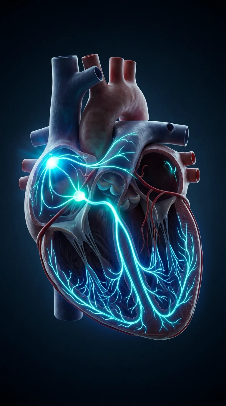
The heart generates and propagates its own electrical impulse through a specialised conduction pathway, and the orderly sequence of that propagation is what the ECG records. The framework rests on five exam-critical ideas: the sinoatrial node is the physiological pacemaker because its membrane drifts up to threshold on the funny current (a sodium inflow through HCN channels) and fires spontaneously; the atrioventricular node deliberately delays conduction (the PR interval) so the atria finish ejecting before the ventricles contract, and conducts slowly and decrementally; the His-Purkinje system then depolarises the ventricles rapidly and synchronously (the QRS); conduction between cells is carried by gap junctions, so disease of the gap junction or of the specialised tissue slows or blocks conduction; and arrhythmia arises by one of three mechanisms — enhanced automaticity, re-entry, or triggered activity — each with drugs and anaesthetic triggers that favour it. Built on the cardiac-conduction and gap-junction biology review (Fujiu 2026), the funny-current structure-function review (Saponaro 2026), the HCN4-in-the-AV-node review (Copier 2025), the HCN4-and-arrhythmia mutations review (Fan 2025), the cardiac ion-channel pharmacology review (Orts 2026), and the sudden-cardiac-death genetics review (Lovric Bencic 2025).
On this page & tools
Your progress
Saved locally on this device.
8 MCQs with explanations
Target exams
Red flags
Why this matters to the anaesthetist
Primary wants pacemaker potentials vs ventricular AP, ion channel phases, conduction pathway timing, and ECG interval meaning. Applied: local anaesthetic/ cardiotoxic drugs, electrolytes, volatile/autonomic effects, pacing concepts.[1]
One-liner: SA node spontaneous phase 4 depolarisation sets HR; AP conducts SA→AV→His–Purkinje→ventricle; ECG PR/QRS/QT map conduction and repolarisation times. [1]
Cardiac cell types
| Cell | AP feature | Key ions |
|---|---|---|
| SA/AV nodal | Slow response; Ca-dependent upstroke; phase 4 automaticity | If (funny Na), Ca, K |
| Atrial/ventricular myocyte | Fast response; Na upstroke; plateau | Na, Ca, K |
| Purkinje | Fast conduction, long AP | Na, Ca, K |
Fast-response AP phases (ventricular)
- Phase 0: rapid Na influx (INa) — dV/dt max determines conduction speed.
- Phase 1: early repolarisation (Ito K).
- Phase 2: plateau — inward Ca (ICa-L) balances outward K.
- Phase 3: repolarisation — K efflux dominates (IKr, IKs, IK1).
- Phase 4: resting ≈ −90 mV (high K permeability). [1]
Refractory periods: absolute then relative — protect against tetany; long QT prolongs vulnerable window for EADs/TdP. [1]
Pacemaker (slow-response) AP
- No stable −90 mV rest.
- Phase 4 prepotential: funny current If (Na/K), T-type Ca, decay of K conductance → threshold.
- Phase 0: mainly L-type Ca (not fast Na) — slower conduction in nodal tissue.
- Autonomic: sympathetic ↑If/Ca → steeper phase 4 → tachycardia; vagal ↑K conductance / ↓Ca → flatter phase 4 → bradycardia. [1]
Conduction pathway and times
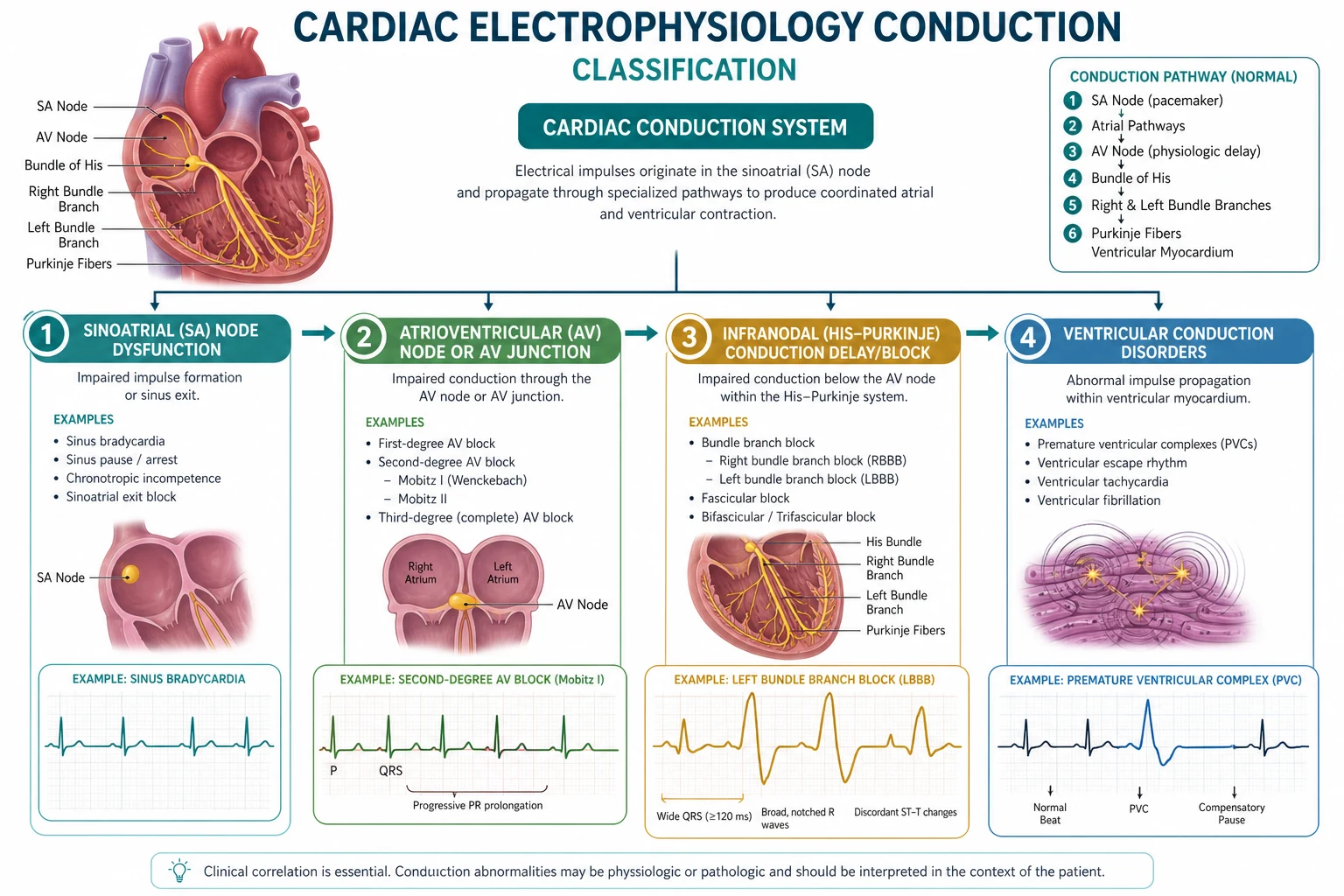
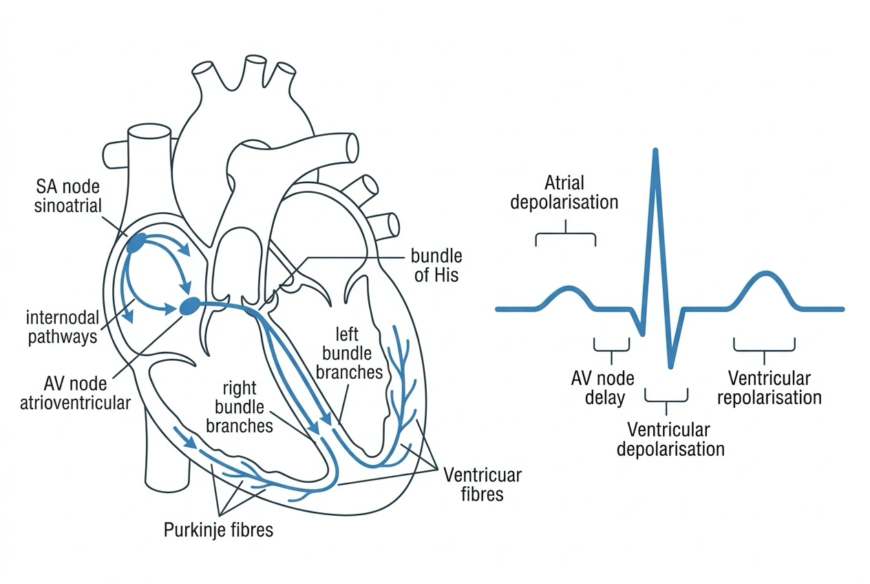
SA node → atrial muscle (P wave) → AV node (delay — PR segment major) → His bundle → bundle branches → Purkinje → ventricular muscle (QRS) → repolarisation (T). [1]
| Interval | Reflects | Typical adult |
|---|---|---|
| PR | Atrial + AV conduction | 120–200 ms |
| QRS | Ventricular depolarisation | <120 ms |
| QTc | Ventricular repolarisation | sex/rate dependent; prolonged risk TdP |
| RR | Cycle length | rate |
Arrhythmia mechanisms (exam trio)
- Enhanced automaticity — steeper phase 4 (ischaemia, catecholamines).
- Triggered activity — EADs (long QT) / DADs (Ca overload, digoxin).
- Re-entry — need unidirectional block + slow conduction + recoverable tissue (AF, AVNRT, VT circuits). [1]
Electrolytes and drugs (hooks)
- K↑: resting potential less negative → inexcitability, peaked T, wide QRS, sine wave.
- K↓: hyperpol / excitability changes, U waves, VT/VF risk.
- Ca↓: long QT; Ca↑: short QT.
- Mg↓: TdP facilitation.
- Local anaesthetic systemic toxicity: Na channel block → wide QRS, arrhythmias, collapse.
- Volatiles: QT effects variable; sevo/halothane historical arrhythmia sensitisation with adrenaline (halothane classic). [1]
Autonomic & anaesthetic rate control
Laryngoscopy → SNS → tachycardia/ischaemia risk. High spinal → unopposed vagal / loss of cardioaccelerator fibres (T1–4) → bradycardia. β-blockers flatten phase 4 and reduce MVO2. Anticholinergics oppose vagal bradycardia. [1]
Numbers board
- SA intrinsic ~60–100/min; AV junction ~40–60; ventricle ~30–40
- AV nodal delay allows atrial kick before ventricular systole
- QRS <120 ms normal conduction [1]
Nodal AP
- Phase 4 automaticity
- Ca upstroke
- ANS sensitive
- Sets HR
Ventricular AP
- Na upstroke
- Plateau phase 2
- Long refractory
- QRS/T on ECG
Viva scripts
Draw ventricular AP and label ions per phase. [1]
Explain sympathetic vs vagal effects on phase 4. [1]
Map PR/QRS/QT to anatomy. [1]
Extended viva dialogue
Examiner: Why is AV nodal conduction slow? [1]
Candidate: Nodal cells have calcium-dependent slow upstrokes and specialised coupling, creating a delay that appears as the PR interval and permits atrial kick. That same slow conduction is a substrate for re-entrant nodal tachycardias. [1]
Examiner: How do local anaesthetics cause cardiovascular collapse in LAST? [1]
Candidate: Sodium channel blockade slows phase 0 and conduction, widens QRS, impairs contractility and can trigger malignant arrhythmias — treat with airway, seizures control, and lipid emulsion protocols. [1]
Clinical synthesis: Electrophysiology is ion phases + wiring diagram + three arrhythmia mechanisms — attach every drug and electrolyte to those. [1]
Vaughan-Williams sketch (link drugs to channels)
- Class I: Na channel block (LA-like; Ic flecainide use-dependence).
- Class II: β-block → ↓phase 4 slope.
- Class III: K block → prolong repolarisation/QT.
- Class IV: Ca block → nodal slowing. [1]
Anaesthetists use amiodarone, β-blockers, calcium blockers — map them. [1]
Defibrillation vs cardioversion physiology
Shock depolarises critical mass → extinguishes re-entry → allows pacemaker takeover. Synchronised shocks avoid T-wave vulnerable period (R-on-T). [1]
Worked SAQ
SAQ: Describe the ionic basis of the ventricular action potential (8 marks)
Phase 0 is rapid sodium influx through voltage-gated Na channels. Phase 1 early repolarisation involves transient outward potassium current. Phase 2 plateau balances inward calcium through L-type channels against outward potassium currents. Phase 3 repolarisation is dominated by potassium efflux as calcium channels inactivate. Phase 4 resting potential is maintained by high potassium permeability and the Na/K-ATPase preserving gradients. [1]
Pacemaker hierarchy and overdrive suppression
SA node fastest intrinsic rate dominates; subsidiary pacemakers suppressed. If SA fails, atrial escape then junctional then ventricular. Anaesthesia and drugs shift automaticity (volatiles, opioids, β-blockers, atropine). [1]
Gap junctions and conduction velocity
Connexins allow local circuit current between myocytes. Ischaemia closes gap junctions → slow conduction → re-entry substrate. Hyperkalaemia also slows conduction via Na channel inactivation. [1]
WPW physiology one-liner
Accessory AV pathway bypasses nodal delay → short PR, delta wave (pre-excitation); can support macro re-entry (AVRT) and dangerously rapid AF conduction — avoid AV nodal blockers as sole therapy in pre-excited AF. [1]
Extended viva add-on
Examiner: Explain re-entry prerequisites. [1]
Candidate: A circuit, unidirectional block, and conduction slow enough that tissue ahead recovers excitability when the wavefront arrives. Ischaemia and accessory pathways create these conditions; defibrillation depolarises the circuit mass and terminates re-entry. [1]
Primary exam expansion — dense examiner pack
Ventricular myocyte AP — phase-by-phase ions
| Phase | Name | Dominant currents | Drugs that act here |
|---|---|---|---|
| 0 | Upstroke | INa fast | Class I antiarrhythmics, LA toxicity |
| 1 | Early repol | Ito (K) | — |
| 2 | Plateau | ICa-L inward vs K outward | CCB (verapamil/diltiazem), volatiles reduce ICa |
| 3 | Final repol | IK (Kr, Ks) | Class III (amiodarone, sotalol) |
| 4 | Rest | IK1, pumps | Digoxin (Na/K ATPase) |
Plateau prolongs refractory period — prevents tetany and limits re-entry windows when normal. [1]
Pacemaker AP (SA node)
No true stable phase 4: funny current If (Na/K, cAMP modulated) + T-type then L-type Ca → threshold → Ca-dependent upstroke (not fast Na) → repolarisation K currents. Autonomic control: sympathetic ↑If and Ca currents (faster rate); vagus ↑K conductance and opposes If (slower rate). Why atropine works; why beta-blockers slow SA node. [1]
Conduction pathway anatomy-physiology
SA node → atrial myocardium / internodal tracts → AV node (decremental conduction, delay allows atrial kick) → His bundle → right/left bundles → Purkinje → ventricle. Conduction velocity: Purkinje fastest; AV node slowest (protective). PR interval reflects atrial + AV nodal time; QRS ventricular conduction; QT ventricular AP duration. [1]
ECG intervals as physiology
| Interval | Physiology | Anaesthetic relevance |
|---|---|---|
| PR | AV conduction | Long with beta-block, CCB, dig, high vagal |
| QRS | Ventricular conduction | Wide: bundle branch block, hyperK, TCA/LA toxicity |
| QT / QTc | Ventricular repolarisation | Long QT → TdP risk (drugs, electrolytes) |
| RR | Cycle length | Rate control targets |
Arrhythmia mechanisms (three pillars)
- Automaticity abnormal (ischaemia, catecholamines, hypokalaemia). 2. Triggered activity (EADs in long QT → TdP; DADs with dig toxicity/Ca overload). 3. Re-entry needs circuit, unidirectional block, slow conduction so tissue recovers when wavefront returns — ischaemia, accessory pathways (WPW), atrial flutter isthmus. [1]
WPW essentials
Accessory AV pathway → pre-excitation (short PR, delta wave). Orthodromic AVRT uses AV node anterograde; antidromic uses accessory anterograde (wide complex). AF + WPW: avoid AV nodal blockers (verapamil, digoxin, adenosine caution) that favour accessory conduction — treat unstable with cardioversion; procainamide/expert pathways for stable wide irregular. [1]
Autonomic and anaesthetic effects
Laryngoscopy → SNS surge → tachyarrhythmia. High spinal → unopposed vagal → bradycardia/asystole risk. Volatiles: various QT and contractility effects; sevoflurane QT mild prolongation teaching. Local anaesthetic systemic toxicity: Na channel block → wide QRS, bradycardia, asystole. Hyperkalaemia: peaked T, flat P, wide QRS, sine wave. [1]
Defibrillation and pacing physiology one-liners
Defibrillation depolarises critical mass → extinguishes re-entry. Cardioversion synchronised to avoid R-on-T. Temporary pacing captures refractory tissue when automaticity fails or blocks. [1]
SAQ: ionic basis of ventricular AP (8 marks)
Phases 0–4 with ions → plateau purpose → refractory periods → one drug class per key phase → link to ECG QT/QRS. [1]
Viva
Q: Why is AV nodal delay useful? A: Allows atrial emptying before ventricular contraction; also filters rapid atrial rates. Q: Re-entry prerequisites? A: Circuit, unidirectional block, sufficiently slow conduction relative to refractory period. Q: Why is ventricular AP long? A: Plateau maintains contraction window and long refractory period preventing tetanic contraction. [1]
High-yield viva battery and numbers lock-in
Phase–ion–drug cheat grid
| Phase | Ion | Drug class example |
|---|---|---|
| 0 | Na in | Class I, LA toxicity |
| 2 | Ca in | Non-dihydropyridine CCB |
| 3 | K out | Class III |
| 4 pacemaker | If / Ca | Beta-blockers, ivabradine teaching |
Re-entry conditions (repeat until automatic)
- Available circuit (anatomical or functional). 2. Unidirectional block. 3. Conduction time around circuit longer than refractory period of tissue ahead. Therapy either breaks the circuit (ablation, some drugs slowing/blocking path) or prolongs refractory period (class III) or suppresses triggers. [1]
Autonomic effects on ECG/rate
Sympathetic: ↑If, ↑Ca currents → sinus tach, ↑AV conduction. Vagal: opposite + ↑K conductance → sinus bradycardia, AV delay. High spinal: unopposed vagal → severe bradycardia. Laryngoscopy: sympathetic surge → tachyarrhythmia/ischaemia in CAD. [1]
Full viva dialogue (additional)
Examiner: Why is the AV node both a blessing and a problem in AF? [1]
Candidate: Decremental conduction limits the number of impulses reaching the ventricle, protecting against extreme ventricular rates — that is the blessing. The problem is that rate may still be too fast for diastolic filling, and AV nodal blocking drugs are dangerous if an accessory pathway is conducting in pre-excited AF. [1]
Examiner: Explain the ionic basis of the pacemaker potential. [1]
Candidate: SA nodal cells lack a stable phase 4 resting potential. Funny current If slowly depolarises the membrane, T-type then L-type calcium channels carry the upstroke, and potassium currents repolarise. Autonomic transmitters alter the slope of phase 4 and thereby heart rate. [1]
Exam traps
- Saying ventricular upstroke is calcium-driven (that is nodal).
- Treating AF+WPW with verapamil/digoxin.
- Ignoring hyperkalaemia as a wide-complex cause.
- Equating QT prolongation with a single drug class only. [1]
Examiner synthesis paragraph
Cardiac electrophysiology vivas reward a clean chain: pacemaker funny-current automaticity at the SA node, decremental AV nodal delay, His–Purkinje rapid conduction, then the ventricular myocyte phases with Na upstroke, Ca plateau and K repolarisation. Layer on the three arrhythmia mechanisms — automaticity, triggered activity and re-entry — and you can explain AF, flutter, VT and WPW without memorising endless eponyms. Always close with an anaesthetic hook: laryngoscopy sympathetic surge, high-spinal bradycardia, local-anaesthetic sodium-channel toxicity widening the QRS, or hyperkalaemia marching from peaked T waves to a sine-wave arrest rhythm. [1]
References
- [1]Fujiu K, et al. Cardiac Conduction in Physiology and Disease - Gap Junction Biology, Immune Modulation, and Computational Electrophysiology Circ J, 2026.PMID 41535054
- [2]Saponaro A. Structure mirroring function: What's the 'matter' with the funny current? J Physiol, 2026.PMID 40013960
- [3]Copier JS, et al. HCN4 in the atrioventricular node Heart Rhythm, 2025.PMID 39988103
- [4]Fan W, et al. HCN4 and arrhythmias: Insights into base mutations Mutat Res Rev Mutat Res, 2025.PMID 39922561
- [5]Orts DJB, et al. Chemically induced cardiotoxicity: Role of voltage dependent ion channels Curr Top Membr, 2026.PMID 42082304
- [6]Lovric Bencic L, et al. Genetics of Sudden Cardiac Death Diseases, 2025.PMID 41590224