Anaes · Applied cardiovascular & respiratory physiology
Control of ventilation
Also known as Respiratory centres · Central chemoreceptors · Peripheral chemoreceptors · Carotid body · CO2 ventilatory drive · Hering-Breuer reflex
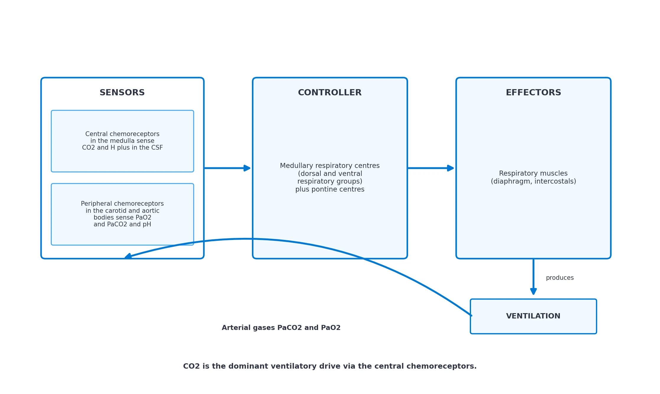
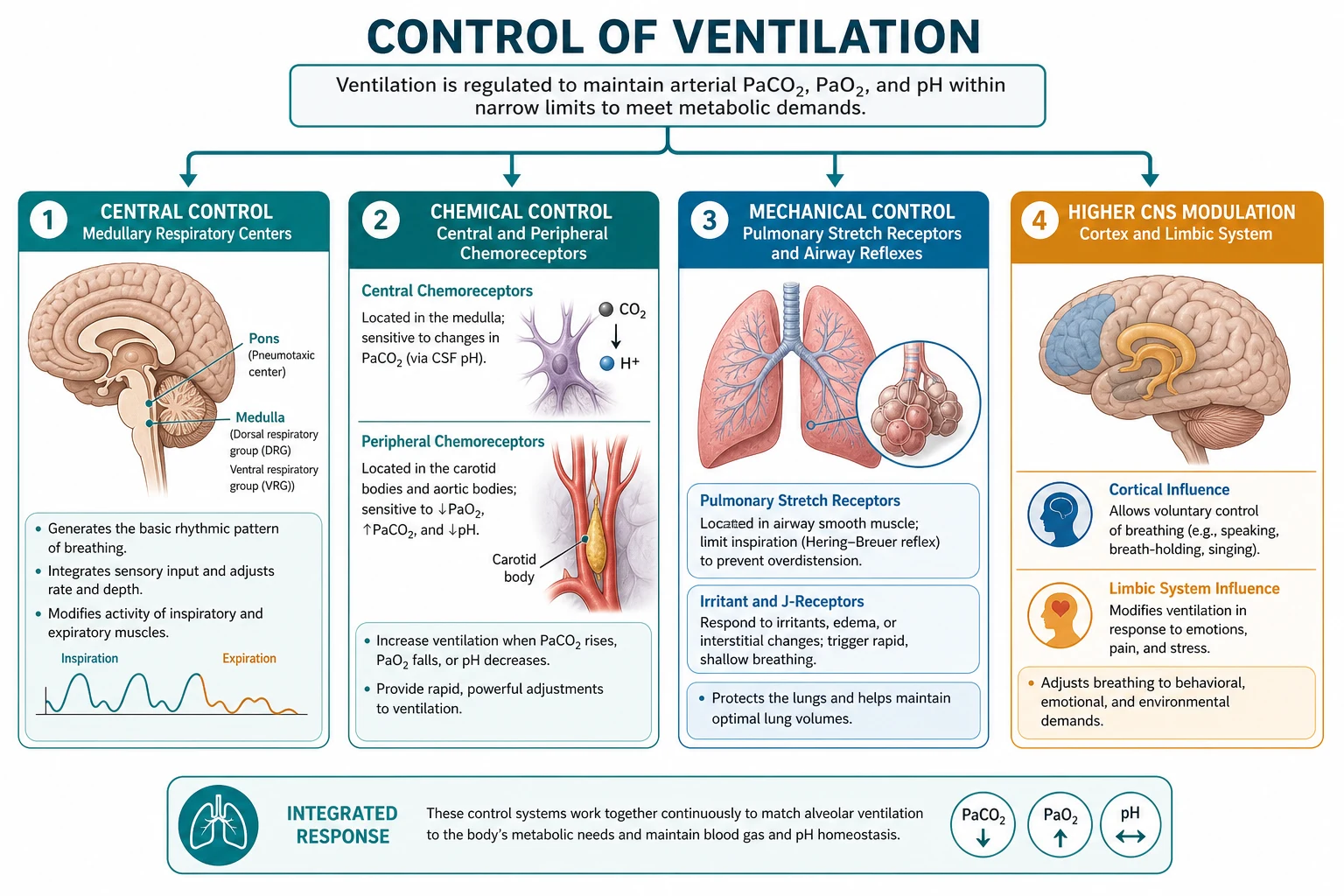
Ventilation is controlled automatically by a brainstem network that matches alveolar ventilation to metabolic demand, and it does so chiefly by tracking arterial carbon dioxide. The framework rests on five exam-critical ideas: the automatic rhythm of breathing is generated in the medullary respiratory centres (dorsal and ventral respiratory groups), shaped by pontine pneumotaxic and apneustic centres; the dominant ventilatory drive is carbon dioxide, sensed by central chemoreceptors on the ventral medulla that respond to the hydrogen ion concentration of cerebrospinal fluid (which CO2 crosses the blood-brain barrier to set); the peripheral chemoreceptors in the carotid and aortic bodies are the principal sensors of arterial oxygen (and also respond to CO2 and pH), and they drive the hypoxic ventilatory response; ventilation is a negative-feedback loop — sensors feed the controller, which drives the effectors, which set the arterial gases that feed back; and anaesthesia, opioids and the volatile agents depress both the carbon dioxide and the hypoxic ventilatory responses, which is why the anaesthetised and the opioid-dosed patient hypoventilate. Built on the carotid body and ventilatory acclimatisation study (MacDonald 2026), the carotid body amino-acid modulation study (Gold 2026), the NTS-to-medulla GABAergic study (Shao 2026), the acid-sensing ion channel central chemoreception study (Zhu 2024), the hypoxia central circuits study (Wang 2026), and the ventilatory CO2 response study (Ekman 2025).
On this page & tools
Your progress
Saved locally on this device.
8 MCQs with explanations
Target exams
Red flags
Why this matters to the anaesthetist
Every anaesthetic and opioid depresses ventilatory control. Recovery of spontaneous breathing, postoperative desaturation on the ward, opioid-induced respiratory depression, the CO2 retainer on uncontrolled oxygen, and the apnoeic threshold under anaesthesia are all manifestations of the same feedback system. Primary candidates must draw the control loop, state the sensors, write the CO2-response relationship, and explain how drugs shift the curves [5][6].
The respiratory rhythm generator
Automatic rhythm arises in the medulla oblongata: [1]
- Dorsal respiratory group (DRG) in the nucleus tractus solitarius (NTS): predominantly inspiratory, integrates visceral afferents (chemoreceptor, lung stretch).
- Ventral respiratory group (VRG): rostral inspiratory and caudal expiratory neurones; includes the pre-Bötzinger complex, the putative kernel of inspiratory rhythm generation.
- Pontine centres: the pneumotaxic centre (upper pons, including Kölliker–Fuse) limits inspiration and fine-tunes rate; the apneustic centre (lower pons) promotes prolonged inspiration if unopposed. Classic lesion physiology (apneusis after mid-pontine cut with vagi cut) is still examined [3].
Voluntary control from cortex can override automatic rhythm (speech, breath-holding) via corticospinal projections to respiratory motor neurones, but chemoreflex drive eventually breaks through. [1]
Central chemoreceptors — the dominant CO2 drive
The dominant ventilatory drive is carbon dioxide, sensed by central chemoreceptors on the ventral medullary surface (and distributed sites including the retrotrapezoid nucleus). Molecular CO2 crosses the blood–brain barrier freely; in CSF it is hydrated: [1]
CO2 + H2O ⇌ H2CO3 ⇌ H+ + HCO3− [1]
Central chemoreceptors respond primarily to CSF [H+], not to CO2 itself. Because CSF has little protein buffer (mainly bicarbonate), a small rise in PaCO2 produces a relatively large fall in CSF pH and a brisk increase in ventilation. Over hours to days, choroid plexus adjusts CSF [HCO3−], resetting the central drive (chronic respiratory acidosis or alkalosis compensation) [4].
Key quantitative anchors: [1]
- Resting PaCO2 ≈ 40 mmHg (5.3 kPa).
- Ventilation rises approximately linearly with PaCO2 above the apnoea threshold.
- Slope of the CO2-response curve ≈ 1.5–3 L·min−1 per mmHg PaCO2 in awake adults (classically ~2 L/min/mmHg).
- Apnoea threshold: the PaCO2 below which spontaneous ventilation ceases in the absence of other drives — typically a few mmHg below resting PaCO2 when awake; rises under anaesthesia/opioids (so apnoea occurs at a higher absolute PaCO2). [1]
Peripheral chemoreceptors — O2, CO2 and pH
The carotid bodies (bifurcation of common carotid; afferents via CN IX) are the primary arterial oxygen sensors; aortic bodies (CN X) play a lesser role in humans. They respond to: [1]
- Fall in PaO2 (not oxygen content — anaemia and CO poisoning do not strongly stimulate them despite low content).
- Rise in PaCO2 / fall in pH (faster, smaller contribution than central CO2 drive at rest).
- Reduced arterial flow / stagnation. [1]
Hypoxic ventilatory response (HVR): little change until PaO2 falls below ~60 mmHg (~8 kPa), then a steep hyperbolic rise in ventilation. The carotid body mediates acute HVR and the ventilatory acclimatisation to altitude. Sustained hypoxia produces a biphasic pattern: initial rise, then hypoxic ventilatory decline, then further acclimatisation over days [1][2].
Viva trap: Pulse oximetry SpO2 of ~90% corresponds roughly to PaO2 ~60 mmHg on the steep part of the ODC — the point at which HVR becomes clinically important. [1]
The CO2–ventilation response curve (must describe)
Plot minute ventilation (VE) on the y-axis against PaCO2 on the x-axis: [1]
- Above the apnoea point the relationship is nearly linear.
- Slope = CO2 sensitivity (chemoreflex gain).
- X-intercept / apnoea threshold = the PaCO2 at which VE would be zero if extrapolated.
- Anaesthesia, volatiles, opioids, benzodiazepines, and deep sleep shift the curve down and to the right: lower slope (blunted gain) and higher apnoea threshold. For any given PaCO2, ventilation is less; for any given ventilation, PaCO2 is higher.
- Metabolic acidosis shifts the curve left (higher ventilation at any PaCO2 — respiratory compensation).
- Metabolic alkalosis shifts right.
- Hypoxia multiplies CO2 sensitivity (interaction at peripheral chemoreceptors). [1]
Hypoxic ventilatory response in detail
VE versus PaO2 is hyperbolic, not linear. Interaction with CO2: at higher PaCO2 the HVR is steeper (multiplicative). Under anaesthesia both curves are depressed; the residual drive that restarts breathing after induction apnoea is often rising PaCO2 once the apnoea threshold is crossed — not hypoxia (which takes longer to develop if preoxygenated). [1]
Other receptors modulating breathing
- Pulmonary stretch receptors (Hering–Breuer): vagal afferents inhibit inspiration at high lung volume; more important in infants than resting adults.
- Irritant receptors: cough, bronchoconstriction, shallow rapid breathing.
- J receptors (juxtacapillary): activated by interstitial oedema → dyspnoea, tachypnoea (classic in pulmonary oedema).
- Muscle and joint proprioceptors: contribute to exercise hyperpnoea.
- Upper airway receptors: maintain dilator muscle tone; loss under anaesthesia → obstruction.
- Thermoregulatory and behavioural drives: fever, anxiety, pain increase VE. [1]
The closed-loop model
Sensors → controller (medulla/pons + cortical override) → effectors (diaphragm, intercostals, accessory muscles, upper airway dilators) → alveolar ventilation → PaCO2/PaO2/pH → sensors. Dead space, CO2 production (VCO2), and cardiac output modify the relationship between VE and PaCO2: [1]
PaCO2 ≈ K × VCO2 / VA [1]
where VA = VE − VD. Raised VCO2 (sepsis, MH, shivering) or raised VD/VT (PE, low tidal volume) elevates PaCO2 unless VE rises accordingly. [1]
Effect of anaesthesia and opioids
- Induction agents and volatiles: dose-dependent depression of CO2 response and HVR; FRC falls; upper airway collapsibility rises.
- Opioids: classic right/down shift of CO2 curve; rate falls more than tidal volume initially; irregular breathing; blunted HVR; risk peaks with co-sedation and in OSA/elderly.
- Residual neuromuscular block: effector failure — drive present, pump weak; high PaCO2 with distress if conscious.
- Spinal/epidural: high thoracic block can impair intercostals; phrenic (C3–5) usually spared unless cervical. [1]
Postoperative respiratory depression is the clinical expression of a still-shifted CO2 curve plus opioids plus residual anaesthesia plus possible residual block [6].
Special clinical situations
COPD / chronic CO2 retention. Chronic hypercapnia resets central chemoreception (higher CSF [HCO3−]). In a subset of severe retainers, removing hypoxic drive with high FiO2 can worsen hypercapnia via: (1) reduced ventilation, (2) loss of HPV → increased V/Q mismatch, (3) Haldane effect (oxygenated Hb carries less CO2). Target controlled oxygen (e.g. SpO2 88–92% in known retainers) rather than unrestricted high-flow O2. [1]
Altitude. Fall in PIO2 → HVR → hyperventilation → respiratory alkalosis → renal HCO3− excretion over days (acclimatisation). Carotid body is essential [1].
Exercise. VE rises in proportion to VCO2 so PaCO2 stays nearly constant until anaerobic threshold, then falls as lactic acidosis adds drive. [1]
Pregnancy. Progesterone increases VE; PaCO2 falls to ~30 mmHg — important for obstetric acid–base and apnoea tolerance. [1]
Raised ICP / head injury. CO2 reactivity of cerebral vessels: hyperventilation lowers PaCO2 → cerebral vasoconstriction → lower CBV/ICP (temporary bridge only). [1]
Anaesthetic relevance — practical
- Preoxygenation extends the time from apnoea to desaturation by raising PAO2 and denitrogenating FRC.
- After induction, expect apnoea until PaCO2 rises above the new, higher threshold.
- EtCO2 underestimates PaCO2 by the a–ET gradient (dead space); do not assume equality.
- Naloxone reverses opioid depression of the CO2 curve but is short-acting (renarcotisation risk).
- Monitor respiratory rate and sedation after opioid PCA; OSA patients need extended observation. [1]
Central chemoreceptors
- Ventral medulla / RTN
- Sense CSF [H+] from CO2
- Dominant resting drive
- Reset by chronic CSF HCO3− change
Peripheral chemoreceptors
- Carotid body (CN IX)
- Sense PaO2, PaCO2, pH
- HVR steep below ~60 mmHg
- Key at altitude and some COPD retainers
Viva traps
- Draw VE–PaCO2 and VE–PaO2 curves; mark apnoea threshold and the effect of morphine.
- "Do central chemoreceptors sense arterial H+ directly?" No — BBB limits H+/HCO3−; they sense CSF H+ generated from CO2.
- "Does anaemia stimulate the carotid body?" Little — they sense PO2, not content.
- State PaCO2 ≈ K·VCO2/VA and define VA = VE − VD.
- Explain why EtCO2 can be normal while the patient is hypoventilating if sampling or calibration is wrong — always correlate with clinical ventilation. [1]
Quantitative relationships and worked examples
Mass balance for CO2: PaCO2 = 863 × V̇CO2 / VA (when gas volumes are BTPS and partial pressures in mmHg; constant form varies by unit system). [1]
Clinically: if V̇CO2 is constant, halving alveolar ventilation doubles PaCO2. If a patient produces more CO2 (malignant hyperthermia, sepsis, hyperalimentation with high RQ, shivering), the same VE yields higher PaCO2. [1]
Dead-space fraction (Bohr): VD/VT = (PaCO2 − PECO2) / PaCO2 where PECO2 is mixed expired PCO2. Raised VD/VT (PE, low cardiac output, excessive PEEP, COPD) means a larger share of each breath is wasted; PaCO2 rises unless VE increases. [1]
Worked example — opioid hypoventilation: Awake: VE 6 L/min, PaCO2 40 mmHg. After morphine, CO2 curve shifts; at the same chemical drive the patient now ventilates 4 L/min. If V̇CO2 unchanged and VD similar, PaCO2 rises toward ~60 mmHg. The patient may not feel dyspnoeic if cortical perception and drive are both depressed — silent hypoventilation. [1]
Worked example — apnoea after induction: Preoxygenated patient, PaCO2 starts ~35 mmHg after hyperventilation on mask. CO2 rises ~3–6 mmHg/min during apnoea (rough exam figure; higher if high V̇CO2). If the apnoea threshold under anaesthesia is 45–50 mmHg, spontaneous efforts may not return for several minutes — plan manual ventilation. [1]
Neural anatomy beyond the headline nuclei
- NTS: first central synapse for peripheral chemoreceptor and baroreceptor afferents.
- Retrotrapezoid nucleus (RTN): CO2/H+-sensitive glutamatergic neurones driving breathing — modern anatomic substrate of "central chemoreceptors."
- Botzinger / pre-Bötzinger: expiratory modulation and inspiratory rhythm.
- Phrenic motor neurones (C3–C5): final common path to diaphragm; vulnerable in cervical spine injury and high neuraxial block.
- Hypoglossal motor neurones: genioglossus tone; reduced under anaesthesia and in REM sleep → obstruction. [1]
Interaction of O2 and CO2 drives
Peripheral chemoreceptor discharge is a function of both PaO2 and PaCO2/pH. Graphically, families of VE–PaO2 curves at different fixed PaCO2 show steeper hypoxic responses when PaCO2 is high. Conversely, the CO2 response is steeper under hypoxia. Anaesthetic agents flatten both surfaces of this interaction. [1]
Sleep, OSA and anaesthesia — shared physiology
Sleep, especially REM, reduces upper-airway dilator tone and mildly depresses chemoreflexes. OSA patients already live near the edge of obstruction; residual anaesthesia and opioids push them over. The physiology is the same loop: controller depression + effector (airway) failure + blunted arousal to CO2/hypoxia. [1]
Metabolic acid–base and ventilation
Kussmaul respiration in DKA is the CO2-response curve shifted left by low pH (peripheral and central contributions as H+ and as falling HCO3− alter CSF). Compensation cannot fully normalise pH when the primary disorder is metabolic, but PaCO2 may fall to the teens. Conversely, chronic metabolic alkalosis (diuretics, vomiting) suppresses ventilation and raises PaCO2 — do not "correct" this compensatory hypercapnia by forced hyperventilation without treating the alkalosis. [1]
Control of ventilation during mechanical ventilation
Positive-pressure ventilation bypasses the effector limb. If the patient is apnoeic, PaCO2 is set entirely by the ventilator and V̇CO2: choose minute ventilation to a target EtCO2/PaCO2. During spontaneous breathing trials, the chemoreflex must again drive the pump against the load of resistance, elastance and intrinsic PEEP — failure is as often load/weakness as pure drive. [1]
High-yield comparisons
| Depressant | CO2 curve | HVR | Notes |
|---|---|---|---|
| Volatile anaesthetics | Down/right | Markedly reduced | Dose-dependent |
| Propofol | Down/right | Reduced | Also airway collapsibility |
| Opioids | Down/right | Reduced | Rate falls; irregular pattern |
| Benzodiazepines | Mild–moderate | Variable | Synergistic with opioids |
| N2O | Mild | Less than volatiles | Still not neutral in combination |
Central pattern generation — exam narrative
Describe breathing as a three-phase cycle: inspiration (phrenic and external intercostal ramp), post-inspiration (laryngeal braking, declining phrenic), and expiration (passive at rest; active abdominal muscles when minute ventilation is high). The pre-Bötzinger complex generates inspiratory bursts; inhibitory networks sculpt the phases. Opioids act partly on medullary µ-receptors to slow rhythm and blunt chemoreflex gain — hence low rate and long expiratory pauses. [1]
Clinical measurement of drive and response
- Occlusion pressure P0.1: mouth pressure 100 ms after airway occlusion at onset of inspiration — index of central drive independent of respiratory mechanics.
- CO2 rebreathing tests (Read method): measure slope of VE vs PETCO2.
- Hypoxic challenges: careful laboratory tests of HVR. In theatre we use surrogates: respiratory rate, tidal volume, EtCO2, SpO2, and clinical airway obstruction. A normal SpO2 on supplemental O2 does not mean drive is intact. [1]
SAQ-ready description of the CO2 response curve
"Minute ventilation is plotted against PaCO2. Above an apnoea threshold the relationship is approximately linear with a slope of about two litres per minute per millimetre of mercury in the awake subject. The slope is chemoreflex gain; the extrapolated intercept is the apnoea point. Opioids and general anaesthesia reduce the slope and increase the apnoea threshold, shifting the curve down and to the right. Metabolic acidosis shifts the curve to the left so that ventilation is higher at any given PaCO2. Hypoxia increases the slope. These curve shifts explain why a patient may be apnoeic after induction until carbon dioxide rises, why postoperative opioids cause hypercapnia, and why the acidotic patient hyperventilates." [1]
Integration with mechanics
Drive (neural output) must overcome resistive and elastic loads. A patient with high drive but severe bronchospasm may still hypoventilate (load > pump). Conversely, residual neuromuscular block is pump failure with intact or high drive — distress with rising CO2 if conscious. Separate drive, pump, and load in every hypoventilation differential. [1]
Neonatal and paediatric notes
Infants have stronger Hering–Breuer reflexes, risk of periodic breathing, and different hypoxic responses (biphasic). Premature infants may become apnoeic with hypoxia. Not the focus of adult Primary answers but a bonus link. [1]
High-altitude pathophys sequence
Low PIO2 → carotid body → hyperventilation → low PaCO2 → respiratory alkalosis → left-shifted ODC and reduced central drive until renal HCO3− excretion restores CSF pH toward normal → further ventilatory rise (acclimatisation). Acetazolamide accelerates renal HCO3− loss and is used to prevent AMS — applied ventilatory-control pharmacology. [1]
Red flags
- CO2 is the dominant ventilatory drive, sensed centrally via CSF hydrogen ion.
- Peripheral chemoreceptors are the oxygen sensors; HVR becomes major below PaO2 ~60 mmHg.
- Anaesthesia and opioids depress CO2 and hypoxic responses — basis of perioperative respiratory depression.
- In CO2-retaining COPD, hypoxic drive and V/Q effects of oxygen matter; over-oxygenation can raise PaCO2.
- The apnoea threshold rises under anaesthesia. [1]
References
- [1]MacDonald TJA, et al. Going with the respiratory flow: The carotid body mediates human ventilatory acclimatization during high-altitude sojourn J Physiol, 2026.PMID 42210543
- [2]Gold OMS, et al. Amino acid modulation of the carotid body selectively modulates peripheral chemoreceptor respiratory reflex J Physiol, 2026.PMID 42139065
- [3]Shao L, et al. GABAergic Inhibition from the Nucleus Tractus Solitarius to Ventrolateral Medulla Phox2b Neurons Modulates Central Respiratory Chemoreflex and Ventilatory Homeostasis Neurosci Bull, 2026.PMID 41588273
- [4]Zhu Y, et al. Acid-sensing ion channel 1 in nucleus tractus solitarii neurons contributes to the enhanced CO(2)-stimulated cardiorespiratory effect in spontaneously hypertensive rats Life Sci, 2024.PMID 38889841
- [5]Wang X, et al. Molecular changes in hypoxia-induced central neural circuits and nuclei Med Gas Res, 2026.PMID 41964599
- [6]Ekman L, et al. Increased ventilatory response to carbon dioxide after dive training Undersea Hyperb Med, 2025.PMID 41429036