Anaes · Applied cardiovascular & respiratory physiology
Coronary & cerebral circulation
Also known as Coronary blood flow · Cerebral blood flow · Cerebral autoregulation · Coronary perfusion pressure · CO2 reactivity · Special circulations
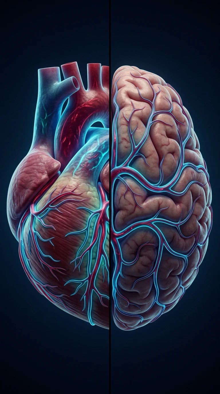
The coronary and cerebral circulations are the two most vital special circulations, and each is defended by its own tightly-regulated autoregulation. The framework rests on five exam-critical ideas: the coronary circulation is unusual in that flow occurs predominantly in DIASTOLE, because the contracting myocardium in systole compresses the intramural vessels, so coronary perfusion pressure is diastolic blood pressure minus the left-ventricular end-diastolic pressure; coronary flow is matched to myocardial oxygen demand chiefly by metabolic vasodilation (the heart extracts oxygen near-maximally at rest, so increased demand must be met by increased flow); the cerebral circulation autoregulates, holding cerebral blood flow constant across a cerebral perfusion pressure range of roughly 50 to 150 mmHg, with the curve shifted right in chronic hypertension; cerebral blood flow is exquisitely sensitive to arterial carbon dioxide (hypercarbia increases, hypocarbia decreases it); and cerebral perfusion pressure equals mean arterial pressure minus intracranial pressure, so raised intracranial pressure threatens cerebral perfusion. Built on the cerebrovascular-autoregulation study (Soule 2026), the cerebrovascular-function MRI study (Walsh 2026), the intraoperative cerebral zero-flow-pressure study (Murakami 2026), the flow-mediated epicardial-vasodilation study (Tribhuvan 2026), the perioperative LVAD study (Bottiroli 2026), and the coronary-flow-interventions study (Shah 2026).
On this page & tools
Your progress
Saved locally on this device.
13 MCQs with explanations
Target exams
Red flags
Why this matters to the anaesthetist
These two special circulations appear in almost every Final stem involving CAD, aortic stenosis, neurosurgery, carotid disease, or deliberate hypotension. Primary wants equations, diastolic coronary flow, autoregulation curve, CO2 reactivity. Speak in graphs and numbers.[5][3]
One-liner: CPP_cor = DBP − LVEDP (mostly diastolic flow); CBF autoregulated ~50–150 mmHg CPP and rises/falls with PaCO2; CPP_brain = MAP − ICP (or CVP if higher). [1]
Shared design principles
- High baseline O2 extraction (heart extreme; brain high).
- Flow must rise when metabolism rises (limited extraction reserve).
- Autoregulation over a perfusion pressure range.
- Vulnerable watershed regions when pressure or vessel lumen fails. [1]
Coronary circulation
Anatomy in one breath
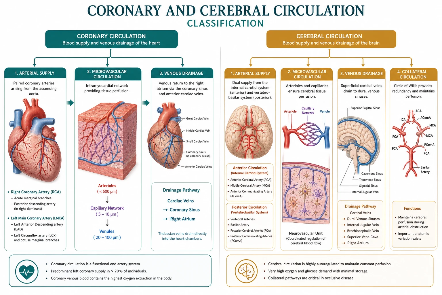
Left main → LAD + circumflex; RCA supplies RV and usually inferior wall/SA or AV node variants. Venous return mainly coronary sinus to RA. Subendocardium is most ischaemia-prone (highest wall stress, compressive forces). [1]
Flow is predominantly diastolic (LV)
Systolic contraction compresses intramural vessels → LV coronary flow occurs mainly in diastole. RV faces lower wall tension so more systolic flow possible. [1]
Coronary perfusion pressure (LV) ≈ DBP − LVEDP [1]
Anything that lowers DBP, raises LVEDP, or shortens diastole (tachycardia) reduces supply, especially subendocardial. [1]
Demand determinants (and therapy targets)
MVO2 rises with: [1]
- Heart rate (most expensive often)
- Contractility
- Wall stress ∝ (P × r)/h (Laplace) — afterload and dilation matter [1]
Supply–demand mismatch = ischaemia. Anaesthetic strategy in CAD: avoid tachycardia, maintain DBP, control pain/SNS, careful anaemia correction, normoxia. [1]
Metabolic regulation
Basal O2 extraction ~60–75% — near maximal → flow must increase for demand. Metabolites (adenosine, K+, H+, CO2, NO, endothelium-dependent dilation) couple flow to work. α-constriction can compete epicardially in SNS surge; β2 and metabolic dilation usually win in healthy vessels. Endothelial dysfunction (atheroma) impairs dilation.[4][6]
Anaesthetic coronary hooks
- Aortic stenosis: fixed output, need sinus rhythm and DBP for coronary drive.
- Pure vasodilators that drop DBP can steal supply.
- Anaemia + tachycardia is a classic supply–demand trap.
- Coronary steal discussions with some vasodilators — know the concept. [1]
Cerebral circulation
Anatomy / BBB / Monro–Kellie
ICA + vertebrobasilar → circle of Willis. Capillary tight junctions = BBB (lipid-soluble agents cross; polar drugs less). Rigid skull: V_brain + V_blood + V_CSF = constant → ↑blood volume or mass lesion raises ICP. [1]
Cerebral blood flow numbers
- Global CBF ~50 mL/100 g/min (~15% of CO; ~750 mL/min).
- CMRO2 ~3–3.5 mL O2/100 g/min.
- CPP = MAP − ICP (or MAP − CVP if CVP > ICP).
- Ischaemia risk when CPP falls below autoregulatory lower limit or vessels are stenosed. [1]
Autoregulation curve (describe/draw)

- Plateau: myogenic + metabolic arteriolar adjustments hold CBF steady.
- Below lower limit: pressure-passive fall → ischaemia.
- Above upper limit: pressure-passive rise → oedema/haemorrhage risk.
- Chronic hypertension: right shift — “normal” MAP may be too low for that patient; do not casually normalise BP perioperatively without thought.[1][2]
Impaired autoregulation: TBI, volatile high-dose, hypercapnia extremes, some critical illness. [1]
CO2 and O2 reactivity
- PaCO2: main acute lever — roughly ~2–4% CBF change per mmHg PaCO2 (teaching). Hypercapnia ↑CBF/CBV (±↑ICP); hypocapnia ↓CBF/CBV ( temporises ICP but risks ischaemia if extreme/prolonged).
- PaO2: little effect until severe hypoxaemia (<~50 mmHg) then dilation.
- Flow–metabolism coupling: active regions dilate; propofol lowers CMRO2 and CBF; volatiles tend to raise CBF relative to CMRO2 (uncoupling risk at higher MAC). [1]
ICP and venous drainage
ICP rises with masses, oedema, CSF obstruction, high CBV (vasodilation, venous obstruction). Head-down, tight ETT ties, coughing, high PEEP/CVPcan impede venous outflow. CPP defence = lower ICP + adequate MAP. [1]
Side-by-side compare (exam gold)
| Feature | Coronary | Cerebral |
|---|---|---|
| Key pressure | DBP − LVEDP | MAP − ICP |
| Phase | Diastolic LV flow | Continuous with pulse |
| Extraction reserve | Very low | Low–moderate |
| Key gas | Local metabolites | PaCO2 dominant acute |
| Autoregulation | Present | Classic 50–150 curve |
| Anaesthetic threat | Tachycardia + low DBP | Low MAP, high ICP, low PaCO2 extremes |
Anaesthetic strategy board
Protect myocardium: HR control, maintain DBP, blunt SNS to laryngoscopy, optimise O2 delivery (Hb, SaO2, CO), treat pain. [1]
Protect brain: adequate CPP, head midline/up if ICP, normocapnia as default, brief hypocapnia only for herniation bridge, choose agents thoughtfully for ICP, avoid venous obstruction, treat seizures/fever that raise CMRO2. [1]
Numbers board
- Coronary extraction ~70%
- CBF ~50 mL/100 g/min
- Autoregulation ~50–150 mmHg
- CO2 reactivity ~2–4%/mmHg
- Brain ~15% of CO; heart ~5% of CO at rest but highest O2 extraction [1]
Coronary threat
- Tachycardia
- Low DBP
- High LVEDP
- Anaemia + SNS
Cerebral threat
- MAP below auto limit
- High ICP/CVP
- Extreme hypocapnia
- Hypoxaemia/seizure/fever
Graph scripts
Draw coronary flow vs time in cardiac cycle (LV diastolic predominance). [1]
Draw CBF vs CPP plateau with right-shifted dashed curve for hypertension. [1]
Draw CBF vs PaCO2 near-linear rising relationship in physiologic range. [1]
Extended viva dialogue
Examiner: Give the coronary perfusion pressure equation and its implications. [1]
Candidate: Approximately diastolic blood pressure minus LV end-diastolic pressure. Tachycardia shortens diastole; hypotension lowers DBP; heart failure raises LVEDP — all three reduce subendocardial perfusion. [1]
Examiner: How does PaCO2 change cerebral blood flow? [1]
Candidate: Hypercapnia dilates cerebral arterioles and increases CBF about 2 to 4 percent per mmHg; hypocapnia does the opposite. That is why ventilation is an ICP tool and also an ischaemia risk if overdone. [1]
Clinical synthesis: For the heart, guard diastole and DBP; for the brain, guard CPP and CO2. Same anaesthetic, two equations. [1]
Coronary steal concept (one paragraph)
Dilating resistance vessels in well-perfused myocardium can divert flow away from collateral-dependent ischaemic zones if those arterioles are already max-dilated. Exam concept more than everyday drug label — still fair game with dipyridamole/adenosine imaging physiology. [1]
Neuroanaesthesia agent summary
| Agent | CBF | CMRO2 | ICP tendency |
|---|---|---|---|
| Propofol | ↓ | ↓ | ↓ |
| Volatiles (dose-dep) | ↑ relative | ↓ | ↑ possible |
| Ketamine | ↑ classic | ↑ classic | ↑ classic (context nuances) |
| Opioids | minimal direct | minimal | OK if ventilated |
Worked SAQ
SAQ: Define coronary perfusion pressure and factors reducing supply (7 marks)
Coronary perfusion pressure for the left ventricle approximates aortic diastolic pressure minus LV end-diastolic pressure, because LV coronary flow is predominantly diastolic. Supply falls with diastolic hypotension, elevated LVEDP (failure, overload), and tachycardia that shortens diastole. Anaemia and hypoxia reduce oxygen content; fixed stenoses limit flow reserve when demand rises. [1]
Primary exam expansion — dense examiner pack
Coronary flow essentials
LV myocardium: ~80% flow in diastole because systolic compression of intramural vessels. RV less systolic limitation. CPP ≈ aortic diastolic pressure − LVEDP (or RAP if higher). Tachycardia shortens diastole → less coronary perfusion time — bad for LV ischaemia. Normal coronary blood flow ~250 mL/min (~5% CO); high O2 extraction at rest (~60–70%) so ↑ demand met mainly by ↑ flow (metabolic vasodilation: adenosine, NO, KATP, hypoxia). [1]
Determinants of myocardial O2 supply vs demand
| Supply | Demand |
|---|---|
| CPP (ADP − LVEDP) | HR (major) |
| Coronary patency / spasm | Contractility |
| Diastolic time | Wall tension (preload/afterload, Laplace) |
| CaO2 (Hb, SaO2) | Basal metabolism |
| Autoregulation / collaterals | — |
Anaesthetic ischaemia avoidance: control HR, maintain diastolic BP, avoid extreme LVEDP rise (fluid overload, outflow obstruction), maintain O2 content, reverse causes of spasm. [1]
Coronary autoregulation and steal concepts
Autoregulation over wide pressure range if vessels healthy. Fixed stenoses: distal beds dilated at rest → cannot dilate further → pressure-dependent flow. Coronary steal teaching: arteriolar dilators may divert blood from collateral-dependent myocardium — clinical importance debated for many drugs; still viva concept. Subendocardium most vulnerable (highest wall tension, most systolic compression). [1]
Cerebral blood flow (CBF) determinants
Normal CBF ~50 mL/100 g/min (~15% CO). CMRO2 ~3.5 mL/100 g/min. Coupling: ↑ metabolism → ↑ flow. Autoregulation MAP ~50–150 mmHg (shifts up in chronic hypertension). Outside range: pressure-passive. [1]
PaCO2, PaO2, haematocrit, temperature
| Factor | CBF effect | Magnitude teaching |
|---|---|---|
| PaCO2 | Linear 20–80 mmHg range | ~3–4% CBF change per mmHg CO2 |
| PaO2 | Little until below 50 mmHg then rises | Hypoxaemic vasodilation |
| Temperature | ↓T → ↓CMRO2 → ↓CBF | 6–7% per °C teaching |
| Haematocrit | Viscosity extremes | Optimal O2 delivery trade-off |
| Volatiles | Direct vasodilation vs CMRO2↓ | Uncoupling risk at high dose |
| Propofol/barbiturate | ↓CMRO2 and ↓CBF | Coupled reduction |
| Ketamine | May ↑CBF/CMRO2 | Context dependent |
ICP and CPP relationship
CPP = MAP − ICP (or CVP if higher). Raised ICP → lower CPP → ischaemia → further swelling (danger loop). Cushing response: ischaemia → hypertension + bradycardia. Anaesthetic goals in neuro: adequate CPP, avoid obstructing venous drainage, control CO2, osmotherapy when indicated, temperature, seizures. [1]
Circle of Willis and regional notes
Collateral potential variable; not all patients complete. Focal stenosis: regional ischaemia risk despite global MAP. Steal and vasospasm (SAH) clinical cousins. [1]
Compare coronary vs cerebral one-liner table
| Coronary | Cerebral | |
|---|---|---|
| Extraction | High at rest | Moderate |
| Main metabolic controller | Local metabolites | CO2 / H+ / coupling |
| Systolic flow | Limited in LV | Continuous more |
| Autoregulation | Yes | Yes (classic teaching curves) |
SAQ: determinants of coronary blood flow (8 marks) or CBF (8 marks)
Pick one and structure: anatomy/timing → equation CPP → metabolic control → autonomic → pathology stenosis → anaesthetic implications. [1]
Viva
Q: Why avoid tachycardia in coronary disease? A: Raises demand and cuts diastolic supply time. Q: Effect of hyperventilation on CBF? A: Lower PaCO2 constricts cerebral vessels → lower CBF/ICP (and risk ischaemia if extreme). Q: CPP formula brain? A: MAP − ICP. [1]
High-yield viva battery and numbers lock-in
Coronary supply–demand grid (fill in every viva)
Supply: CPP (ADP−LVEDP), diastolic time, vessel patency, CaO2, collateral flow. Demand: HR, contractility, wall tension (preload/afterload), basal metabolic rate. Anaesthetic ischaemia usually from tachycardia + hypotension + anaemia combinations. [1]
CBF CO2 rule of thumb
Between roughly PaCO2 20–80 mmHg, CBF changes about 3–4% per mmHg PaCO2. Hyperventilation lowers ICP via vasoconstriction but risks ischaemia if excessive; hypoventilation raises CBF and ICP. Always couple CO2 strategy to metabolic needs and blood pressure. [1]
Neuroanaesthesia CPP defence sentence
"I set a CPP target based on the clinical context, measure or estimate ICP when available, keep the head neutral with venous drainage free, control PaCO2 in the normal–low normal range unless herniation crisis demands temporary hyperventilation, maintain oxygen content, and avoid agents or depths that steal autoregulation without benefit." [1]
Full viva dialogue (additional)
Examiner: Why is left ventricular subendocardium vulnerable? [1]
Candidate: It faces the highest wall tension, is compressed most during systole so its flow is predominantly diastolic, and sits farthest along the perforating arterial path. Tachycardia and raised LVEDP both hit it hard — one cuts supply time, the other cuts CPP. [1]
Examiner: Compare effects of propofol and a high-dose volatile on CBF–CMRO2 coupling. [1]
Candidate: Propofol reduces CMRO2 and CBF in a roughly coupled way, often lowering ICP. Potent volatiles reduce CMRO2 but cause direct cerebral vasodilation at higher doses, which can uncouple flow from metabolism and raise CBF/ICP — dose and agent matter. [1]
Exam traps
- CPP = MAP − CVP always when ICP higher — wrong; use higher of ICP/CVP.
- Treating hypotension in CAD with pure inotrope tachycardia.
- Extreme hyperventilation as prolonged ICP therapy.
- Forgetting diastolic hypotension as coronary supply threat. [1]
References
- [1]Soule Z, et al. Microcirculation and Cerebrovascular Autoregulation in Patients With Mechanical Circulatory Support Devices J Stroke, 2026.PMID 42237618
- [2]Walsh HJ, et al. Multi-modal magnetic resonance imaging to assess cerebrovascular function in patients with atrial fibrillation J Cereb Blood Flow Metab, 2026.PMID 42345329
- [3]Murakami S, et al. In Reply to: Comment on Effect of intraoperative position on the zero flow pressure of the cerebral circulation under propofol anesthesia: a prospective observational study J Anesth, 2026.PMID 42313123
- [4]Tribhuvan M, et al. Comment on Prevalence of impaired flow-mediated epicardial vasodilation among different types of coronary flow regulation Int J Cardiovasc Imaging, 2026.PMID 42337215
- [5]Bottiroli M, et al. Perioperative Management of Patients With Durable and Temporary Left Ventricular Assist Devices Undergoing Non-Cardiac Surgery: A Comprehensive Review J Cardiothorac Vasc Anesth, 2026.PMID 42350177
- [6]Shah S, et al. Effects of pharmacologic, non-pharmacologic and exercise-based interventions on coronary collateral circulation: A scoping review Cardiology, 2026.PMID 42319860