Anaes · Applied cardiovascular & respiratory physiology
Haematology & coagulation cascade physiology
Also known as Coagulation cascade · Haemostasis · Clotting factors · Fibrinolysis · Anticoagulants · Platelet function
Haemostasis is a precisely balanced process of clot formation (primary platelet plug, secondary coagulation cascade) and clot dissolution (fibrinolysis), and the anaesthetist intervenes in it with anticoagulants, antiplatelets, transfusion and neuraxial technique. The framework rests on five exam-critical ideas: primary haemostasis is the platelet plug (von Willebrand factor mediates platelet adhesion, then activation releases agonists, and aggregation forms the plug); the coagulation cascade amplifies via the intrinsic (contact) pathway (Factor XII through IX, monitored by the APTT) and the extrinsic (tissue factor) pathway (Factor VII, monitored by the PT and INR), converging on the common pathway (Factor X to thrombin to fibrin, cross-linked by Factor XIII); natural anticoagulants (antithrombin, protein C and S, tissue factor pathway inhibitor) prevent runaway clotting; fibrinolysis (plasmin from plasminogen via tPA) dissolves the clot, producing D-dimer; and the anticoagulant drugs (heparin via antithrombin, warfarin via vitamin K antagonism, DOACs by direct factor inhibition) act at specific points and are monitored by specific tests. Built on the contact-pathway COVID-coagulopathy study (Capecchi 2025), the Factor V deficiency review (Stoj 2026), the SERPINB2-fibrinolysis study (Zhu 2026), the fibrinolytic-DIC review (Iba 2026), the tPA-coagulopathy study (Takenaka 2026), and the paediatric DOAC perioperative-management review (Furman 2024).
On this page & tools
Your progress
Saved locally on this device.
8 MCQs with explanations
Target exams
Red flags
Why this matters to the anaesthetist
Primary science wants primary vs secondary haemostasis, the intrinsic/extrinsic/common pathways (even if cell-based models are more modern), which test covers which path, and natural anticoagulants/fibrinolysis. Final applies the same map to bleeding, VTE, neuraxial timing, and massive transfusion. Do not recite a biochemistry textbook — recite a teachable cascade with clinical hooks.[1]
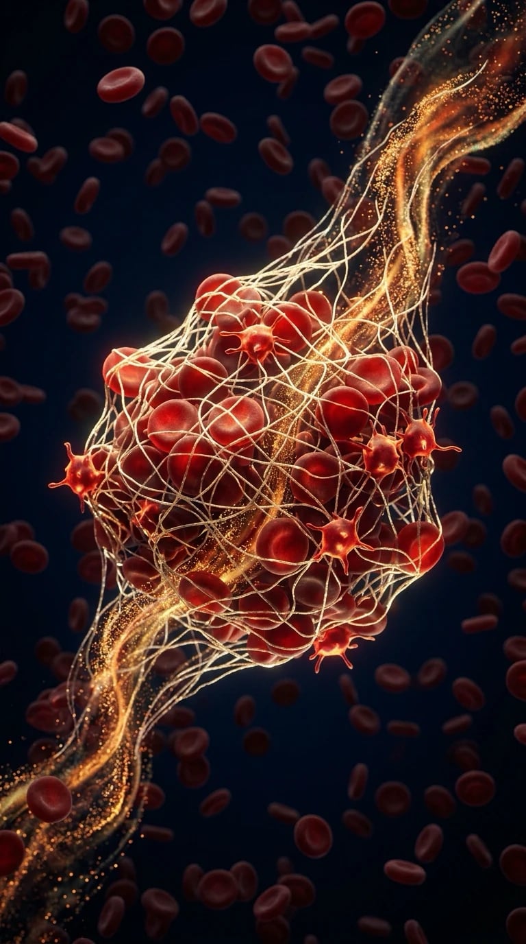
One-liner: Injury → platelet adhesion/activation/aggregation (primary) → tissue factor starts thrombin burst → fibrin mesh (secondary) → antithrombin/protein C/S limit spread → plasmin dissolves clot when the job is done. [1]
Primary haemostasis — the platelet plug
- Endothelial breach exposes collagen and vWF.
- Adhesion: GPIb–vWF–collagen bridge (Bernard–Soulier if GPIb deficient).
- Activation: shape change, release of ADP, TXA2, serotonin; GPIIb/IIIa activation.
- Aggregation: fibrinogen bridges GPIIb/IIIa between platelets (Glanzmann if GPIIb/IIIa deficient).
- Platelet phospholipid surface becomes the platform for coagulation complexes (link to secondary haemostasis). [1]
Drugs on primary haemostasis: aspirin (irreversible COX-1 → ↓TXA2), P2Y12 inhibitors (clopidogrel etc.), GPIIb/IIIa inhibitors, and quantitative thrombocytopenia. [1]
vWF roles: platelet adhesion and carrier for factor VIII (von Willebrand disease: mucocutaneous bleeding ± low VIII). [1]
Secondary haemostasis — classical cascade (exam map)
Extrinsic (tissue factor) pathway
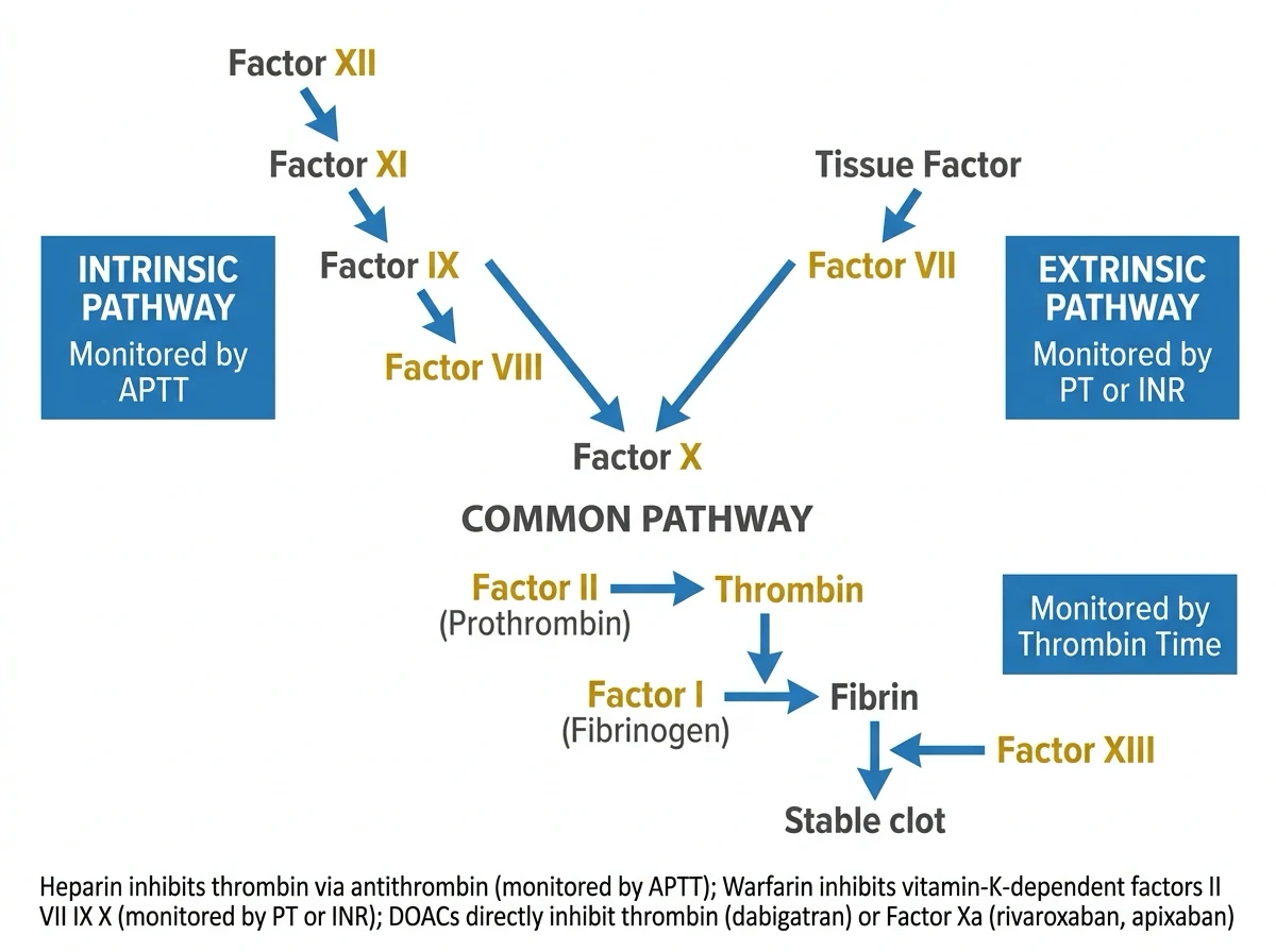
Tissue factor (III) + VIIa → activates X (and IX). PT/INR screens this path (VII short half-life — first to fall with warfarin/vitamin K deficiency). [1]
Intrinsic (contact) pathway
XII → XI → IX (+ VIII as cofactor) → X. APTT screens this path (and heparin, lupus anticoagulant, VIII/IX/XI deficiency). XII deficiency prolongs APTT without clinical bleeding — classic trap. [1]
Common pathway
X (+ V) → prothrombin (II) → thrombin → fibrinogen (I) → fibrin monomers → polymer; XIII cross-links fibrin (stabilises clot; XIII not well seen on PT/APTT). [1]
Thrombin’s many jobs (viva gold): cleaves fibrinogen; activates V, VIII, XI, XIII; potent platelet activator; when bound to thrombomodulin, activates protein C (negative feedback). [1]
Cell-based model (one paragraph for modern viva)
Initiation on TF-bearing cells → amplification on platelet surfaces → propagation with thrombin burst. Explains why isolated factor tables miss the living process, but you still need the classical map for tests and drug targets. [1]
Natural anticoagulants
| System | Mechanism | Deficiency phenotype |
|---|---|---|
| Antithrombin | Inhibits thrombin, Xa (and others); heparin amplifies | VTE risk; heparin resistance if very low |
| Protein C / S | Activated protein C with S inactivates Va, VIIIa | VTE; warfarin skin necrosis risk if protein C low |
| TFPI | Inhibits TF–VIIa–Xa | Less day-to-day exam focus |
| Endothelium | NO, PGI2, ADPase, thrombomodulin | Healthy vessels prevent inappropriate clotting |
Fibrinolysis
Plasminogen → plasmin (tPA, uPA). Plasmin degrades fibrin → D-dimers/FDP. TAFI, PAI-1, α2-antiplasmin restrain fibrinolysis. Hyperfibrinolysis (trauma, liver disease, thrombolysis) → bleeding; antifibrinolytics (tranexamic acid) block plasminogen activation/binding — major anaesthetic drug. [1]
Laboratory tests — what they do and do not tell you
| Test | Watches | Misses |
|---|---|---|
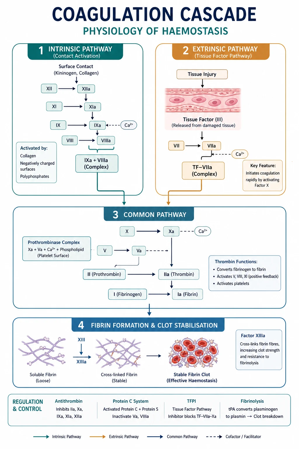
| PT/INR | Extrinsic/common; warfarin | Platelet function, XIII, mild defects |
| APTT | Intrinsic/common; heparin | Same; can be long without bleeding (XII, lupus) |
| TT / fibrinogen | Fibrin formation | — |
| Platelet count | Number | Function (aspirin) |
| PFA / aggregometry | Function (specialised) | Not routine everywhere |
| TEG/ROTEM | Global viscoelastic | Needs protocolised interpretation |
| Anti-Xa | Heparin/LMWH activity | — |
| D-dimer | Fibrin turnover | Non-specific when positive |
Anticoagulant and antiplatelet physiology targets
- Warfarin: inhibits vitamin K epoxide reductase → ↓II, VII, IX, X, protein C/S.
- Heparin: AT-dependent anti-IIa/Xa; APTT (UFH) or anti-Xa.
- LMWH/fondaparinux: preferential anti-Xa.
- DOACs: direct Xa or IIa inhibitors.
- Aspirin/P2Y12: primary haemostasis.
- Thrombolytics: drive plasmin generation. [1]
Neuraxial timing tables are applied pharmacology on top of this physiology — know half-lives/offset conceptually and follow ASRA/national guidance in clinical leaves. [1]
Anaesthetic relevance board
- Surgical haemostasis needs platelets + factors + fibrinogen + controlled fibrinolysis.
- Massive transfusion: dilute factors/platelets/fibrinogen; citrate; TXA early in trauma protocols.
- Liver failure: synthetic failure (factors, AT, proteins C/S, fibrinogen) + variable fibrinolysis + portal thrombocytopenia/splenomegaly.
- DIC: consumption + fibrinolysis — prolonged times, low platelets/fibrinogen, high D-dimer.
- Obstetric: hypercoagulable baseline; fibrinogen critical in PPH.
- CPB: heparin + AT, then protamine; platelet dysfunction from circuit. [1]
Numbers board
- Normal platelet count ~150–400 × 10⁹/L; many procedures tolerate lower with context.
- Fibrinogen critical often discussed around <1.5–2.0 g/L in bleeding (context-specific).
- Factor VII half-life short (~4–6 h) — INR rises first on warfarin.
- Factor II half-life long (~60 h) — full warfarin effect delayed. [1]
PT/INR prolonged
- VII/common path
- Warfarin, vit K def
- Liver synthetic failure
- DIC, rare factor def
APTT prolonged
- Intrinsic/common
- Heparin, VIII/IX/XI
- Lupus anticoagulant
- XII (no bleed)
Viva draw / SAQ scripts
Draw Y-shaped cascade with APTT and PT limbs labelled and heparin/warfarin marks. [1]
Explain why warfarin needs overlap with heparin when treating acute VTE (protein C short half-life → early hypercoagulable window). [1]
Cell-based vs cascade: initiation/amplification/propagation in three sentences. [1]
TXA mechanism and trauma timing principle. [1]
Extended viva dialogue
Examiner: Walk me from vessel injury to a stable clot. [1]
Candidate: Platelets adhere via vWF and collagen receptors, activate and aggregate to form a primary plug. Tissue factor generates small thrombin, which amplifies on platelet surfaces to a thrombin burst converting fibrinogen to fibrin; factor XIII cross-links fibrin. Antithrombin and protein C systems limit spread; later tPA-driven plasmin remodels the clot. [1]
Examiner: Which tests for heparin and warfarin? [1]
Candidate: Unfractionated heparin mainly prolongs APTT (or use anti-Xa); warfarin prolongs PT/INR because factor VII falls quickly. Neither test assesses platelet function. [1]
Clinical synthesis: If you own primary plug, thrombin burst, inhibitors, fibrinolysis, and what each test sees, you can reason any coagulopathy stem. [1]
Cell-based model — three stages for modern viva
- Initiation: TF-bearing cells + VIIa generate small Xa/thrombin.
- Amplification: thrombin activates platelets, V, VIII, XI on platelet surfaces.
- Propagation: tenase and prothrombinase complexes on activated platelets create thrombin burst → fibrin. [1]
This explains why haemophilia (VIII/IX) bleeds despite intact “extrinsic” initiation — amplification/propagation on platelets fails. [1]
Vitamin K cycle
Reduced vitamin K is cofactor for γ-carboxylation of factors II, VII, IX, X and proteins C/S. Warfarin inhibits epoxide reductase → depleted reduced vitamin K. Vitamin K and PCC/FFP reverse depending on urgency. [1]
Worked SAQ
SAQ: Describe primary haemostasis and name two drug targets (7 marks)
Endothelial injury exposes collagen and vWF. Platelets adhere via GPIb–vWF, activate, release ADP and TXA2, and aggregate via GPIIb/IIIa–fibrinogen bridges, forming a primary plug and providing phospholipid surface for coagulation. Aspirin inhibits platelet COX-1 reducing TXA2; P2Y12 inhibitors block ADP amplification; GPIIb/IIIa inhibitors block aggregation. [1]
Primary exam expansion — dense examiner pack
Haemostasis phases (modern cell-based model + classic cascade)
- Endothelial injury / vasoconstriction. 2. Primary haemostasis: platelet adhesion (GPIb–vWF), activation, aggregation (GPIIb/IIIa–fibrinogen). 3. Secondary haemostasis: thrombin burst generation of fibrin. 4. Stabilisation FXIIIa cross-links fibrin. 5. Regulation/fibrinolysis: protein C/S, antithrombin, TFPI; plasminogen → plasmin. [1]
Classic intrinsic (XII, XI, IX, VIII) vs extrinsic (VII + tissue factor) converge at X → Xa + V → prothrombinase → thrombin (II) → fibrinogen to fibrin. Cell-based model emphasises tissue factor–bearing cells and platelet surfaces — better explains why XII deficiency does not bleed much in vivo. [1]
Platelet physiology
Lifespan ~7–10 days; count 150–400 × 10^9/L teaching. Dense granules ADP/serotonin; alpha granules vWF/fibrinogen/factor V. Drugs: aspirin irreversible COX-1 (TXA2↓); clopidogrel P2Y12; GPIIb/IIIa inhibitors; desmopressin releases vWF (useful in mild haemophilia A/vWD selected cases). [1]
Lab tests mapping
| Test | Pathway | Prolonged in |
|---|---|---|
| PT/INR | Extrinsic/common | Warfarin, vit K def, VII, liver synthetic failure |
| APTT | Intrinsic/common | Heparin, VIII/IX/XI def, lupus anticoagulant (thrombosis paradox) |
| TT / fibrinogen | Fibrin formation | Hypofibrinogenaemia, thrombin inhibitors, high FDPs |
| Anti-Xa | Heparin/LMWH activity | Monitoring LMWH/UFH |
| ACT | Global high-dose heparin | CPB |
| TEG/ROTEM | Viscoelastic whole blood | Trauma/obstetric algorithms |
| D-dimer | Fibrin degradation | Thrombosis/fibrinolysis marker (nonspecific) |
Vitamin K dependent factors
II, VII, IX, X + proteins C/S. Warfarin inhibits vitamin K epoxide reductase → falls first in shortest half-life (VII, protein C) — transient hypercoagulable risk → heparin bridge contexts. Reversal: vitamin K, PCC (preferred emergency), FFP if PCC unavailable. [1]
Heparin and antithrombin
UFH potentiates antithrombin against IIa and Xa; LMWH anti-Xa > IIa; fondaparinux pure anti-Xa. HIT type II: antibodies to PF4–heparin → thrombosis paradox — stop heparin, use alternative anticoagulant. Protamine reverses UFH (partly LMWH); risk anaphylactoid/pulmonary hypertension especially prior sensitisation/NPH insulin history teaching. [1]
Hypercoagulable states and periop VTE physiology
Virchow: stasis, endothelial injury, hypercoagulability. Surgery adds all three. Prophylaxis mechanical + pharmacological balance bleeding risk. Neuraxial timing with anticoagulants — sister safety topic. [1]
DIC physiology
Widespread TF drive → consumption of platelets/factors → thrombosis microvasculature + bleeding. Causes: sepsis, trauma, amniotic fluid embolism, malignancy, mismatched transfusion. Labs: ↓platelets, ↑PT/APTT, ↓fibrinogen, ↑D-dimer/FDPs. Treat cause; support components; heparin rare selected thrombotic phenotypes. [1]
Massive haemorrhage physiology
Lethal triad: hypothermia, acidosis, coagulopathy. Damage-control resuscitation: limit crystalloid, balanced ratios, early fibrinogen, calcium (citrate bind), tranexamic acid antifibrinolysis (time-critical trauma/obstetric evidence base). Viscoelastic-guided algorithms increasingly standard. [1]
SAQ: describe coagulation pathways and point of action of heparin and warfarin (10 marks)
Cascade/cell-based outline (4). Heparin–antithrombin (2). Warfarin–vitamin K factors (2). Monitoring PT vs APTT/ACT (2). [1]
Viva
Q: Why does warfarin raise INR? A: Depletes factor VII (and others) — PT sensitive to extrinsic path. Q: Why can HIT cause thrombosis? A: Antibody-mediated platelet activation. Q: Which factor deficiency prolongs APTT but little bleeding? A: Factor XII (contact pathway) teaching contrast with VIII/IX. [1]
High-yield viva battery and numbers lock-in
Factor half-life teaching (why INR rises first on warfarin)
Factor VII shortest among vitamin K factors → PT/INR prolongs early. Full anticoagulant effect needs II, IX, X decline. Protein C also short → early transient hypercoagulability possible. [1]
Trauma coagulopathy pillars
Tissue injury TF exposure; shock/hypoperfusion-activated protein C pathway acute traumatic coagulopathy; dilution from crystalloid; hypothermia; acidosis; hypocalcaemia from citrate; hyperfibrinolysis in some phenotypes — TXA logic early. [1]
Viscoelastic trace interpretation skeleton
Prolonged R/CT → clotting factor problem/heparin; low α angle/k → fibrinogen problem; low MA/MCF → platelets (±fibrinogen); high LY30/LI30 → fibrinolysis. Algorithms guide plasma, cryoprecipitate/fibrinogen concentrate, platelets, TXA. [1]
Full viva dialogue (additional)
Examiner: Where does heparin act and how do we reverse it? [1]
Candidate: Unfractionated heparin potentiates antithrombin, accelerating inactivation of thrombin and factor Xa. We monitor with APTT or ACT depending on dose context. Protamine forms an inactive complex with heparin; dose roughly matches circulating heparin, and excess protamine can itself anticoagulate mildly and cause pulmonary hypertension or anaphylactoid reactions. [1]
Examiner: Why is the intrinsic/extrinsic cascade incomplete as a story? [1]
Candidate: It explains PT and APTT well but not biology: factor XII deficiency markedly prolongs APTT without severe bleeding, and in vivo initiation is largely tissue-factor driven on cell surfaces with amplification on platelets — the cell-based model. [1]
Exam traps
- Equating INR with full bleeding risk in cirrhosis.
- Giving platelets for all high INR.
- Forgetting calcium in massive transfusion.
- Using cryoprecipitate thinking without fibrinogen level/ROTEM. [1]
References
- [1]Capecchi M, et al. Involvement of the contact pathway in COVID-19 coagulopathy Intern Emerg Med, 2025.PMID 41206422
- [2]Stoj KM. Factor V Deficiency 2026.PMID 35593819
- [3]Zhu Y, et al. Epithelial SERPINB2 Overexpression Contributes to Impaired Fibrinolysis in Chronic Rhinosinusitis With Nasal Polyps via tPA Downregulation Front Biosci (Landmark Ed), 2026.PMID 42216541
- [4]Iba T, et al. Redefining Fibrinolytic Insufficiency in Sepsis-Associated DIC Semin Thromb Hemost, 2026.PMID 41956121
- [5]Takenaka R, et al. Prolonged low-dose tPA ameliorates coagulopathy and organ injury in an LPS-induced rat DIC model Int J Hematol, 2026.PMID 41801558
- [6]Furman K, et al. A review of the perioperative management of direct oral anticoagulants for pediatric anesthesiologists Paediatr Anaesth, 2024.PMID 39244715