Anaes · Applied cardiovascular & respiratory physiology
Lung volumes & capacities
Also known as Tidal volume · Functional residual capacity · Closing capacity · Vital capacity · Dead space · Spirometry
The lung holds characteristic volumes of air at each phase of breathing, and the way those volumes change with position, anaesthesia and disease explains both gas exchange and atelectasis. The framework rests on five exam-critical ideas: the four lung volumes (tidal volume, inspiratory reserve, expiratory reserve, residual volume) combine into four capacities (inspiratory capacity, functional residual capacity, vital capacity, total lung capacity); the functional residual capacity (about 2.5 to 3 L) is the oxygen reservoir that buffers against hypoxaemia during apnoea and the volume at which the lung rests with the least work; closing capacity rises with age and, when it exceeds functional residual capacity, small airways close during tidal breathing causing atelectasis and shunt; dead space (anatomical, alveolar and physiological) is the volume that is ventilated but does not take part in gas exchange, governed by the Bohr equation; and anaesthesia and the supine position reduce functional residual capacity by about 10 to 20 percent, bringing closing capacity closer to it and predisposing to atelectasis. Built on the patient-positioning study (Harris 2026), the high-flow nasal preoxygenation study (Bouvet 2026), the pulmonary-function-testing consensus (CACP Task Force 2026), the Global Lung Initiative 2022 spirometry reference-equation study (Al-Qerem 2026), the spirometric lung-function study (Tabbah 2026), and the non-invasive-ventilation-distribution study (Chidini 2025).
On this page & tools
Your progress
Saved locally on this device.
8 MCQs with explanations
Target exams
Red flags
Why this matters to the anaesthetist
You must define every volume/capacity, state typical adult values, explain spirometry vs helium dilution/body box for RV, and reason how posture, anaesthesia, pregnancy, obesity and disease move FRC relative to closing capacity.[1]
One-liner: TV+IRV+ERV+RV make TLC; FRC = ERV+RV is the resting volume where elastic recoil of lung and chest wall balance; under anaesthesia FRC falls toward CC → airway closure and shunt. [1]
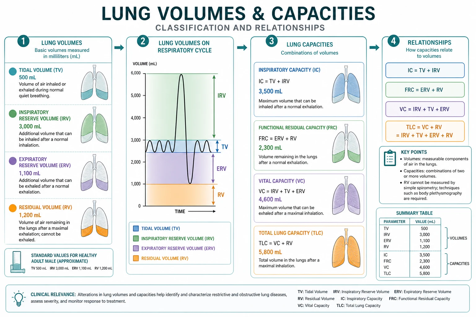
The four volumes
| Volume | Definition | Typical 70 kg adult |
|---|---|---|
| TV tidal volume | Quiet breath | ~500 mL (6–8 mL/kg IBW) |
| IRV inspiratory reserve | Extra inhale from end-inspiration | ~2500 mL |
| ERV expiratory reserve | Extra exhale from end-expiration | ~1500 mL |
| RV residual volume | Gas left after maximal exhale | ~1200 mL |
The four capacities (sums)
| Capacity | Sum | Typical |
|---|---|---|
| IC inspiratory capacity | TV + IRV | ~3 L |
| FRC functional residual capacity | ERV + RV | ~2.5–3 L |
| VC vital capacity | IRV + TV + ERV | ~4.5–5 L |
| TLC total lung capacity | VC + RV | ~6 L |
FRC — the star of anaesthesia
Definition: volume at end of quiet expiration; equilibrium between lung inward recoil and chest wall outward recoil. [1]
Why it matters: [1]
- O2 store during apnoea (with VC preoxygenation you fill FRC with O2).
- Keeps airways open if FRC > closing capacity.
- Falls with: supine posture, anaesthesia (especially GA + muscle relaxation), obesity, pregnancy, abdominal surgery, ascites, laparoscopic insufflation.
- Rises with: PEEP/CPAP, upright posture, some obstructive disease (air trapping). [1]
Closing capacity / closing volume
Closing capacity (CC) = closing volume + RV — volume at which dependent airways begin to close. [1]
CC rises with age and smoking; FRC falls when supine/anaesthetised → CC may exceed FRC → cyclic airway closure → absorption atelectasis, shunt, impaired gas exchange. [1]
Measurement methods
| Method | Measures | Notes |
|---|---|---|
| Spirometry | TV, VC, ERV, IRV, FEV1, FVC | Cannot measure RV/FRC/TLC alone |
| Helium dilution | FRC | Underestimates gas behind severe obstruction |
| Nitrogen washout | FRC | Similar limitations |
| Body plethysmography | FRC/TLC including trapped gas | Gold for obstruction |
| DLCO | Gas transfer | Separate from volumes |
Obstructive pattern: ↑RV, ↑TLC possible, ↓FEV1/FVC. Restrictive: ↓TLC, ↓VC, FEV1/FVC normal/high. [1]
Minute ventilation and dead space links
VE = TV × RR. Alveolar ventilation VA = (TV − VD) × RR. Volumes set the stage for V/Q leaf content — mention anatomic VD ~2 mL/kg. [1]
Factors shifting volumes (exam table)
| Factor | FRC | Notes |
|---|---|---|
| Supine | ↓ | Abdominal contents cephalad |
| GA + NMB | ↓ ~20% | Atelectasis common |
| PEEP | ↑ | Recruits; watch CO |
| Obesity | ↓ | High CC relative risk |
| Pregnancy | ↓ | Especially late; ERV falls |
| Emphysema | ↑ | Air trapping |
| Pulmonary fibrosis | ↓ TLC | Restrictive |
Apnoea oxygenation teaching
Preoxygenate to FETO2 > 0.9 fills FRC with O2 → prolongs safe apnoea. High-flow nasal oxygen can extend apnoea by providing ongoing O2 to FRC (THRIVE concepts). Desaturation faster in children/obese/pregnant/ill because O2 store small and VO2 high. [1]
Numbers board
- TV ~500 mL; RV ~1200 mL; FRC ~30 mL/kg; TLC ~6 L
- VC ≥15 mL/kg classic extubation/ventilatory reserve teaching threshold (context-dependent)
- FEV1/FVC <0.7 suggests obstruction (guideline nuances exist) [1]
Obstructive
- ↑RV/FRC often
- ↓FEV1/FVC
- Air trapping
- He dilution may err
Restrictive
- ↓TLC
- VC reduced
- Ratio normal/high
- Fibrosis, NM weakness
Graph scripts
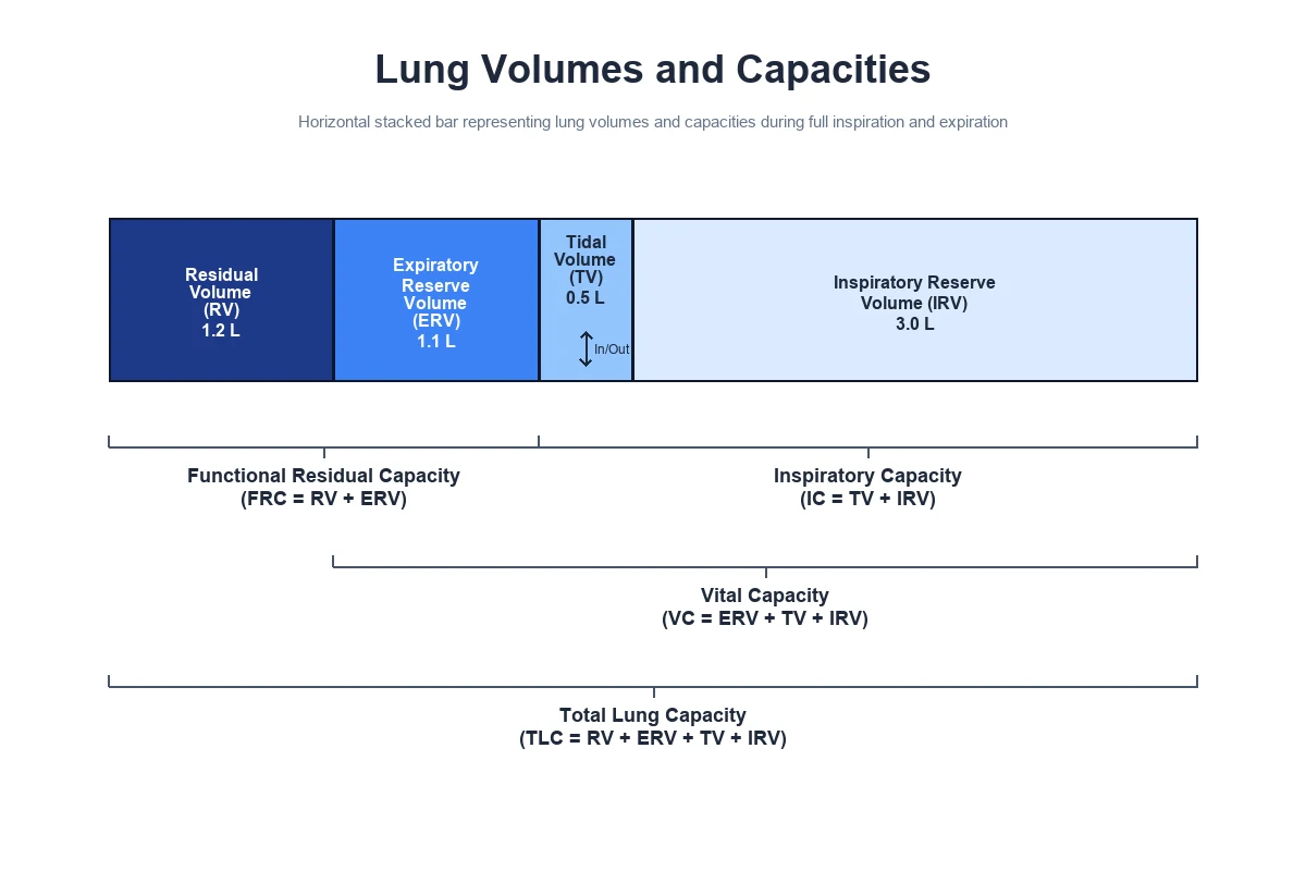
Draw stacked volume bar and bracket capacities. [1]
Draw FRC vs CC with age — lines cross in older supine patients. [1]
Spirometry traces: obstructive scooped exhalation vs restrictive small volumes. [1]
Extended viva dialogue
Examiner: Define FRC and give two reasons it falls under anaesthesia. [1]
Candidate: FRC is the lung volume at end of quiet expiration, ERV plus RV. It falls when supine because the abdomen displaces the diaphragm, and under general anaesthesia with muscle relaxation the chest wall and diaphragm lose tone so the equilibrium volume drops. [1]
Examiner: Why does that cause hypoxia? [1]
Candidate: If FRC falls below closing capacity, dependent airways close, alveolar gas is absorbed, atelectasis and shunt increase, and the oxygen store for apnoea shrinks. [1]
Clinical synthesis: If you can draw the stack and reason FRC versus CC, you own lung volume vivas. [1]
Helium dilution principle
Subject breathes known He concentration in closed circuit; final He dilution calculates FRC (C1V1 = C2V2). Does not see gas in bullae poorly communicating — plethysmography includes trapped gas (Boyle’s law box). [1]
Preoxygenation maths intuition
FRC ~30 mL/kg O2-filled after denitrogenation; VO2 ~3 mL/kg/min → rough apnoea time scales with store/consumption, shortened by shunt, low FRC, high VO2 (child, pregnant, septic). [1]
Worked SAQ
SAQ: Define FRC and explain changes under general anaesthesia (7 marks)
FRC is the lung volume at end of quiet expiration, equal to ERV plus RV, set by balance of lung and chest wall recoil. Under general anaesthesia, especially with muscle relaxation and supine posture, FRC falls toward or below closing capacity, promoting airway closure, atelectasis, shunt and faster desaturation during apnoea. PEEP and head-up positioning help restore end-expiratory volume. [1]
Body plethysmography Boyle’s law
Patient pants against closed shutter; box and alveolar pressure–volume changes estimate intrathoracic gas volume including trapped gas: P1V1 = P2V2. Why TLC in emphysema is higher by box than by dilution. [1]
Dynamic hyperinflation link
In obstruction, incomplete emptying raises end-expiratory volume above relaxed FRC — intrinsic PEEP. Anaesthesia and PPV interact with this physiology in COPD. [1]
Extended viva add-on
Examiner: Why can’t spirometry measure FRC? [1]
Candidate: Spirometry only measures exhaled or inhaled volumes from the breathing path. Residual volume remains in the lung after maximal expiration and cannot be seen without gas dilution or plethysmography; FRC includes residual volume. [1]
Primary exam expansion — dense examiner pack
Definitions with spirometry pattern (commit)
| Volume/capacity | Definition | Typical adult male order |
|---|---|---|
| TV | Tidal volume | 500 mL |
| IRV | Inspiratory reserve | 2500 mL |
| ERV | Expiratory reserve | 1500 mL |
| RV | Residual volume (cannot spirometry-exhale) | 1200 mL |
| IC | IRV+TV | 3000 mL |
| FRC | ERV+RV | 30 mL/kg ≈ 2.5–3 L |
| VC | IRV+TV+ERV | 4.5–5 L |
| TLC | VC+RV | 6 L |
| FEV1/FVC | Dynamic | >0.7 young normal teaching |
RV and FRC need helium dilution, N2 washout, or body plethysmography — spirometry alone cannot measure them. [1]
FRC — the anaesthetic centre of gravity
FRC is volume at end of quiet expiration: balance of inward lung recoil and outward chest wall recoil. Oxygen store for apnoea; determines atelectasis propensity; alters airway resistance and compliance (volumes on steep part of curve if low). Factors reducing FRC: GA induction, supine/Trendelenburg, obesity, pregnancy, laparoscopy, abdominal compartment, ascites, thoracic banding, inadequate PEEP. [1]
Closing capacity and airway closure
Closing capacity (CC) = closing volume + RV. When FRC falls below CC, dependent airways close in normal tidal breathing → shunt-like effect, impaired gas exchange. CC rises with age; FRC falls when supine — elderly supine spontaneously may encroach; GA makes it worse. Why PEEP/CPAP and head-up help. [1]
Preoxygenation maths intuition
After denitrogenation, alveolar O2 fraction high; FRC store ≈ FRC × FAO2. Consumption VO2 ≈ 3 mL/kg/min. Apnoea time roughly store/VO2, shortened by low FRC (obesity, pregnancy), high VO2 (sepsis, pregnancy, child), airway obstruction, and inadequate denitrogenation. High-flow nasal oxygen / THRIVE extends safe apnoea by ongoing O2 delivery and some CO2 clearance — sister clinical topics. [1]
GA effects on volumes (classic list)
- Loss of diaphragmatic/end-expiratory muscle tone → FRC ↓ ~20%. 2. Atelectasis in dependent lung within minutes on 100% O2. 3. Cephalad diaphragm in supine. 4. Blood volume shift. 5. Airway closure tendency ↑. Recruitment: PEEP, sigh, head-up, avoid prolonged pure O2 when safe, NM blockade carefully with ventilation strategy. [1]
Obstructive versus restrictive patterns
| Pattern | TLC | RV | FEV1/FVC | Example |
|---|---|---|---|---|
| Obstructive | Normal/↑ | ↑ | ↓ | Asthma, COPD |
| Restrictive | ↓ | ↓/N | Normal/↑ | Fibrosis, NM weakness, obesity mixed |
Flow–volume loops: scooped expiration obstructive; tall narrow restrictive — draw in viva. [1]
Dead space volumes link
Anatomical dead space ~2 mL/kg; apparatus dead space added by connectors. Alveolar ventilation VA = (TV − VD) × RR. Small TV high RR strategies may ventilate mostly dead space — EtCO2 and PaCO2 diverge with high VD/VT (shock, PE). [1]
Body plethysmography Boyle link
Patient pants against closed shutter; P1V1=P2V2 estimates intrathoracic gas volume including trapped gas — may exceed dilution FRC in obstruction (trapped gas detected). [1]
SAQ: FRC under general anaesthesia (7–8 marks)
Define FRC (1). Determinants recoil balance (1). List factors reducing FRC under GA and positioning (3). Consequences for oxygenation/atelectasis (2). Mitigation PEEP/position/FiO2 (1). [1]
Viva
Q: Why measure FRC not just VC? A: Gas exchange and apnoea store depend on end-expiratory volume including RV component. Q: Why do elderly desaturate faster when supine under GA? A: CC approaches/exceeds FRC; plus lower FRC from GA. Q: Can spirometry give RV? A: No — needs dilution/plethysmography methods. [1]
High-yield viva battery and numbers lock-in
Capacity arithmetic
- IC = TV + IRV
- FRC = ERV + RV
- VC = IRV + TV + ERV
- TLC = VC + RV = IC + FRC
- Minute ventilation = TV × RR
- Alveolar ventilation = (TV − VD) × RR [1]
FRC modifiers list (memorise 8)
GA, supine, Trendelenburg, obesity, pregnancy, laparoscopy/↑IAP, abdominal pain/binders, inadequate muscle tone/paralysis without PEEP, pulmonary fibrosis (low volumes overall), emphysema (high FRC/TLC — contrast). [1]
Preoxygenation and apnoea extension tools
Tight mask FiO2 1.0, end-tidal O2 >0.9 target teaching, head-up if possible, CPAP/NIV in obese, nasopharyngeal oxygen/THRIVE during apnoea, avoid premature suction desaturation cycles, careful RSI timing. [1]
Full viva dialogue (additional)
Examiner: Why does FRC fall under general anaesthesia? [1]
Candidate: Loss of end-expiratory diaphragmatic and chest wall muscle tone allows the balance of recoils to settle at a lower volume; the supine posture and cephalad diaphragm shift compound this, and absorption atelectasis develops especially with high FiO2, further reducing ventilated FRC. [1]
Examiner: Define closing capacity and its anaesthetic importance. [1]
Candidate: Closing capacity is the lung volume at which dependent airways begin to close during expiration. If FRC falls below closing capacity during tidal breathing, airways close, V/Q mismatch and shunt increase, and oxygenation falls — common in elderly supine patients under anaesthesia. [1]
Exam traps
- Claiming spirometry measures RV/FRC alone.
- Forgetting units on 30 mL/kg FRC estimate.
- Saying PEEP only 'opens alveoli' without FRC language.
- Mixing obstructive high TLC with restrictive low TLC patterns. [1]
Examiner synthesis paragraph
Lung volumes become anaesthetic the moment you treat FRC as the oxygen store and the atelectasis set-point rather than a spirometry curiosity. Under general anaesthesia FRC falls, closing capacity encroaches, dependent airways close, and shunt rises — faster still in the elderly, obese and pregnant. Preoxygenation fills FRC with oxygen; PEEP, head-up posture and recruitment defend it; apnoea time is store divided by consumption. Quote the capacity arithmetic, know that RV and FRC need dilution or plethysmography, and contrast obstructive high TLC with restrictive low TLC on a flow–volume loop. [1]
References
- [1]Harris BR. The Impact of Patient Positioning on the Compromised Respiratory System Respir Care, 2026.PMID 42300995
- [2]Bouvet L, et al. Effect of high-flow nasal preoxygenation on anesthetic preparation time for general anesthesia in emergency caesarean delivery: a prospective randomized cross-over simulation study Minerva Anestesiol, 2026.PMID 42206896
- [3]Task Forces on Pulmonary Function Testing (CACP). [Expert consensus on optimizing the multidisciplinary clinical pathways and management models for pulmonary function testing] Zhonghua Jie He He Hu Xi Za Zhi, 2026.PMID 41912385
- [4]Al-Qerem W, et al. Applicability of the Global Lung Initiative 2022 Reference Equations on a Sample of Healthy Adolescents in Jordan Children (Basel), 2026.PMID 42194139
- [5]Tabbah K, et al. Spirometric Lung Function Among Smokers and Non-Smokers: A Cross-Sectional Study in University Students Int J Environ Res Public Health, 2026.PMID 42354240
- [6]Chidini G, et al. Effects of Noninvasive Respiratory Support on Ventilation Distribution During Spontaneous Breathing Sedation in Preschool/School-Aged Children: An Electrical Impedance Tomography Study Paediatr Anaesth, 2025.PMID 40119601