Anaes · Applied cardiovascular & respiratory physiology
Microcirculation & control of blood flow
Also known as Microcirculation · Capillary exchange · Starling forces · Autoregulation · Local blood flow control · Hypoxic pulmonary vasoconstriction
The microcirculation — the arterioles, capillaries and venules — is where blood flow is actually distributed and where exchange with the tissues occurs. The framework rests on five exam-critical ideas: local blood flow is controlled chiefly at the arteriolar and precapillary sphincter level, by metabolic feedback (CO2, hydrogen ion, potassium, adenosine, lactate) that matches flow to demand, by myogenic tone, and by endothelial factors (nitric oxide, prostaglandins, endothelin); capillary exchange is governed by the Starling forces — the balance of hydrostatic and oncotic pressures across the capillary wall — with filtration at the arterial end and reabsorption at the venous end; oedema results from a disturbance of one of four factors (raised hydrostatic pressure, low plasma oncotic pressure, raised capillary permeability, lymphatic obstruction); the lung is the exception that proves metabolic control, constricting rather than dilating in response to hypoxia (hypoxic pulmonary vasoconstriction); and microcirculatory dysfunction — a leaky, leaky-flow state with shunting — explains why a normal macroscopic blood pressure does not guarantee tissue oxygenation in sepsis. Built on the microvascular-failure-in-septic-shock review (Popovich 2026), the carbon-dioxide-vasodilator review (Duse 2026), the transvascular-exchange/revised-Starling review (Alamilla-Sanchez 2023), the capillary-hydration study (Pstras 2022), the perioperative-renal-microcirculation review (Li 2026), and the pulmonary-vascular-tone study (Maier 2026).
On this page & tools
Your progress
Saved locally on this device.
8 MCQs with explanations
Target exams
Red flags
Why this matters to the anaesthetist
Shock resuscitation is not only MAP — it is microvascular perfusion and exchange. Primary wants Poiseuille resistance, autoregulation mechanisms, Starling forces (including revised glycocalyx ideas at a light level), and local vs neural control.[1]
One-liner: Arterioles control R and distribution (Q = ΔP/R); capillaries exchange gases/nutrients/fluid via Starling forces; metabolic autoregulation matches flow to demand; SNS sets global tone. [1]
Architecture
- Arteriole → metarteriole → capillary → venule.
- Capillaries: continuous, fenestrated, sinusoidal — permeability differs by organ.
- Precapillary sphincters / arteriolar tone control how many capillaries are perfused (recruitment).
- Endothelium: NO, PGI2, endothelin, glycocalyx barrier. [1]
Flow physics (exam equations)
Ohm: organ flow Q = (P_a − P_v)/R [1]
Poiseuille (laminar): R = 8ηL / πr⁴ → radius rules. 50% radius cut → resistance ×16. [1]
Series vs parallel: organs in parallel off aorta; within organ arterioles parallel. Total SVR is integrated arteriolar tone. [1]
Reynolds: turbulence when Re high (stenosis, high flow) — murmurs/bruits. [1]
Viscosity η: rises with Hct and low shear (low flow); polycythaemia increases SVR work; extreme haemodilution lowers O2 content despite easier flow. [1]
Control of blood flow
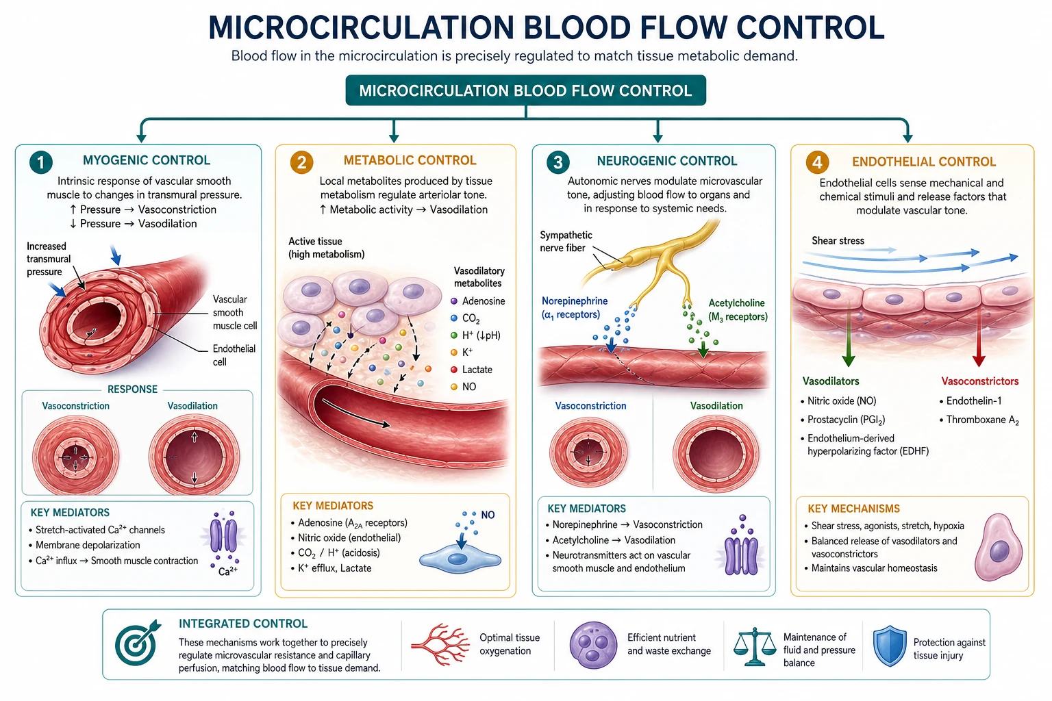
Local metabolic (dominant in heart, brain, working muscle)
↑Adenosine, K+, H+, CO2, lactate, low O2 → arteriolar dilation. Active hyperaemia and reactive hyperaemia after occlusion. [1]
Myogenic
↑Transmural pressure → stretch → constriction (Bayliss) — autoregulation component (kidney, brain, heart). [1]
Endothelial
Shear stress → NO → dilation (flow-mediated). Dysfunction in atherosclerosis/diabetes/sepsis. [1]
Neural
SNS α1 constriction (skin, splanchnic, kidney); β2 dilation in some beds; parasympathetic specialised (e.g. genital, some cranial). Baroreflex redistributes CO. [1]
Humoral
Adrenaline, Ang II, AVP, ANP/BNP, bradykinin, histamine — systemic modifiers of micro tone and leak. [1]
Starling forces at the capillary
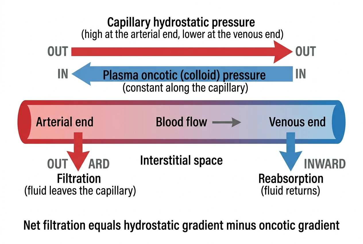
Net pressure ≈ (P_c − P_i) − σ(π_c − π_i) [1]
- P_c capillary hydrostatic (drives out)
- P_i interstitial hydrostatic
- π_c plasma oncotic (holds in) — mainly albumin
- π_i interstitial oncotic
- σ reflection coefficient (1 = impermeable to protein) [1]
Classic exam story: filtration arterial end, reabsorption venous end; slight net filtration returned by lymphatics. [1]
Clinical oedema mechanisms: ↑Pc (heart failure, venous obstruction), ↓πc (hypoalbuminaemia), ↑permeability/low σ (inflammation, sepsis — protein leak), lymphatic failure. [1]
Glycocalyx note (modern viva): endothelial surface layer makes effective oncotic gradients more complex; large crystalloid volumes can degrade barrier function — reason for judicious fluid and interest in albumin/plasma in critical care debates without overselling. [1]
Oxygen exchange and diffusion
Fick diffusion: flux ∝ area × diffusivity × ΔP / thickness. Oedema thickens path; low capillary density reduces area; stagnant flow lowers effective delivery. Diffusion hypoxia of N2O is a different alveolar topic — do not confuse. [1]
Special microcirculations (pointers)
- Coronary/cerebral: dense autoregulation (other leaf).
- Skin: SNS thermoregulatory arteriovenous anastomoses.
- Splanchnic: large SNS-mediated reservoir (other leaf).
- Pulmonary: HPV hypoxic pulmonary vasoconstriction — opposite systemic hypoxic dilation pattern. [1]
Shock at the micro level
| Shock type | Macro | Micro theme |
|---|---|---|
| Hypovolaemic | Low preload/CO | Low Pc then ischaemic injury/leak |
| Cardiogenic | Low CO high filling | Congestion + low delivery |
| Distributive | Low SVR | Maldistribution, weak autoregulation, leak |
| Obstructive | Impeded CO | Downstream hypoperfusion |
Lactate, mottling, oliguria, ScvO2/SvO2 are crude windows into adequacy of micro delivery vs demand. [1]
Anaesthetic relevance
- Hypotension below organ autoregulatory limits → supply dependence.
- Excessive vasopressors may restore MAP but over-constrict some beds — watch urine, lactate, perfusion signs.
- Anaemia + hypocapnia + low flow compound tissue hypoxia.
- Sepsis: endothelial injury, shunting, impaired extraction. [1]
Numbers board
- Resistance ∝ 1/r⁴
- FF kidney ~0.2 is organ-level Starling cousin
- Oncotic pressure plasma ~25 mmHg teaching
- Lymph returns ~2–4 L/day net filtrate teaching order [1]
Raise flow
- Metabolic dilators
- NO
- ↓SNS to bed
- ↑driving pressure
Cause oedema
- ↑Pc
- ↓πc
- Leaky σ↓
- Lymph failure
Viva scripts
Write Poiseuille and state clinical implication. [1]
List Starling forces and four oedema mechanisms. [1]
Contrast hypoxic responses of systemic vs pulmonary arterioles. [1]
Extended viva dialogue
Examiner: Why does arteriolar dilation increase capillary filtration? [1]
Candidate: Dilation raises capillary hydrostatic pressure transmission from arteries, increasing Pc and net filtration unless concurrent changes oppose it. Venous congestion raises Pc from the downstream side. [1]
Examiner: What is reactive hyperaemia? [1]
Candidate: After a period of occlusion, metabolites accumulate and arterioles dilate so that when flow is restored there is a transient overshoot in blood flow until the debt is repaid. [1]
Clinical synthesis: Macro haemodynamics set the head pressure; microcirculation decides whether cells actually get oxygen and whether the tissues drown in oedema. [1]
Revised Starling / glycocalyx (exam-aware paragraph)
The endothelial glycocalyx forms a protein-poor exclusion zone; filtration may occur with less reabsorption than classical arterial/venous cartoons suggest, making lymphatics critical for returning interstitial fluid. Large crystalloid volumes and inflammation can degrade the glycocalyx, promoting leak. Use this as nuance after stating classical Starling forces. [1]
Autoregulation lower limits under anaesthesia
Volatile agents and hypercapnia can impair autoregulation; hypotension then linearly cuts organ flow. Why “permissive hypotension” needs a brain and heart context, not a universal MAP 50 for everyone. [1]
Worked SAQ
SAQ: State Starling forces and four causes of oedema (7 marks)
Net filtration depends on capillary hydrostatic pressure favouring filtration, plasma oncotic pressure opposing it, and interstitial hydrostatic/oncotic pressures, modified by the reflection coefficient. Oedema follows raised capillary pressure (heart failure, venous obstruction), low plasma oncotic pressure (hypoalbuminaemia), increased permeability (inflammation, sepsis), or lymphatic obstruction. [1]
Critical closing pressure and vascular waterfall
Vessels may collapse when surrounding pressure exceeds intraluminal pressure — relevant to West zones of the lung and to compartment syndromes. Driving pressure for flow can become P_arterial − P_critical rather than P_arterial − P_venous. [1]
Haematocrit, viscosity and DO2 trade-off
Raising Hct raises CaO2 but increases viscosity and resistance, especially at low shear in the microcirculation. Optimal Hct for DO2 is a balance — physiology behind transfusion thresholds debates without claiming a single magic number. [1]
Extended viva add-on
Examiner: How does sepsis disrupt the microcirculation? [1]
Candidate: Endothelial injury and glycocalyx degradation cause leak and oedema; heterogeneous arteriolar tone creates perfused and unperfused capillaries; shunting and impaired mitochondrial utilisation mean normal ScvO2 can coexist with cellular hypoxia; vasopressors restore MAP but may worsen microvascular flow if overused without volume and inotropic balance. [1]
Primary exam expansion — dense examiner pack
Hierarchy of vessels and functions
| Segment | Function | Smooth muscle |
|---|---|---|
| Arteries | Conductance | Yes |
| Arterioles | Resistance (major SVR site) | Rich |
| Metarterioles / precapillary sphincters | Local on–off | Yes |
| Capillaries | Exchange | No (endothelium) |
| Venules/veins | Capacitance / return | Yes |
SVR primarily arteriolar; venous tone governs preload reservoir. [1]
Determinants of flow (Poiseuille link)
Q = ΔP × πr^4 / (8ηL). Radius dominates (fourth power). Haematocrit raises viscosity; hypothermia raises viscosity; laminar vs turbulent (Reynolds) at constrictions/valves. Fahraeus–Lindqvist effect: apparent viscosity falls in small vessels — microcirculation optimises. [1]
Local control mechanisms
- Metabolic: ↑ adenosine, K+, H+, lactate, CO2, low O2 → vasodilation (active hyperaemia). 2. Myogenic: ↑ stretch → constriction (Bayliss) — autoregulation backbone in brain/kidney. 3. Endothelial: NO (vasodilator), prostacyclin, endothelin (constrictor), EDHF. 4. Neural: SNS α1 constriction; some β2 dilator beds; cholinergic specialised. 5. Humoral: angiotensin II, vasopressin, ANP/BNP, bradykinin, histamine. [1]
Autoregulation ranges (teaching)
| Bed | Approx autoregulation range (MAP) | Notes |
|---|---|---|
| Brain | 50–150 mmHg | Shifts in chronic hypertension |
| Kidney | 80–180 mmHg | Protects GFR |
| Heart | Wide; metabolic dominant | Diastole critical for LV |
| Skin | Poor autoregulation | Thermoregulatory priority |
Below lower limit: flow becomes pressure-passive → ischaemia risk. [1]
Starling forces and oedema
Net filtration ≈ Kf [(Pc − Pi) − σ(πc − πi)]. Raised Pc (heart failure, venous obstruction), low πc (hypoalbuminaemia), ↑ permeability (inflammation, sepsis — σ falls), lymphatic failure. Pulmonary oedema when pulmonary capillary pressure high or permeability injury (ARDS). [1]
Oxygen extraction at tissue level
DO2 vs VO2: extraction ratio rises when flow falls until critical DO2. Microvascular shunting and heterogeneity in sepsis: adequate total flow but weak exchange — explains why ScvO2 and lactate can disagree. [1]
Anaesthetic and critical care levers
Vasopressors raise MAP but may over-constrict gut/skin microcirculation — balance macro targets with lactate, urine, skin perfusion, mental state. Vasodilators and neuraxial block open capacitance/resistance vessels. Hyperoxia effects on microcirculation debated; hypoxia dilates most beds (lung HPV opposite — sister leaf). Hypothermia left-shifts ODC and slows metabolism. [1]
Endothelial glycocalyx (modern viva spice)
Layer protecting endothelial integrity; degraded by sepsis, hypervolaemia, hyperglycaemia, surgery — capillary leak. Supports careful fluid therapy narrative (not dogma). [1]
SAQ: control of blood flow in microcirculation (8 marks)
Poiseuille/radius (2). Metabolic and myogenic (2). Endothelial mediators (2). Neural/humoral (1). Clinical example autoregulation failure (1). [1]
Viva
Q: Where is SVR mainly set? A: Arterioles. Q: Why does small radius change dominate flow? A: Fourth power in Poiseuille. Q: Why lactate in sepsis with normal MAP? A: Microcirculatory heterogeneity/extraction failure despite macro resuscitation. [1]
High-yield viva battery and numbers lock-in
Poiseuille consequences clinically
Halving radius → flow falls 16-fold for same pressure gradient — why arteriolar tone dominates SVR and why microvascular spasm or capillary dropout devastates tissue oxygen delivery even if macro MAP looks acceptable. Viscosity: polycythaemia increases resistance; extreme haemodilution decreases O2 content despite easier flow. [1]
Autoregulation failure states
MAP below lower limit; extreme hypertension above upper limit (breakthrough oedema in brain); volatile-agent impairment of autoregulation at high dose; trauma/inflammation fixed dilated beds; stenotic vessels already maximally dilated distal to lesion. [1]
Sepsis microcirculation paragraph
Endothelial activation, glycocalyx shedding, microthrombi, heterogeneous capillary perfusion, impaired oxygen extraction — patients may have high CO and low SVR yet high lactate. Resuscitation targets macro variables first but ultimate goal is tissue perfusion; excess vasoconstrictors can blanch microcirculation. [1]
Full viva dialogue (additional)
Examiner: List endothelial vasodilators and vasoconstrictors. [1]
Candidate: Dilators include nitric oxide, prostacyclin and endothelium-derived hyperpolarising factors. Constriction is mediated by endothelin-1 and by removal of dilator tone; sympathetic α1 agonists constrict vascular smooth muscle directly. Inflammation can tip the balance toward leak and dilation. [1]
Examiner: Explain reactive hyperaemia. [1]
Candidate: After a period of occlusion, accumulated metabolic vasodilator substances and myogenic relaxation produce a transient overshoot in blood flow when perfusion is restored, repaying oxygen debt — a demonstration of local metabolic control. [1]
Exam traps
- Equating MAP with microcirculatory perfusion always.
- Forgetting fourth-power radius relationship.
- Ignoring viscosity and temperature.
- Saying skin always autoregulates like brain. [1]
Examiner synthesis paragraph
Microcirculation answers should start with Poiseuille and the fourth-power radius effect at arterioles, then name local metabolic, myogenic and endothelial controllers before neural and humoral overlays. Autoregulation keeps brain, kidney and heart flow stable across a MAP window; below the lower limit flow is pressure-passive and ischaemia begins. Starling forces explain oedema, and sepsis reminds examiners that a normal macro blood pressure can still hide heterogeneous capillary perfusion and failing oxygen extraction. Vasopressors restore MAP but can bleach the periphery — balance numbers against lactate, urine and skin perfusion. [1]
References
- [1]Popovich I, et al. On the inevitability of microvascular failure in septic shock and other vasodilatory conditions Crit Care, 2026.PMID 42343425
- [2]Duse DA. Carbon dioxide is a triple vasodilator Cardiovasc Res, 2026.PMID 42334380
- [3]Alamilla-Sanchez ME, et al. Advances in the Physiology of Transvascular Exchange and A New Look At Rational Fluid Prescription Int J Gen Med, 2023.PMID 37408844
- [4]Pstras L, et al. Vascular refilling coefficient is not a good marker of whole-body capillary hydraulic conductivity in hemodialysis patients: insights from a simulation study Sci Rep, 2022.PMID 36088359
- [5]Li S, et al. Pathological triad of perioperative acute kidney injury: renal microcirculatory hypoxia, mitochondrial damage, and immuno-metabolic reprogramming Front Immunol, 2026.PMID 42338604
- [6]Maier LE, et al. The influence of metaboreflex activation on pulmonary pressure with combined chemoreflex activation in acute and chronic hypoxia J Physiol, 2026.PMID 42306962