Anaes · Applied cardiovascular & respiratory physiology
Neonatal physiology
Also known as Neonatal circulation · Fetal circulation · Circulatory transition at birth · Ductus arteriosus · Foramen ovale · Neonatal pharmacokinetics
The neonate undergoes the most dramatic physiological transition of any human — from placental dependence to independent air-breathing — and the anaesthetist managing the newborn must understand the fetal circulation, the transition at birth, and the pharmacokinetic and thermoregulatory differences that make neonatal anaesthesia a distinct discipline. The framework rests on five exam-critical ideas: fetal circulation shunts blood past the lungs via the foramen ovale (right-to-left atrial) and the ductus arteriosus (pulmonary artery to aorta) because pulmonary vascular resistance is high; at birth the first breaths lower pulmonary vascular resistance, pulmonary blood flow rises, left atrial pressure rises (closing the foramen ovale), and the ductus arteriosus closes (oxygen and falling prostaglandins) — converting to the adult pattern; neonatal thermoregulation is precarious (large surface area to mass ratio, brown fat non-shivering thermogenesis, limited glycogen); neonatal pharmacokinetics differ (higher total body water, lower protein binding, immature hepatic metabolism, more permeable blood-brain barrier); and the neonatal airway and cardiovascular response to hypoxia (bradycardia, not tachycardia) make rapid desaturation and bradycardia during induction the defining hazards. Built on the paediatric sedation behaviour study (Nikula 2026), the dexmedetomidine pharmacokinetics study (Tsai 2026), the paediatric TIVA review (Quintao 2026), the maternal-neonatal circulation study (Piani 2026), the neonatal resuscitation review (Krishnaprasadh 2026), and the neonatal left-ventricle study (Sehgal 2023).
On this page & tools
Your progress
Saved locally on this device.
8 MCQs with explanations
Target exams
Red flags
Why this matters to the anaesthetist
Neonatal physiology is high-yield Primary and essential Final for paediatric lists, NLS, congenital heart disease, and the ex-prem. Know fetal shunts and transitional circulation, respiratory mechanics, HbF/ODC, thermogenesis, and PK differences.[1]
One-liner: Fetus: placenta + FO/DA/DV shunts; first breath ↓PVR, cord clamp removes low-resistance placenta, FO/DA functionally close; neonate has high O2 demand, low FRC reserve, obligatory nose breathing, HbF, brown fat heat, immature liver/kidney/BBB. [1]
Fetal circulation (draw this)
- Umbilical vein (oxygenated from placenta) → ductus venosus bypasses liver partly → IVC → RA.
- Preferential stream through foramen ovale → LA → LV → brain/coronaries.
- SVC/deoxygenated blood → RA → RV → PA; high PVR → most flow through ductus arteriosus → descending aorta → umbilical arteries back to placenta.
- Parallel circulations; placenta is the gas exchanger. [1]
Transitional circulation at birth
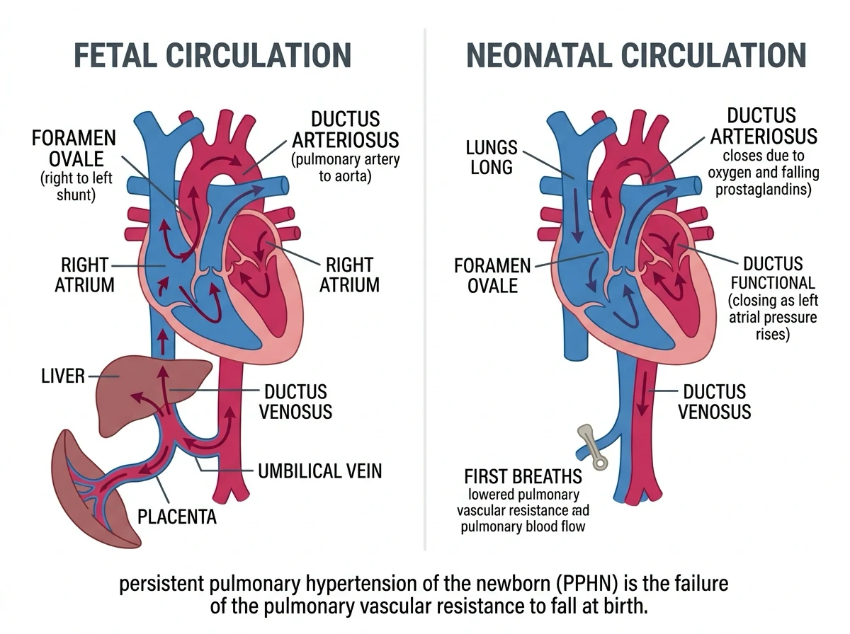
Sequence teaching: [1]
- Lung expansion + rising PaO2 → PVR falls dramatically; pulmonary flow rises.
- Cord clamp → sudden rise in SVR (placenta removed).
- ↑pulmonary venous return → ↑LA pressure → functional FO closure (anatomical later).
- ↑PaO2 + ↓prostaglandins → DA constriction (functional hours; anatomical weeks).
- DV closes with cord. [1]
Failure modes / PPHN: hypoxia, acidosis, cold, sepsis keep PVR high → right-to-left shunt via FO/DA → vicious hypoxaemia. Treatment physiology: oxygen, ventilation, normothermia, correct acidosis, iNO/inotropes as indicated — avoid the spiral. [1]
Respiratory physiology of the neonate
- High O2 consumption (~6–8 mL/kg/min vs adult ~3–4).
- FRC low relative to closing capacity → atelectasis tendency; prefer PEEP/CPAP thinking.
- Compliant chest wall + less compliant lungs → easy to distort chest, hard to keep volume.
- Diaphragm more type IIc/fatigue-prone fibres in neonate; horizontal ribs → less bucket-handle.
- Preferential nasal breathing — nasal obstruction matters.
- Small airways → resistance rises steeply with radius (Poiseuille).
- Apnoea risk especially ex-prem (immature control) — caffeine, monitoring. [1]
Surfactant from ~34 weeks mature enough generally; deficiency → RDS (preterm). [1]
Cardiovascular
- CO is HR-dependent (limited SV recruitment; less compliant ventricle).
- Bradycardia → precipitous CO fall (and hypoxia often causes bradycardia via vagal pathways — treat oxygen/ventilation first in NLS algorithms).
- Blood volume ~80–90 mL/kg teaching; transfusion/bolus calc on weight.
- Autonomic balance more parasympathetic responsive. [1]
Haematology / oxygen transport
- HbF: left-shifted ODC relative to adult HbA context; higher O2 affinity aids placental uptake; after birth 2,3-DPG and HbA rise over months.
- Neonatal Hb often high at birth then physiological anaemia of infancy.
- Vitamin K–dependent factors low → VKDB prevention with vitamin K.
- Immature immune function → infection vulnerability. [1]
Thermoregulation
- High surface area:mass → rapid heat loss (radiation, convection, evaporation, conduction).
- Non-shivering thermogenesis via brown fat (β3, UCP1) — oxygen-costly.
- Cold → ↑O2 use, pulmonary vasoconstriction, coagulopathy risk, apnoea.
- Warm environment, dry, hats, radiant warmer — core anaesthetic skill. [1]
Renal, hepatic, metabolic, neuro
- GFR low at birth, matures over months → prolonged renally cleared drugs.
- Immature Phase I/II hepatic metabolism → prolonged many drugs (morphine, midazolam caution).
- Low glycogen stores especially preterm → hypoglycaemia risk.
- Immature BBB and higher mg/kg local anaesthetic risk → LAST vigilance; dose by weight carefully.
- Higher MAC in infants (peaks in infancy then falls) — age-specific volatile needs. [1]
Airway anatomy hooks (physiology-adjacent)
Large tongue, high larynx, long epiglottis, narrowest point classically cricoid in young children (debated but exam-persistent), obligatory careful tube sizing — see paediatric airway leaves. [1]
Anaesthetic implications board
| Domain | Implication |
|---|---|
| Induction | Rapid desaturation — preoxygenation limited, hard mask seal |
| Ventilation | Small TV, fast RR, PEEP, careful PIP |
| CVS | Avoid bradycardia; weight-based adrenaline/fluids |
| Fluids/glucose | Maintenance with glucose awareness in neonates |
| Drugs | Weight-based; immature clearance; air bubble vigilance IV |
| Temperature | Active warming mandatory |
| PPHN risk | Avoid hypoxia/hypercarbia/acidosis/cold |
Numbers board
- VO2 neonate ~6–8 mL/kg/min
- Blood volume ~80–90 mL/kg
- Fetal shunts: FO, DA, DV
- DA functional closure: hours; anatomical: weeks
- Glucose vigilance continuous in neonates [1]
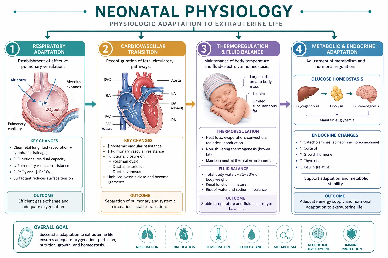
Fetal pattern
- Placenta gas exchange
- High PVR
- FO/DA right-to-left
- Parallel circuits
Neonatal pattern
- Lung gas exchange
- Low PVR
- Shunts closed
- Series circuits
Viva draw scripts
Draw fetal circulation with arrows through FO and DA. [1]
List five reasons neonates desaturate fast. [1]
Explain ductal closure triggers. [1]
Extended viva dialogue
Examiner: What closes the foramen ovale? [1]
Candidate: Functional closure when pulmonary venous return raises left atrial pressure above right atrial pressure after PVR falls and umbilical venous return ceases. Anatomical fusion occurs later; probe patent FO can persist. [1]
Examiner: Why is heat loss so dangerous? [1]
Candidate: High surface area causes rapid cooling; brown fat thermogenesis increases oxygen consumption; cold promotes pulmonary hypertension, coagulopathy and apnoea — so thermal care is resuscitation. [1]
Clinical synthesis: Neonates are transitional circulation plus small FRC plus cold physics plus immature clearance — plan for all four every case. [1]
Persistent pulmonary hypertension spiral
Hypoxia, hypercarbia, acidosis, cold → ↑PVR → R→L shunt FO/DA → worse hypoxaemia → further PVR rise. Break with oxygen, ventilation, warmth, correct acidosis, pulmonary vasodilators (iNO) when indicated. [1]
Pharmacokinetic neonatal bullets
- Higher ECF fraction → larger Vd for hydrophilic drugs (higher mg/kg loading sometimes).
- Immature CYP/UGT → prolonged midazolam, morphine caution.
- Low BuChE possible → sux duration variable.
- Immature GFR → aminoglycoside interval extension.
- Higher MAC in infants than neonates extremes and adults — age curve matters. [1]
Worked SAQ
SAQ: Describe the circulatory changes at birth (8 marks)
Lung expansion and rising PaO2 reduce pulmonary vascular resistance, increasing pulmonary blood flow and left atrial return. Cord clamping removes the low-resistance placenta, raising systemic vascular resistance. Left atrial pressure exceeds right, functionally closing the foramen ovale. Rising oxygen tension and falling prostaglandins constrict the ductus arteriosus. The ductus venosus closes with cessation of umbilical venous flow, establishing series neonatal circulation. [1]
Primary exam expansion — dense examiner pack
Transition at birth — cardiopulmonary rewrite
In utero: high PVR, low SVR (placenta), right-to-left shunts at foramen ovale and ductus arteriosus, gas exchange placental. At birth: lung aeration → PVR falls dramatically; cord clamp → SVR rises; FO functional close as LA pressure > RA; DA closes functionally (PaO2 rise, prostaglandin fall) then anatomically. Persistent pulmonary hypertension of newborn (PPHN): PVR stays high → extrapulmonary shunt → hypoxaemia — iNO, oxygen, avoid acidosis/hypothermia, support. [1]
Airway and respiratory differences (exam table)
| Feature | Neonate | Adult |
|---|---|---|
| Larynx level | C3–4 | C6 |
| Narrowest point (exam) | Cricoid | Glottis |
| Epiglottis | Large, U-shaped | Leaf-like |
| Tongue/occiput | Relatively large | Proportionate |
| Ribs/chest | Horizontal, compliant | Rigid lever |
| FRC | Low relative; closes airways easily | Higher reserve |
| O2 consumption | 6–8 mL/kg/min | ~3 mL/kg/min |
| Alveolar ventilation | High for size | Lower relative |
| Type I fibres diaphragm | Fewer | More fatigue-resistant |
| Response to hypoxia | Biphasic; can depress | Sustained increase more robust |
Desaturate fast: high VO2 + low FRC + high closing capacity tendency. [1]
Cardiovascular neonate
CO is rate-dependent — limited stroke volume recruitability (less compliant ventricle). Bradycardia = low CO = bad (hypoxia-driven via vagus classic in neonates). HbF: left-shifted ODC relative, higher Hb concentration in utero context. Blood volume ~80–90 mL/kg. Transfusion thresholds lower absolute volumes matter. PDA issues in preterms: overcirculation vs systemic steal. [1]
Thermoregulation
High surface area:mass, thin skin, little fat, brown fat non-shivering thermogenesis (β3, uncoupling protein) — inhibited by anaesthetics. Cold → ↑O2 consumption, pulmonary vasoconstriction, coagulopathy risk, delayed recovery. Active warming mandatory; theatre temp; warmed fluids. [1]
Glucose and liver
Low glycogen reserves especially preterm → hypoglycaemia risk with fasting/stress. Immature gluconeogenesis. Check glucose perioperatively. Immature hepatic enzymes → prolonged drug effects (different from adult per-kg dosing blindly). [1]
Renal and fluid
Low GFR at birth rises over weeks–months; impaired concentrating ability; hyponatraemia risk with free water; careful fluid calculation (mL/kg); insensible losses high with radiant heat/open surgery. [1]
Haematology
Physiological anaemia of infancy later; vitamin K deficiency bleeding risk if not given at birth; HbF→HbA switch. Vitamin K IM at birth standard public health. [1]
Pharmacology principles neonates
- Higher TBW → larger Vd for water-soluble drugs (aminoglycosides teaching).
- Lower fat/muscle → redistribution patterns differ.
- Immature CYP/conjugation → prolonged midazolam etc.
- Immature BBB / higher brain mass fraction → sensitivity.
- MAC higher in infants than neonates extremes teaching curve (neonates different from 1–6 month peak).
- Minimum alveolar concentration and induction uptake: high VA/FRC → faster volatile uptake. [1]
Apnoea risk post-anaesthesia
Ex-preterm infants: post-op apnoea risk — monitor, caffeine sometimes, regional preference for suitable lower abdominal surgery to reduce GA apnoea risk discussions. [1]
SAQ: why neonates desaturate quickly (7 marks)
High VO2 (2). Low FRC/O2 store (2). Airway anatomy obstruction risk (1). Immature control of breathing (1). Clinical mitigation: preoxygenation limits, careful airway, monitoring, warming, glucose (1). [1]
Viva
Q: Why is bradycardia catastrophic in neonates? A: CO nearly linear with HR; hypoxia→bradycardia→spiral. Q: Why warm aggressively? A: Prevent ↑VO2, PPHN tendency, coagulopathy, delayed drug metabolism. Q: Narrowest airway point in infant? A: Cricoid (standard exam answer). [1]
High-yield viva battery and numbers lock-in
Numbers neonates love in vivas
- VO2 ≈ 6–8 mL/kg/min (≈ double adult per kg)
- Blood volume ≈ 80–90 mL/kg
- Larynx C3–4; narrowest cricoid (exam)
- Tube depth ≈ 3 × ID (cm oral teaching)
- Uncuffed ID ≈ age/4 + 4 (older formula); cuffed age/4 + 3.5
- HbF left-shifted ODC relative to adult Hb
- MAC higher in infants than older children extremes of curve teaching [1]
Why bradycardia is the neonatal emergency pathway
Hypoxia → vagal response → bradycardia → CO falls nearly linearly with HR → coronary and cerebral perfusion fall → further hypoxia. Treatment priority: oxygenation/ventilation first, then adrenaline per NLS if needed — not atropine as sole thinking for hypoxic bradycardia. [1]
Preterm extras
Surfactant deficiency RDS; apnoea of prematurity; fragile germinal matrix IVH risk with CO2/BP swings; PDA; retinopathy oxygen toxicity concerns; temperature instability extreme; glucose instability; sepsis risk. Anaesthesia: meticulous CO2 and BP control, temperature, glucose, intercostal/diaphragmatic fatigue awareness. [1]
Full viva dialogue (additional)
Examiner: Explain transitional circulation and persistent pulmonary hypertension. [1]
Candidate: At birth lung expansion lowers pulmonary vascular resistance and cord clamping raises systemic resistance, reversing fetal shunts and closing the foramen ovale functionally and the ductus as oxygen rises and prostaglandins fall. If pulmonary vascular resistance remains high, right-to-left shunting persists at ductal and foramen levels, causing severe hypoxaemia — persistent pulmonary hypertension — treated by oxygen, avoiding acidosis and hypothermia, supporting blood pressure, and inhaled nitric oxide when indicated. [1]
Examiner: Why do neonates cool so quickly? [1]
Candidate: High surface-area-to-mass ratio, thin skin, little subcutaneous fat, and reliance on brown-fat non-shivering thermogenesis which anaesthetic agents impair. Cooling raises oxygen consumption and pulmonary vascular resistance and worsens coagulopathy and apnoea risk. [1]
Exam traps
- Adult airway assumptions in infants.
- Ignoring glucose in long neonatal cases.
- Treating neonatal bradycardia as primary cardiac without ventilating.
- Overheating or over-oxygenating preterms carelessly. [1]
References
- [1]Nikula A, et al. Behavioral Changes in Children Are Uncommon 4 Weeks After Procedural Sedation and Analgesia With Intranasal Dexmedetomidine and Nitrous Oxide Paediatr Neonatal Pain, 2026.PMID 42311919
- [2]Tsai YF, et al. Dexmedetomidine Dosing Strategies in Sedation and Anesthesia: Pharmacokinetics, Safety, and Clinical Applications - A Narrative Review Drug Des Devel Ther, 2026.PMID 42232093
- [3]Quintao VC, et al. Update on total intravenous anesthesia in children Curr Opin Anaesthesiol, 2026.PMID 41817234
- [4]Piani F, et al. Linking maternal and neonatal circulation in preeclampsia Am J Physiol Heart Circ Physiol, 2026.PMID 41525138
- [5]Krishnaprasadh D. Pediatric and Neonatal Resuscitation 2026.PMID 34283435
- [6]Sehgal A, et al. The left ventricle in well newborns versus those with perinatal asphyxia, haemodynamically significant ductus arteriosus or fetal growth restriction Transl Pediatr, 2023.PMID 37814715