Anaes · Applied cardiovascular & respiratory physiology
Neuromuscular junction physiology
Also known as Neuromuscular junction · Acetylcholine receptor · End-plate potential · Nicotinic receptor · Acetylcholinesterase · Excitation-contraction coupling
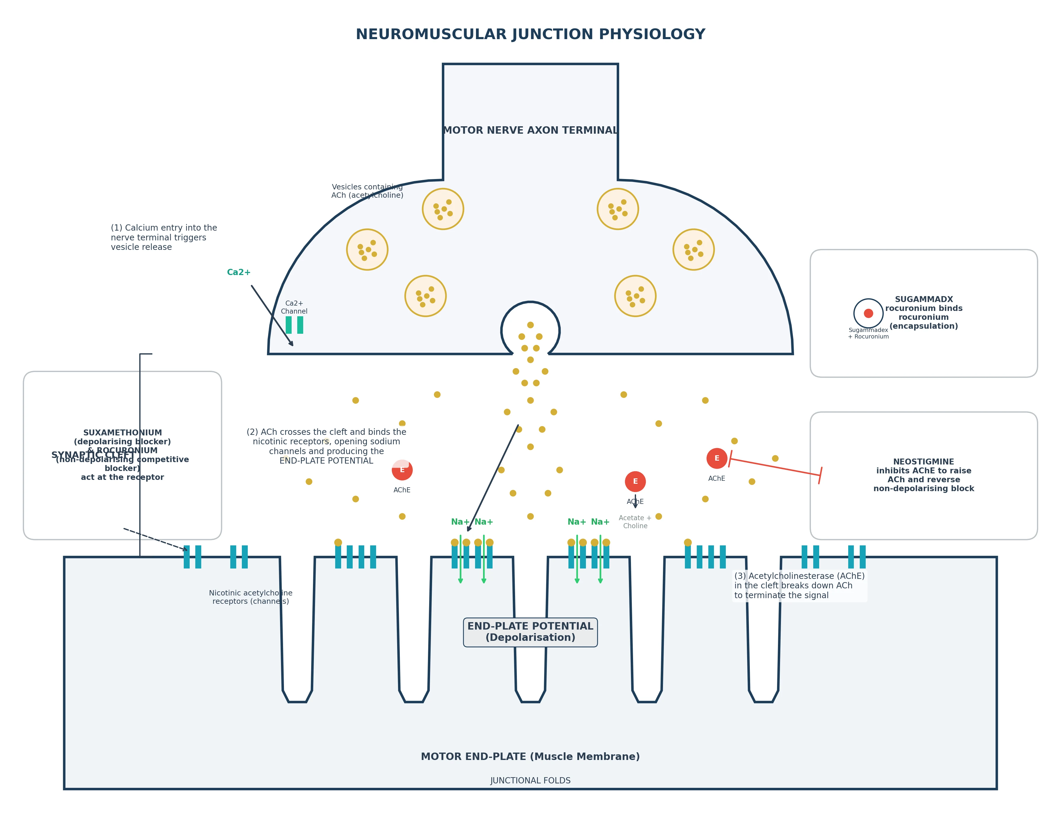
The neuromuscular junction converts the motor nerve action potential into muscle contraction, and it is the site of action of the muscle relaxants and their reversal agents. The framework rests on five exam-critical ideas: the motor nerve action potential opens voltage-gated calcium channels in the terminal, and the calcium entry drives vesicle release of acetylcholine into the synaptic cleft; acetylcholine binds the nicotinic (muscle-type) receptor on the motor end-plate, a ligand-gated cation channel whose opening produces the end-plate potential; the end-plate potential reaches threshold and triggers a muscle action potential, after which acetylcholinesterase in the cleft terminates the signal; the muscle action potential is coupled to contraction by T-tubules, the dihydropyridine and ryanodine receptors, and calcium release from the sarcoplasmic reticulum; and the junction is the target of the neuromuscular blockers (depolarising suxamethonium, non-depolarising rocuronium), their reversal (neostigmine inhibiting acetylcholinesterase, sugammadex encapsulating rocuronium), and of disease (myasthenia gravis, Lambert-Eaton). Built on the miR-206/ACh-receptor NMJ study (Jiang 2026), the neostigmine-bronchospasm report (Sato 2026), the efgartigimod-myasthenia review (Jiang 2026), the eculizumab-myasthenia report (Li 2026), and the sugammadex-anaphylaxis and airway-oedema reports (Ishihara 2025, Habib 2026).
On this page & tools
Your progress
Saved locally on this device.
8 MCQs with explanations
Target exams
Red flags
Why this matters to the anaesthetist
Every muscle relaxant, every reversal agent, and every TOF monitor is applied NMJ physiology. Myasthenia, Lambert–Eaton, burns, denervation, and magnesium all change junctional behaviour. Primary candidates must describe ACh synthesis/release, nAChR structure, EPP vs AP, AChE, depolarising vs non-depolarising block, and monitoring patterns. [1]
NMJ anatomy
The α-motor neurone terminal apposes the motor end-plate of skeletal muscle. Features: synaptic vesicles (ACh quanta), active zones aligned opposite junctional folds packed with nicotinic ACh receptors, basal lamina containing acetylcholinesterase, and Schwann cell covering. The synaptic cleft is ~50 nm. Safety factor: released ACh and receptor density normally produce an end-plate potential far above threshold. [1]
Acetylcholine synthesis and release
Choline + acetyl-CoA → (choline acetyltransferase) → ACh → vesicular ACh transporter packages quanta (~5,000–10,000 ACh molecules per vesicle). [1]
Release: nerve AP → P/Q-type voltage-gated Ca2+ channels at active zones → Ca2+ microdomains → SNARE-mediated exocytosis of many quanta (adult evoked release ~50–100 quanta). Spontaneous single quanta produce miniature EPPs (mEPPs). [1]
Mg2+ antagonises Ca2+-dependent release — high Mg (tocolysis, pre-eclampsia treatment) potentiates non-depolarising block. Aminoglycosides and some other drugs also impair release. [1]
The nicotinic ACh receptor (muscle-type)
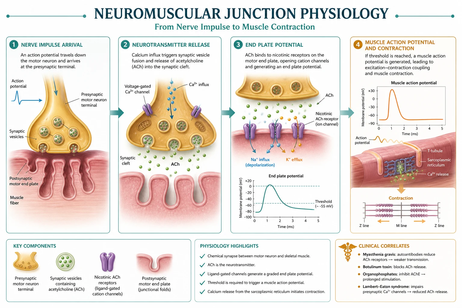
Pentameric ligand-gated cation channel: adult α2βδε (fetal/denervated α2βδγ). Two ACh molecules bind α–δ and α–ε (or α–γ) interfaces → channel opens → Na+ (and Ca2+) in, K+ out → local depolarisation (EPP). [1]
Non-depolarising NMBAs are competitive antagonists at these sites. Suxamethonium is a partial agonist causing prolonged depolarisation. [1]
End-plate potential and acetylcholinesterase
EPP must reach threshold to trigger a muscle action potential via nearby voltage-gated Na+ channels. AChE in the cleft hydrolyses ACh in <1 ms, terminating the signal and providing choline for reuptake. AChE inhibitors (neostigmine, edrophonium, pyridostigmine) raise ACh concentration and duration — reverse competitive block but can worsen depolarising block / cholinergic crisis. [1]
Excitation–contraction coupling (brief)
Muscle AP → T-tubules → DHPR–ryanodine receptor activation → SR Ca2+ release → troponin C → cross-bridge cycling → contraction. Malignant hyperthermia is a coupling disease (RYR1), not a junctional transmission disease — but it is in the differential of rigidity after suxamethonium. [1]
Neuromuscular blockers
Depolarising (suxamethonium): binds nAChR → persistent depolarisation → inactivation of Na channels → flaccid paralysis (phase I). Fasciculations precede. Metabolised by butyrylcholinesterase (plasma cholinesterase), not AChE. Duration short unless deficiency/atypical enzyme (dibucaine number). Side effects: hyperkalaemia (upregulated extrajunctional receptors in burns/denervation — contraindicated), myalgia, bradycardia (especially kids, second dose), ↑IOP/IGP, MH trigger, anaphylaxis. [1]
Phase II block: prolonged exposure to depolarising agent → features resembling non-depolarising block (fade appears). [1]
Non-depolarising (aminosteroids: rocuronium, vecuronium; benzylisoquinoliniums: atracurium, cisatracurium): competitive antagonists. Fade on TOF/tetanus from pre- and post-synaptic effects (presynaptic nAChR block impairs mobilisation). Hofmann elimination / ester hydrolysis for atracurium/cisatracurium — useful in organ failure (laudanosine historically for atracurium). [1]
Reversal
- Neostigmine + glycopyrrolate/atropine: AChE inhibition; ceiling effect; ineffective if block is too deep; muscarinic side effects require anticholinergic.
- Sugammadex: γ-cyclodextrin encapsulates rocuronium (and vecuronium) in plasma → diffusion gradient away from NMJ. Works at deep block; dose by TOF/PTC; renally excreted complex; binds oral contraceptives (advice); rare anaphylaxis; interferes with some steroid assays. [1]
Myasthenia gravis vs Lambert–Eaton
| Myasthenia gravis | Lambert–Eaton (LEMS) | |
|---|---|---|
| Site | Post-synaptic nAChR (autoimmune) | Pre-synaptic P/Q Ca channels |
| Strength with use | Fatigues | May improve transiently |
| Reflexes | Normal usually | Reduced |
| Non-depolarising sensitivity | High (resistant to sux sometimes) | Variable; often sensitive |
| Associated | Thymoma | Small-cell lung cancer |
Monitoring
Train-of-four (TOF): 2 Hz × 4 stimuli. Count and ratio (T4/T1). Fade defines non-depolarising block. TOF ratio ≥0.9 with quantitative monitoring before extubation is modern standard to avoid residual paralysis. Double burst, tetanic fade, post-tetanic count for deep block. Prefer ulnar nerve–adductor pollicis for recovery; facial muscles recover earlier and mislead. [1]
Depolarising block
- Agonist at nAChR
- Fasciculations, no fade (phase I)
- BuChE metabolism
- Hyperkalaemia risk if receptors upregulated
Non-depolarising block
- Competitive antagonists
- Fade on TOF
- Reverse with neostigmine or sugammadex
- Potentiated by Mg, volatiles, acidosis
Quantal transmission maths (exam flavour)
If one quantum produces an mEPP of ~0.5 mV and threshold needs ~15 mV, many quanta must summate — and they do, with margin. Competitive blockers reduce the effective quantal response until summation fails intermittently (fade, fade of tetanus). [1]
Drug interactions that deepen block
Volatiles, magnesium, local anaesthetics (high dose), calcium channel blockers (variable), aminoglycosides, acidosis, hypothermia, electrolyte disorders — all potentiate non-depolarising block. Always recheck TOF before reversal. [1]
Viva traps
- AChE is in the cleft — neostigmine works there, not by "more vesicles."
- Sugammadex does not act on the receptor.
- Phase I vs phase II sux block — fade appears in phase II.
- Myasthenia is post-synaptic; LEMS pre-synaptic.
- Dibucaine number tests quality of BuChE, not quantity alone. [1]
SAQ: compare depolarising and non-depolarising block
"Suxamethonium is an agonist at the muscle nicotinic receptor and causes persistent end-plate depolarisation with sodium-channel inactivation, producing flaccid paralysis after fasciculation. Phase I block shows reduced twitch without fade. It is metabolised by plasma cholinesterase. Non-depolarising drugs are competitive antagonists, produce fade on train-of-four because of pre- and post-synaptic receptor effects, and are reversed by acetylcholinesterase inhibitors or, for rocuronium, by sugammadex encapsulation." [1]
TOF interpretation ladder
TOF count 0: deep block (use PTC). Count 1–2: moderate; neostigmine unreliable; sugammadex high dose if roc. Count 3–4 with fade: suitable for neostigmine if ratio still low but recovering. TOF ratio 0.7: still clinically weak airway. Ratio ≥0.9 quantitative: extubation readiness standard. Always state muscle site. [1]
Upregulation of ACh receptors
Burns, denervation, prolonged ICU immobility, and some upper motor neurone lesions increase extrajunctional nAChR. Suxamethonium then opens vast potassium channels → hyperkalaemic arrest risk. Onset after injury ~24–48 h, peaks weeks, may last months. Avoid sux in that window. [1]
Anaphylaxis ranking note
NMBAs are leading perioperative anaphylaxis triggers in many series; sugammadex also reported. Physiology viva may pivot to recognition and adrenaline management — stay structured. [1]
Primary exam expansion
Presynaptic nicotinic receptors and fade
Non-depolarising blockers inhibit presynaptic positive feedback nAChRs that normally sustain ACh mobilisation during high-frequency stimulation. Hence fade on TOF and tetanic fade — not explained by postsynaptic competition alone. [1]
Butyrylcholinesterase genetics
Homozygous atypical BuChE (dibucaine-resistant) prolongs suxamethonium and mivacurium for hours. Dibucaine number: percent inhibition by dibucaine — normal ~80, atypical ~20. Heterozygotes intermediate. Acquired low quantity: pregnancy, liver disease, malnutrition — milder prolongation. [1]
Hofmann elimination chemistry
Atracurium/cisatracurium undergo spontaneous Hofmann elimination (base-catalysed) and ester hydrolysis. Rate increases with alkalosis and temperature; slows with acidosis/hypothermia. Organ-independent pathway is the exam selling point for ICU renal/hepatic failure (cisatracurium preferred). [1]
Sugammadex dosing framework (physiology of free drug)
Free rocuronium in plasma is bound by sugammadex; NMJ drug diffuses back to plasma down gradient. Deeper block needs more encapsulator. Residual free NMBA determines residual block. This is binding equilibrium physiology, not AChE. [1]
Electrolytes and the junction
Hypokalaemia and hypermagnesaemia potentiate non-depolarising block. Hyperkalaemia risks with sux as above. Hypocalcaemia impairs release. Always check the biochemical milieu when block is unexpectedly prolonged. [1]
ICUAW and neuromuscular transmission
Critical illness polyneuropathy/myopathy may coexist with residual block. TOF may recover while weakness persists — transmission is only one part of the strength pathway (nerve, muscle membrane, contractile proteins). [1]
Structured reversal algorithm narrative
Monitor quantitatively → if roc and deep, sugammadex dose by depth → if intermediate and no sugammadex, wait until TOF count ≥3–4 then neostigmine with glycopyrrolate → recheck ratio ≥0.9 → only then extubate. Never reverse "on the clock" alone. [1]
Extended viva dialogue
Examiner: Describe neuromuscular transmission from nerve impulse to twitch. [1]
Candidate: A motor-nerve action potential opens presynaptic calcium channels; acetylcholine quanta are released into the cleft; acetylcholine binds muscle nicotinic receptors; the end-plate depolarises; if threshold is reached a muscle action potential propagates; calcium is released from the sarcoplasmic reticulum; cross-bridges cycle. Acetylcholinesterase hydrolyses acetylcholine within a millisecond to terminate the signal. [1]
Examiner: Compare suxamethonium and rocuronium at receptor level. [1]
Candidate: Suxamethonium is an agonist causing prolonged depolarisation and sodium-channel inactivation — phase I depolarising block without fade. It is metabolised by butyrylcholinesterase. Rocuronium is a competitive antagonist at the same receptor, produces fade, and is reversed by sugammadex encapsulation or, when lighter, by acetylcholinesterase inhibition with neostigmine plus an anticholinergic. [1]
Examiner: How do you monitor block and when is extubation safe? [1]
Candidate: Quantitative train-of-four at the adductor pollicis is preferred. Fade indicates non-depolarising block. A TOF ratio of at least 0.9 is the modern target before extubation. Clinical tests alone miss residual paralysis. Facial muscles recover earlier and can mislead. [1]
Examiner: Myasthenia versus Lambert–Eaton? [1]
Candidate: Myasthenia is postsynaptic receptor antibody disease with fatigable weakness and high sensitivity to non-depolarising blockers. Lambert–Eaton is presynaptic P/Q calcium channel disease, often paraneoplastic, with reduced reflexes and strength that may improve with use. [1]
Clinical synthesis: Residual block is a failure of safety factor. Measure it; reverse it; only then remove the tube. [1]
Worked SAQ model answers
SAQ: Describe the physiology of the neuromuscular junction and how muscle relaxants act (10 marks)
The neuromuscular junction transmits from α-motor neurone to skeletal muscle. Acetylcholine is synthesised from choline and acetyl-CoA, packaged into vesicles, and released in quanta when an action potential opens presynaptic calcium channels. Acetylcholine binds the muscle-type nicotinic receptor, a pentameric ligand-gated cation channel (adult stoichiometry α2βδε), opening a sodium-permeable pore. The end-plate potential triggers a muscle action potential if threshold is reached. Acetylcholinesterase in the cleft hydrolyses acetylcholine within a millisecond. [1]
Depolarising blockers such as suxamethonium act as agonists, causing prolonged depolarisation and sodium-channel inactivation (phase I block) after fasciculations. Metabolism is by plasma butyrylcholinesterase. Non-depolarising blockers are competitive antagonists and produce fade on train-of-four stimulation because of pre- and postsynaptic receptor effects. [1]
Reversal of non-depolarising block uses acetylcholinesterase inhibitors such as neostigmine, always with an anticholinergic to block muscarinic effects, or sugammadex which encapsulates rocuronium in plasma and creates a diffusion gradient away from the junction. Quantitative neuromuscular monitoring with a train-of-four ratio of at least 0.9 should guide extubation. [1]
Disease states alter responses: myasthenia gravis is postsynaptic and highly sensitive to non-depolarisers; Lambert–Eaton is presynaptic; burns and denervation upregulate extrajunctional receptors and risk hyperkalaemia with suxamethonium. [1]
SAQ: Outline neuromuscular monitoring (5 marks)
Peripheral nerve stimulation of the ulnar nerve with recording at adductor pollicis is standard for recovery. Train-of-four at 2 Hz yields a count and ratio; fade indicates non-depolarising block. Post-tetanic count assesses deep block. Quantitative devices are preferred over subjective visual fade. Diaphragm and laryngeal muscles recover earlier than thumb — clinical airway strength can still be inadequate when facial twitches look strong. [1]
Clinical scenario walkthroughs
Scenario 1 — Cannot reverse with neostigmine
Deep block (TOF count 0–1) has almost no free receptors for ACh to compete at; neostigmine has a ceiling. Wait for spontaneous recovery to a countable TOF or use sugammadex if rocuronium/vecuronium was used. Forcing neostigmine early fails and risks cholinergic side effects. [1]
Scenario 2 — Prolonged paralysis after suxamethonium
Consider atypical butyrylcholinesterase, acquired deficiency, phase II block after large doses, or drug interactions. Keep sedated and ventilated until quantitative recovery; send dibucaine number when appropriate; counsel the family if genetic. [1]
Scenario 3 — Unexpected hyperkalaemic arrest after suxamethonium
Burns, denervation, prolonged immobilisation: extrajunctional receptor upregulation. Massive potassium efflux through agonist-opened channels. Prevention is avoidance; treatment is ACLS hyperkalaemia care and calcium. [1]
Scenario 4 — Myasthenic patient for thymectomy
Reduce or avoid non-depolarisers; if needed, tiny titrated doses with quantitative monitoring. Suxamethonium responses are unpredictable (often resistant). Plan postoperative ventilation risk; coordinate with usual pyridostigmine timing. [1]
Additional exam numerical anchors
Numerical and factual anchors: [1]
- Synaptic cleft width ~50 nm; ACh hydrolysis under 1 ms.
- Adult nAChR stoichiometry α2βδε; fetal/denervated α2βδγ (γ subunit).
- TOF frequency 2 Hz; tetanic stimulation classically 50 Hz for 5 seconds in teaching.
- Extubation target TOF ratio ≥0.9 with quantitative monitoring at adductor pollicis.
- Suxamethonium intubating dose classically 1–1.5 mg/kg; onset ~30–60 s; duration ~5–10 min if BuChE normal.
- Rocuronium RSI dose often 1.0–1.2 mg/kg for rapid onset when suxamethonium contraindicated.
- Dibucaine number ~80 normal, ~20 homozygous atypical.
- Magnesium therapeutic ranges in obstetrics can potentiate block — recheck TOF after dosing.
- Residual paralysis incidence is historically high when only clinical tests are used — the reason quantitative monitoring is now expected.
- Anaphylaxis to NMBAs remains a leading cause of perioperative anaphylaxis in multiple national audits — prepare for it when using these drugs. [1]
Closing synthesis for the Primary
Neuromuscular junction physiology is the bridge between membrane electrophysiology and the pharmacology of every relaxant you give. If you can describe quantal acetylcholine release, the structure of the muscle nicotinic receptor, the role of acetylcholinesterase, the difference between agonist-driven depolarising block and competitive non-depolarising block, the meaning of fade, and the modern standard of quantitative reversal to a train-of-four ratio of at least 0.9, you will handle SAQs, MCQs and vivas in this domain. Add disease states (myasthenia, Lambert–Eaton), receptor upregulation after burns or denervation, magnesium interactions, and sugammadex encapsulation versus neostigmine, and the topic is exam-complete. Residual paralysis is not a minor nuisance — it is a failure to apply this physiology at the end of anaesthesia. [1]
One-line take-home
Master the core equations and mechanisms of this topic until they are automatic under viva pressure; clinical anaesthesia is applied primary science, not a separate subject. [1]
Red flags
- Muscle nAChR is the ligand-gated target of relaxants and myasthenia antibodies.
- AChE terminates signal in milliseconds; neostigmine raises ACh (and muscarinic effects).
- Suxamethonium depolarises (phase I) and can raise K+; non-depolarisers compete.
- Sugammadex encapsulates rocuronium — chemical, not receptor, reversal.
- MG fatigues post-synaptically; LEMS is pre-synaptic and may strengthen with use. [1]
References
- [1]Jiang G, et al. The miR-206-3p/Cpeb1 axis delays acetylcholine receptor degradation and preserves neuromuscular junction stability in denervation-induced muscle atrophy Cell Mol Life Sci, 2026.PMID 42334613
- [2]Sato M, et al. Suspected Neostigmine-Associated Bronchospasm Complicated by Pulmonary Edema During General Anesthesia: A Case Report Cureus, 2026.PMID 42311723
- [3]Jiang L, et al. Efgartigimod for generalized myasthenia gravis: a comprehensive review of clinical evidence and future perspectives Front Neurol, 2026.PMID 42358938
- [4]Li YX, et al. Successful rescue therapy with eculizumab for probable tislelizumab-related MMM overlap syndrome with dual positivity for anti-acetylcholine receptor and anti-titin antibodies: a case report and literature review Front Immunol, 2026.PMID 42358974
- [5]Ishihara H, et al. Anaphylaxis After a Third Exposure to Sugammadex Anesth Prog, 2025.PMID 41905390
- [6]Habib R, et al. Sugammadex-Associated Delayed Laryngeal and Upper Airway Edema: A Novel Adverse Effect J Investig Med High Impact Case Rep, 2026.PMID 42113672