Anaes · Applied cardiovascular & respiratory physiology
Oxygen cascade & hypoxic pulmonary vasoconstriction
Also known as Oxygen cascade · Alveolar gas equation · Alveolar oxygen · Hypoxic pulmonary vasoconstriction · HPV · One-lung ventilation
Oxygen steps down in partial pressure at each stage from the dry inspired air to the mitochondrion — the oxygen cascade — and the lung matches blood flow to that oxygen by a mechanism unique to the pulmonary circulation: hypoxic pulmonary vasoconstriction. The framework rests on five exam-critical ideas: the oxygen cascade falls from about 159 mmHg in dry inspired air at sea level, through 149 in humidified tracheal air, about 100 in alveolar gas, about 95 in arterial blood, about 40 in capillaries, to about 1 to 3 mmHg in the mitochondria; the alveolar gas equation (PAO2 equals FiO2 times the pressure of Patm minus PH2O minus PaCO2 over the respiratory quotient) gives the alveolar oxygen that sets the top of the arterial step; the alveolar-to-arterial difference is normally small (under about 15 mmHg breathing air) and rises with shunt, mismatch and diffusion impairment; hypoxic pulmonary vasoconstriction constricts pulmonary arterioles in response to ALVEOLAR (not mixed venous) hypoxia, diverting perfusion to better-ventilated lung and so defending V/Q matching; and HPV is impaired by volatile anaesthetics, systemic vasodilators and a low systemic vascular resistance, which is the mechanism of hypoxaemia during one-lung ventilation. Built on the alveolar-gas-equation review (Heymer 2026), the lung-isolation study (Brenn 2024), the microRNA-hypoxic-pulmonary-hypertension review (Ma 2026), the high-altitude cardiovascular review (Chacon-Diaz 2026), the respiratory-rate-and-ventilatory-efficiency study (Jung 2026), and the airway-pressure-release-ventilation gas-exchange recommendations (Nieman 2026).
On this page & tools
Your progress
Saved locally on this device.
8 MCQs with explanations
Target exams
Red flags
Why this matters to the anaesthetist
Every oxygenation manoeuvre — raise FiO2, increase alveolar ventilation, recruit alveoli, apply PEEP, protect HPV during one-lung ventilation — is an intervention on the oxygen cascade or on regional pulmonary blood flow. Primary candidates must write the alveolar gas equation from memory, list cascade steps with approximate pressures, define the A–a gradient, and explain HPV mechanism, inhibitors and OLV relevance [1][6].
The oxygen cascade — numbers to recite
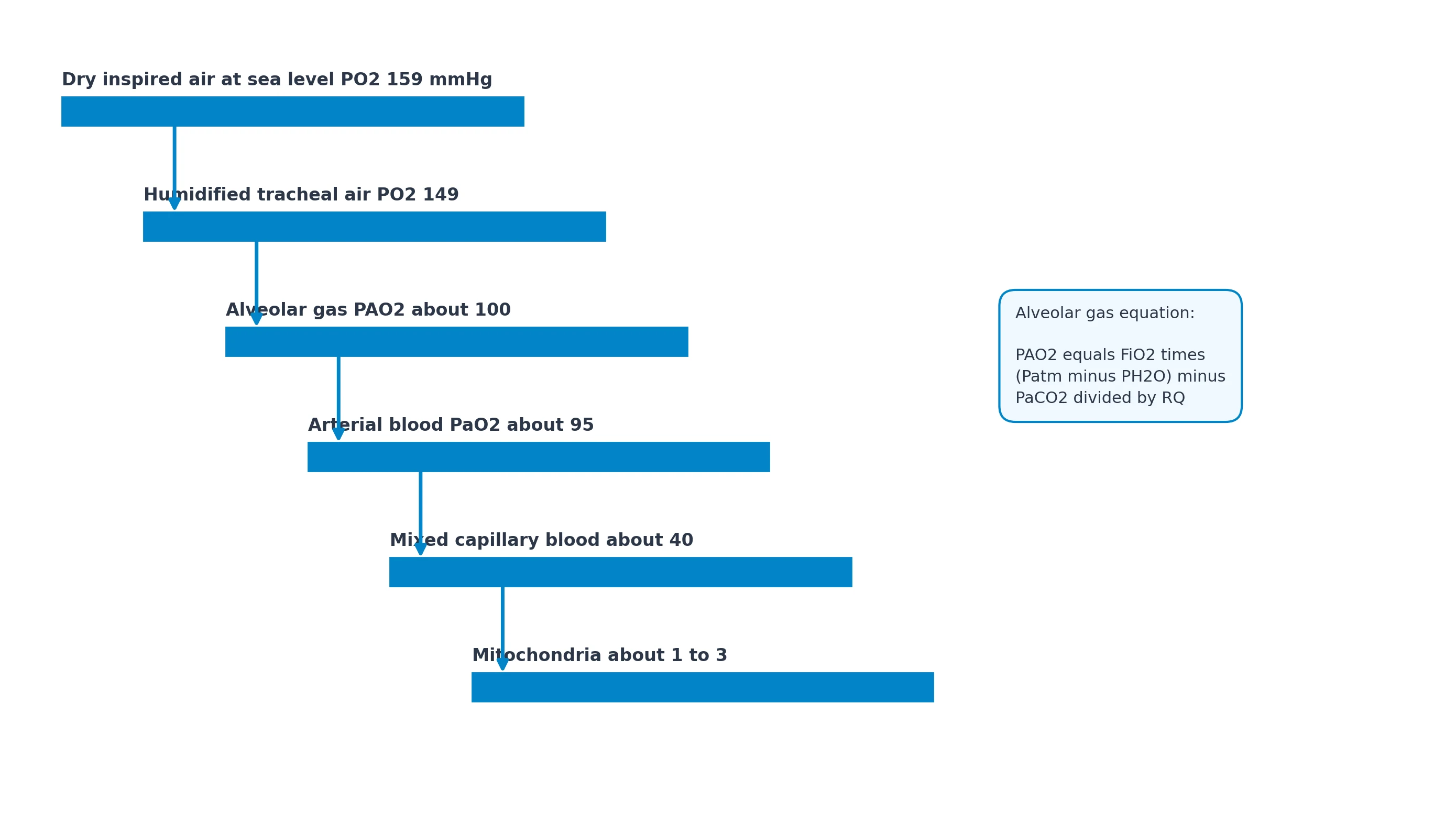
At sea level (Patm = 760 mmHg), dry air has FO2 = 0.2093: [1]
| Step | Approximate PO2 | Mechanism of fall |
|---|---|---|
| Dry inspired air (PIO2 dry) | 159 mmHg (0.2093 × 760) | Atmospheric composition |
| Humidified tracheal gas (37 °C) | ~150 mmHg | Water vapour PH2O = 47 mmHg dilutes O2: 0.2093 × (760 − 47) ≈ 149–150 |
| Alveolar gas (PAO2) | ~100–104 mmHg | Continuous O2 uptake and CO2 addition (alveolar gas equation) |
| Arterial blood (PaO2) | ~95–100 mmHg | Small A–a gradient (V/Q scatter, minor shunt) |
| Mixed venous / end-capillary tissue | ~40 mmHg (PvO2) | Tissue extraction |
| Mitochondrion | ~1–3 mmHg | Diffusion to cytochrome oxidase |
These are classic exam values; individual numbers vary with FiO2, PaCO2, cardiac output and metabolism. [1]
The alveolar gas equation (exact form)
The simplified alveolar gas equation used in every viva: [1]
PAO2 = FiO2 × (Patm − PH2O) − (PaCO2 / R) [1]
where: [1]
- FiO2 = inspired oxygen fraction
- Patm = barometric pressure (760 mmHg at sea level; falls with altitude)
- PH2O = 47 mmHg at 37 °C saturated
- PaCO2 ≈ PACO2 under most conditions
- R = respiratory exchange ratio ≈ 0.8 (VCO2/VO2) on a mixed diet (range ~0.7 fat to 1.0 carbohydrate) [1]
Worked sea-level room air example: [1]
PAO2 = 0.21 × (760 − 47) − (40 / 0.8) = 0.21 × 713 − 50 ≈ 150 − 50 = 100 mmHg. [1]
Levers the anaesthetist uses: [1]
- Raise FiO2 — lifts PIO2 and every downstream step (within shunt limits).
- Lower PaCO2 (increase VA) — reduces the subtracted term, raises PAO2 modestly.
- Raise Patm (hyperbaric oxygen) — increases PIO2 dramatically.
- At altitude, lower Patm collapses the cascade even at FiO2 0.21. [1]
More complete forms include a small correction factor when R ≠ 1: PAO2 = PIO2 − (PACO2/R) + [FIO2 × PACO2 × (1 − R)/R], but the simplified form is what examiners demand first [1].
Humidification and the first step
Inspired gas is saturated with water vapour in the upper airway. Absolute humidity at 37 °C is ~44 mg/L; partial pressure 47 mmHg is independent of Patm (temperature-dependent). Dry medical gases from cylinders lack water; the airway must supply it — evaporative heat and water loss, HME rationale, and the dilution of O2/N2. [1]
The A–a oxygen gradient
A–a gradient = PAO2 − PaO2 [1]
Normal on room air: roughly 5–15 mmHg (increases with age; rule of thumb upper limit ≈ (age/4) + 4 in some texts). On FiO2 1.0, normal A–a can be 30–50+ mmHg because of absorption effects and measurement scatter. [1]
Causes of raised A–a gradient: [1]
- V/Q mismatch
- Right-to-left shunt (true shunt)
- Diffusion limitation (rare at rest; exercise, fibrosis, oedema)
- Increased O2 extraction / low mixed venous O2 amplifies the effect of any shunt fraction on PaO2 [1]
Normal A–a hypoxaemia: low inspired PO2 (altitude) or pure hypoventilation (PAO2 falls with PaCO2 rise, A–a stays normal). [1]
Hypoxic pulmonary vasoconstriction (HPV)
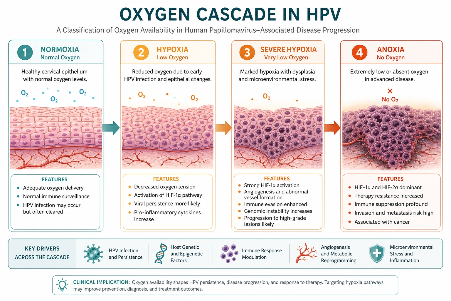
HPV is unique among vascular beds: pulmonary arterioles constrict when alveolar PO2 falls (with a contribution from mixed venous PO2). Purpose: divert blood from poorly ventilated units toward better ventilated units, defending PaO2. [1]
Cellular mechanism (exam outline): O2 sensing in pulmonary artery smooth muscle involves mitochondrial ROS/redox changes → inhibition of voltage-gated K+ channels (KV) → membrane depolarisation → opening of L-type Ca2+ channels → Ca2+ influx → contraction. Endothelium modulates via NO, endothelin and prostacyclin, but HPV can occur in denuded vessels [3].
Stimulus hierarchy: alveolar hypoxia is primary; low PvO2 augments; hypercapnia/acidosis generally enhance HPV; alkalosis attenuates. [1]
Time course: onset within seconds to minutes; biphasic reinforcement over tens of minutes; chronic hypoxia remodels vessels → pulmonary hypertension (high-altitude, COPD cor pulmonale) [3][4].
Factors that inhibit and enhance HPV
Inhibit HPV (worsen hypoxaemia when low-V/Q regions exist): [1]
- Volatile anaesthetics (dose-dependent; more at >1 MAC)
- Direct-acting vasodilators (nitroglycerin, nitroprusside, high-dose calcium channel blockers)
- Prostacyclin / inhaled pulmonary vasodilators if they reach poorly ventilated lung (worsen shunt)
- Very low mixed venous PO2 extremes and extreme atelectasis patterns
- Infection/inflammation in some models
- Systemic hypotension / low HPV stimulus gradients [1]
Enhance / preserve HPV: [1]
- Mild hypercapnia and acidosis
- Almitrine (historical/research)
- Avoidance of high-dose volatiles; total intravenous anaesthesia often preferred conceptually in OLV
- Regional hypoxia confined to diseased lung [1]
HPV and one-lung ventilation
During OLV the non-ventilated lung has alveolar hypoxia → HPV should reduce its blood flow from ~50% of CO toward ~20–30%, limiting shunt. Anything that inhibits HPV or increases blood flow to the non-dependent lung (lateral position, surgical compression of ventilated lung, vasodilators, high volatiles) worsens hypoxaemia. Management hierarchy: check tube position, FiO2, cardiac output, apply CPAP to non-ventilated lung or O2 insufflation, PEEP to ventilated lung carefully, clamp pulmonary artery if open chest extreme — physiology-first [2].
Anaesthetic relevance — cascade interventions
- Preoxygenation: raises PAO2 toward ~660 mmHg on FiO2 1.0 (PAO2 ≈ 713 − 50), filling FRC as O2 store.
- Apnoeic oxygenation: continuous O2 can maintain PAO2 if airway patent; CO2 still rises.
- PEEP/recruitment: reopen atelectatic units → remove alveolar hypoxia stimulus → better V/Q (and less HPV-needed).
- Hyperventilation: modest PAO2 gain via lower PaCO2 term; cost is cerebral vasoconstriction and auto-PEEP risk.
- Altitude / cabin pressure: lower Patm — calculate PAO2 before aeromedical transfer of lung patients. [1]
Raise PAO2
- Increase FiO2
- Increase VA (↓PaCO2)
- Raise Patm (HBO)
- Keep PH2O fixed at body temp
Defend PaO2 via HPV
- Alveolar hypoxia drives constriction
- Diverts blood to ventilated lung
- Volatiles/vasodilators impair
- Critical in atelectasis and OLV
Viva traps
- Derive PIO2 = FiO2(Patm − 47) before writing the full equation.
- "Is HPV triggered by arterial hypoxaemia?" Primarily alveolar hypoxia in the region.
- Calculate PAO2 on FiO2 1.0, PaCO2 40: ≈ 713 − 50 = 663 mmHg.
- Explain normal A–a on pure hypoventilation vs raised A–a on pneumonia.
- List four HPV inhibitors relevant to anaesthesia. [1]
Worked alveolar gas equation set-pieces
Room air, sea level, PaCO2 40, R 0.8: PAO2 = 0.21×(760−47) − 40/0.8 = 149.7 − 50 ≈ 100 mmHg. [1]
FiO2 0.5, PaCO2 40: PAO2 = 0.5×713 − 50 = 356.5 − 50 = 306.5 mmHg. [1]
Hypoventilation, room air, PaCO2 80: PAO2 = 149.7 − 100 ≈ 50 mmHg — hypoxaemia with normal A–a if lungs are otherwise normal. [1]
Altitude example (Patm 380 mmHg, half atmosphere, FiO2 0.21, PaCO2 30 after acclimatisation): PIO2 = 0.21×(380−47) ≈ 70 mmHg; PAO2 ≈ 70 − 30/0.8 = 70 − 37.5 ≈ 32.5 mmHg before further adaptation — explains obligatory hyperventilation and risk of altitude illness. [1]
100% oxygen, PaCO2 40: PAO2 ≈ 713 − 50 = 663 mmHg. PaO2 will be lower by the A–a gradient; a PaO2 of only 200 mmHg on FiO2 1.0 implies a large shunt fraction. [1]
Shunt equation link (interface with V/Q topic)
Classic iso-shunt reasoning: on FiO2 1.0, nitrogen is eliminated and low-V/Q units behave more like shunt. Approximate shunt fraction: [1]
Qs/Qt = (CcO2 − CaO2) / (CcO2 − CvO2) [1]
where CcO2 is end-pulmonary-capillary content estimated from PAO2. HPV reduces Qs/Qt when alveolar hypoxia is regional; global alveolar hypoxia (high altitude) raises pulmonary artery pressure without a "safe" place to divert blood. [1]
Regional HPV versus global hypoxic pulmonary hypertension
| Feature | Regional HPV | Global alveolar hypoxia |
|---|---|---|
| Example | Atelectasis, OLV, pneumonia patch | High altitude, hypoventilation whole lung |
| Effect on PaO2 | Protects PaO2 | Cannot protect by diversion |
| Pulmonary artery pressure | Mild local | Rises globally → RV strain |
| Chronic outcome | Local remodelling | HAPH / cor pulmonale |
Pharmacology of the pulmonary circulation (exam layer)
- NO and prostacyclin: increase cGMP/cAMP → pulmonary vasodilatation; inhaled route preferentially dilates ventilated regions (improves V/Q).
- Endothelin antagonists, PDE5 inhibitors: used in pulmonary hypertension — systemic administration can worsen V/Q in lung disease.
- Volatile agents: inhibit HPV in a dose-related fashion; isoflurane/sevoflurane/desflurane all capable; keep ≤1 MAC if hypoxaemia problematic during OLV.
- Propofol TIVA: relatively HPV-sparing compared with high-dose volatile — one rationale for TIVA in thoracic anaesthesia (practice varies; tube position still dominates). [1]
Oxygen stores and the cascade during apnoea
FRC ~30 mL/kg; after denitrogenation with FiO2 1.0 the O2 store is large. VO2 ~3 mL/kg/min. Rough safe apnoea time scales with O2 content of FRC and Hb stores. Obese, pregnant, paediatric, and septic patients desaturate faster (smaller FRC and/or higher VO2). This is cascade physiology applied to airway management: start higher on the cascade, consume slower, or both (apnoeic oxygenation, THRIVE). [1]
Diffusion and the last cascade steps
From alveolar gas to red-cell Hb, O2 crosses the alveolar–capillary membrane. Equilibration usually completes in about one-third of capillary transit time at rest. Diffusion limitation appears when membrane thickens (fibrosis, oedema), capillary transit shortens (extreme exercise, low mixed venous O2), or FO2 is low (altitude). Mitochondrial PO2 of 1–3 mmHg is the sink that maintains the diffusion gradient; cytochrome oxidase can operate at very low PO2 until delivery fails. [1]
Putting cascade and HPV together in the theatre
Desaturation algorithm physiology:
- Is FiO2 actually being delivered? (machine, disconnect)
- Is VA adequate? (high PaCO2 lowers PAO2)
- Is there new shunt/atelectasis? (recruit, suction, tube position)
- Is mixed venous O2 very low? (low CO amplifies shunt effect)
- Is HPV being inhibited? (volatiles, nitrates)
- Is there diffusion barrier? (oedema, ARDS) [1]
Each question maps to a cascade step or to blood-flow distribution. [1]
Historical and quantitative HPV notes
HPV was characterised in the mid-20th century as a response that optimises regional matching. Magnitude: moderate alveolar hypoxia (PAO2 ~40–60 mmHg) can divert a substantial fraction of local blood flow; extreme hypoxia and vessel remodelling change the dose–response. Acidosis potentiates; volatile anaesthetics produce dose-dependent blunting that is clinically relevant above ~1 MAC. Always restate: HPV senses alveolar gas primarily, which is why bronchial occlusion and atelectasis trigger it even if arterial PO2 is supported by the other lung. [1]
SAQ: write the alveolar gas equation and explain each term
"PAO2 equals the inspired oxygen fraction multiplied by the difference between barometric pressure and water vapour pressure, minus the arterial carbon dioxide tension divided by the respiratory exchange ratio. Water vapour pressure at body temperature is 47 mmHg. The respiratory exchange ratio is about 0.8 on a mixed diet. The equation shows that alveolar oxygen falls if barometric pressure falls, if inspired oxygen fraction falls, or if carbon dioxide rises because alveolar ventilation is inadequate relative to carbon dioxide production. The anaesthetist raises PAO2 chiefly by raising FiO2 and secondarily by lowering PaCO2." [1]
One-lung ventilation physiology paragraph
"When one lung is excluded from ventilation, its alveoli become hypoxic and hypoxic pulmonary vasoconstriction reduces its blood flow, limiting shunt. Lateral positioning sends more blood to the dependent ventilated lung, which helps. Surgical compression, vasodilator drugs, and high concentrations of volatile agent inhibit HPV and increase shunt fraction. Management begins with confirming double-lumen tube position, delivering high FiO2, optimising cardiac output, and applying CPAP to the non-ventilated lung or PEEP to the ventilated lung as appropriate." [1]
Cascade during CPR and low flow
Low cardiac output lowers mixed venous oxygen; any fixed shunt fraction then produces a lower arterial content because the admixture is more desaturated. Raising FiO2 still helps non-shunt units. During low-flow states EtCO2 falls (less CO2 delivery) even if cascade oxygen steps are maximised — another reminder that gas exchange requires perfusion. [1]
Primary exam expansion
This section extends the cascade and HPV discussion to the numerical fluency expected in ANZCA Primary SAQs and vivas.
Inspired gas composition under the anaesthetic machine
Pipeline oxygen is essentially pure O2 at the wall; air is 21% O2; N2O mixtures displace nitrogen and oxygen depending on the rotameter settings. The cascade begins not with "room air" but with whatever FiO2 the machine delivers. Hypoxic mixture delivery (failure of proportioning devices historically) collapses the top of the cascade before the patient is even apnoeic. Modern hypoxic guards and O2 analysers protect the first step of the cascade — physics meeting physiology. [1]
Water vapour is not optional
At 37 °C the saturated vapour pressure of water is fixed at 47 mmHg whether you are at sea level or on a mountain. Therefore the fractional dilution of dry gases by water is greater when Patm is low: PIO2 = FiO2(Patm − 47) shrinks faster than FiO2 × Patm would predict. Altitude physiology is cascade physiology with a smaller first term. [1]
Respiratory exchange ratio nuances
R = V̇CO2/V̇O2. On pure carbohydrate R approaches 1.0; on pure fat ~0.7; mixed diet ~0.8. During unsteady states (onset of exercise, hyperventilation) R can temporarily exceed 1 as CO2 is blown off from stores. In the alveolar gas equation, using R=1 simplifies to PAO2 = PIO2 − PaCO2, which slightly overestimates the CO2 subtraction if true R is 0.8. Examiners usually accept R=0.8. [1]
Diffusion disequilibrium scenarios
At rest, end-capillary PO2 nearly equals PAO2. In severe interstitial disease, extreme exercise, or low mixed venous O2 with short transit, end-capillary PO2 remains below PAO2 — diffusion limitation widens A–a. HPV cannot fix a pure diffusion problem; FiO2 can help by steepening the driving gradient. [1]
HPV versus pulmonary embolism physiology
PE creates high V/Q / dead space; HPV is about low alveolar PO2. Do not say "HPV causes PE hypoxaemia." Hypoxaemia in PE involves low mixed venous O2, some low V/Q from overperfused non-embolised lung, right-to-left shunt through foramen ovale if RA pressure rises, and reduced CO. [1]
Quantitative OLV shunt thinking
If non-ventilated lung still receives 40% of CO and is pure shunt, and the ventilated lung is ideal, arterial content is a mixture: Ca = 0.6×Cc + 0.4×Cv. With Cv substantially below Cc, SaO2 falls into the 80s easily. HPV reducing non-ventilated flow to 20% dramatically improves Ca. That arithmetic is why HPV magnitude matters clinically. [1]
Drug list to recite for HPV inhibition
Volatile anaesthetics (dose-dependent), nitroglycerin, sodium nitroprusside, high-dose calcium channel blockers, prostacyclin if systemic, dobutamine in some settings, and extremes of alkalosis. Preservation strategies: minimise volatile dose or use TIVA, avoid unnecessary vasodilators, maintain reasonable perfusion pressure, and optimise mechanical factors first. [1]
Red flags
- Alveolar gas equation sets PAO2: raise FiO2 and lower PaCO2 to improve oxygenation.
- HPV responds to alveolar hypoxia, diverting perfusion to ventilated lung.
- Volatiles, vasodilators, calcium channel blockers and low SVR impair HPV — OLV hypoxaemia.
- Raised A–a gradient means shunt, V/Q mismatch or diffusion impairment.
- Altitude lowers Patm and the entire cascade. [1]
References
- [1]Heymer J. Alveolar gas equation: a metabolic perspective Intensive Care Med, 2026.PMID 41661294
- [2]Brenn BR, et al. A Comparative Evaluation of Unilateral and Bilateral Sequential Lung Isolation for Vertebral Body Tethering: A Retrospective Propensity Matched Analysis Cureus, 2024.PMID 38854196
- [3]Ma MR, et al. When MicroRNAs meet hypoxic pulmonary hypertension Biomed Pharmacother, 2026.PMID 42166981
- [4]Chacon-Diaz M. The cardiovascular system and high-altitude exposure: from adaptation to disease. Part I Arch Peru Cardiol Cir Cardiovasc, 2026.PMID 42238502
- [5]Jung C, et al. Effects of increasing respiratory rate on ventilatory efficiency and mechanical costs during low-tidal-volume ventilation: a prospective physiological pilot study Ann Intensive Care, 2026.PMID 42306311
- [6]Nieman GF, et al. Expert recommendations for setting and adjusting airway pressure release ventilation based on clinical experience and basic science evidence Front Med (Lausanne), 2026.PMID 41709908