Anaes · Applied cardiovascular & respiratory physiology
Renal: GFR & tubular function
Also known as Glomerular filtration rate · Filtration fraction · Clearance · Tubuloglomerular feedback · Proximal tubule · NHE3
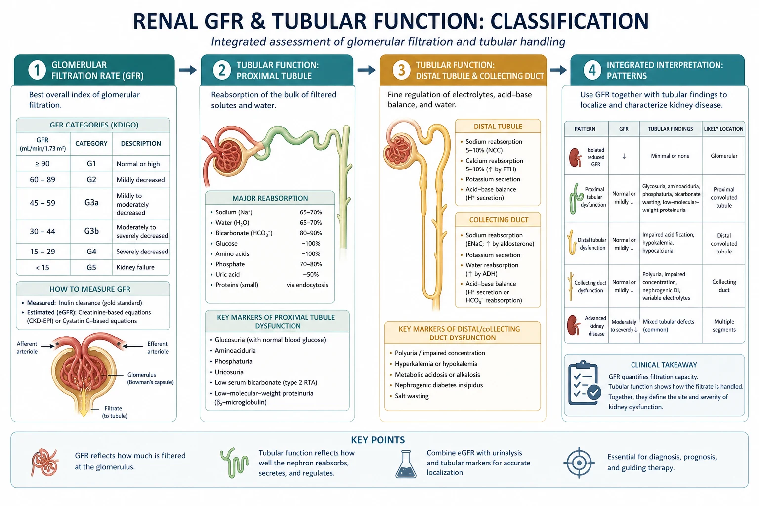
The kidney filters plasma at the glomerulus and then selectively reabsorbs and secretes along the nephron, and the glomerular filtration rate (about 125 mL per minute) is the single best measure of kidney function. The framework rests on five exam-critical ideas: GFR is normally about 125 mL per minute and the filtration fraction (GFR over renal plasma flow) is about 20 percent, set by the balance of Starling forces across the glomerular capillary and the relative tone of the afferent and efferent arterioles; GFR is held constant across a range of blood pressures by autoregulation (myogenic and tubuloglomerular feedback from the macula densa); the clearance of a freely filtered, neither-reabsorbed-nor-secreted solute (inulin) equals GFR, and creatinine clearance approximates it clinically; the nephron segments each specialise — the proximal tubule reabsorbs about 65 percent of the bulk filtrate (including all glucose and amino acids and most bicarbonate and sodium, via the NHE3 sodium-hydrogen exchanger), the loop of Henle is the countercurrent multiplier that concentrates the medulla, and the distal nephron and collecting duct fine-tune sodium (aldosterone) and water (ADH); and the kidney is uniquely vulnerable to anaesthesia and nephrotoxins because it receives a high blood flow and depends on glomerular pressure and tubular energy. Built on the GFR-physiology review (Ayub 2026), the macula densa tubuloglomerular-feedback study (Li 2026), the renal NHE3 study (Nogueira Coelho 2026), the proximal-tubule-dysfunction study (Ikeme 2026), the SGLT2-inhibition study (Gao 2026), and the chloride-and-RAAS study (Adin 2026).
On this page & tools
Your progress
Saved locally on this device.
8 MCQs with explanations
Target exams
Red flags

Why this matters to the anaesthetist
Primary candidates must define GFR and filtration fraction, write determinants of glomerular filtration pressure, explain autoregulation (myogenic + TGF), map nephron segment transport with diuretic targets, and explain creatinine’s non-linear relationship to GFR. Final applies this to AKI prevention and perioperative drug dosing.[1][4]
One-liner: GFR ≈ 125 mL/min from net filtration pressure across glomerular capillaries; afferent constriction or efferent dilation lowers GFR; 65% Na/water reclaimed proximally; loop builds medullary gradient; distal nephron fine-tunes under aldosterone/ADH. [1]
GFR, RBF and filtration fraction
- GFR: volume filtered into Bowman’s space per unit time — normal young adult ~125 mL/min (~180 L/day).
- Renal blood flow (RBF): ~20–25% of CO (~1–1.2 L/min); renal plasma flow (RPF) ≈ RBF × (1 − Hct).
- Filtration fraction (FF) = GFR/RPF ≈ 0.20 (one-fifth of plasma entering kidney is filtered).
- Only ~1% of filtered water is finally excreted (variable). [1]
Clearance concept: C_x = (U_x × V̇)/P_x. For ideal filtration marker (inulin): C = GFR. Creatinine clearance approximates GFR but slight tubular secretion → mild overestimate. eGFR equations use creatinine (± cystatin C). [1]
Creatinine–GFR curve: at high GFR, creatinine is flat; once GFR falls substantially, creatinine rises steeply. A “small” creatinine rise can mean a large relative GFR loss — exam cliché. [1]
Starling forces at the glomerulus
Net filtration pressure ≈ (P_gc − P_bs) − (π_gc − π_bs) [1]
- P_gc (glomerular capillary hydrostatic): main driver; set by arterial pressure and afferent/efferent tone.
- π_gc (oncotic): rises along the capillary as water filters (FF effect).
- P_bs: Bowman hydrostatic; rises if tubular obstruction.
- K_f: filtration coefficient (area × permeability); falls in glomerular disease. [1]
Afferent vs efferent — drug viva table
| Manoeuvre | Afferent | Efferent | GFR effect |
|---|---|---|---|
| NSAIDs (↓PGE2) | Constrict | — | ↓ GFR (especially if volume-depleted) |
| Ang II | Mild constrict | Strong constrict | Supports GFR when perfusion low |
| ACEi/ARB | — | Dilate | ↓ GFR if Ang II-dependent |
| Afferent dilation (low resistance) | Dilate | — | ↑ P_gc / GFR (within limits) |
| Afferent constriction (SNS extreme) | Constrict | — | ↓ RBF and GFR |
Triple whammy: ACEi/ARB + diuretic + NSAID on a dry patient → classic community/perioperative AKI setup.[1]
Autoregulation
GFR and RBF held relatively constant over MAP roughly 80–180 mmHg (teaching range; lower limit rises in chronic hypertension). [1]
- Myogenic: afferent smooth muscle contracts when stretched by pressure.
- Tubuloglomerular feedback (TGF): macula densa senses high NaCl → signals afferent constriction (and modulates renin). Protects from hyperfiltration; contributes to oliguria when tubules injured.[2]
Beyond the lower limit, flow becomes pressure-passive — hypotension causes ischaemic GFR fall. Anaesthesia, SNS surge, and exogenous catecholamines can override autoregulation. [1]
Nephron segment map
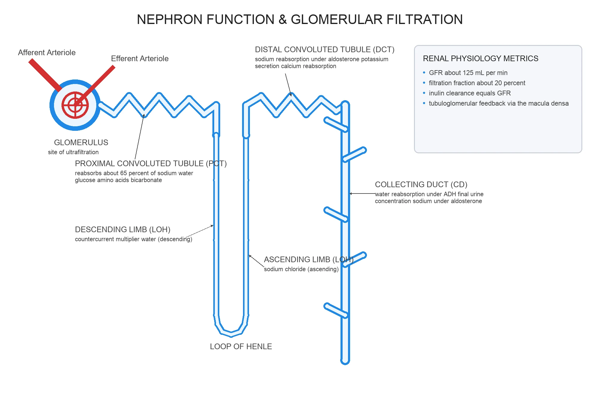
Proximal tubule (~65% Na and water)
- Isosmotic bulk reabsorption.
- NHE3 apical Na/H exchanger; basolateral Na/K-ATPase.
- SGLT2 reclaims virtually all filtered glucose (until threshold ~10 mmol/L plasma teaching value — glycosuria above).
- Bicarbonate reclaim via carbonic anhydrase (acetazolamide target).
- Amino acids, low-molecular-weight proteins reabsorbed. [1]
Loop of Henle
- Thin descending: water out into hypertonic medulla.
- Thick ascending (TAL): NKCC2 reabsorbs Na–K–2Cl; water impermeable → dilute urine + build medullary gradient. Loop diuretics block NKCC2 → lose concentrating ability, increase distal flow (K/Mg wasting). [1]
Distal convoluted tubule
- NCC (thiazide target) reabsorbs NaCl.
- Fine calcium handling interplay (thiazides can spare Ca). [1]
Collecting duct
- Principal cells: ENaC (aldosterone), AQP2 (ADH).
- Intercalated cells: H+/HCO3 handling.
- Amiloride/triamterene block ENaC; spironolactone blocks aldosterone receptor. [1]
Glomerulo-tubular balance
Proximal reabsorption scales with GFR so fractional reabsorption stays roughly stable — prevents massive natriuresis when GFR rises slightly. Ang II and peritubular physical forces contribute. [1]
Measurement and intraoperative thinking
- Oliguria is a late, non-specific signal.
- Perfusion pressure, venous congestion (high CVP/IAP), nephrotoxins, and obstruction are the mechanistic buckets.
- Rhabdomyolysis: myoglobin toxic to tubules + volume depletion.
- Contrast nephropathy risk physiology: vasoconstriction + direct toxicity on background of low flow. [1]
Numbers board
| Parameter | Teaching value |
|---|---|
| GFR | ~125 mL/min |
| FF | ~0.2 |
| RBF share of CO | ~20–25% |
| Proximal Na reabsorb | ~65% |
| Glucose threshold | ~10 mmol/L plasma |
| Autoregulation MAP | ~80–180 mmHg |
Lower GFR via afferent
- NSAIDs
- Strong SNS
- Low MAP below auto
- Afferent disease
Lower GFR via efferent
- ACEi/ARB
- When AngII-dependent
- Bilateral RAS physiology
- Combine with dry+NSAID
Graph viva scripts
Draw afferent/efferent arteriole around glomerulus and show NSAID vs ACEi arrows. [1]
Draw TGF loop: ↑distal NaCl → macula densa → afferent constriction → ↓GFR. [1]
Explain why loop diuretics work in low GFR better than thiazides (teaching: act more proximally in the loop, greater Na delivery effect). [1]
Extended viva dialogue
Examiner: Define filtration fraction. [1]
Candidate: GFR divided by renal plasma flow, normally about 0.2. If efferent constriction rises, FF rises; if afferent constriction dominates, RBF and GFR both fall. [1]
Examiner: How does tubuloglomerular feedback protect the kidney? [1]
Candidate: When NaCl delivery to the macula densa is high, the afferent arteriole constricts, lowering GFR to reduce distal overload. In ATN, debris and impaired reabsorption can inappropriately activate signals that keep GFR low. [1]
Clinical synthesis: GFR physiology is arterioles + Starling forces; tubular physiology is segment transporters. Anaesthesia endangers both via pressure, drugs and toxins. [1]
Creatinine kinetics teaching curve
Plot GFR on x, serum creatinine on y — hyperbolic. Falling from 120 to 60 mL/min may barely move creatinine; falling from 30 to 15 doubles it. Hence “creatinine normal” does not mean GFR normal in the elderly or sarcopenic. [1]
Diuretic map (must draw on nephron)
| Diuretic | Target | Segment |
|---|---|---|
| Acetazolamide | Carbonic anhydrase | Proximal |
| Loop (furosemide) | NKCC2 | TAL |
| Thiazide | NCC | DCT |
| Amiloride | ENaC | CD |
| Spironolactone | MR | CD |
| SGLT2i | SGLT2 | Proximal |
Worked SAQ
SAQ: Explain how NSAIDs and ACE inhibitors reduce GFR (6 marks)
When effective volume is low, afferent dilation depends on prostaglandins and efferent constriction depends on angiotensin II to preserve glomerular capillary pressure. NSAIDs reduce prostaglandin-mediated afferent dilation; ACE inhibitors/ARBs reduce angiotensin II-mediated efferent constriction. Together with diuretic-induced volume loss they can precipitate acute falls in GFR. [1]
Primary exam expansion — dense examiner pack
GFR determinants (write Starling for glomerulus)
GFR = Kf [(Pgc − Pbs) − (πgc − πbs)]. Pgc glomerular capillary hydrostatic pressure (main driver, dependent on arterial pressure and afferent/efferent tone). πgc oncotic rises along capillary as filtration concentrates plasma proteins. Kf surface area × permeability — reduced in glomerular disease. [1]
Normal GFR ~125 mL/min (180 L/day); urine output ~1–2 L/day → >99% reabsorbate. [1]
Clearance concept
Cx = UxV / Px. Inulin clearance = GFR gold standard teaching; creatinine clearance approximates GFR (slight secretion). PAH clearance approximates RPF if fully extracted; filtration fraction = GFR/RPF ~0.2. [1]
Afferent vs efferent tone (classic table)
| Manoeuvre | Afferent | Efferent | GFR | RBF |
|---|---|---|---|---|
| Low dose dopamine teaching historical | dilate | — | variable | ↑ |
| NSAIDs (↓PG) | relative constriction | — | ↓ especially if renin-high | ↓ |
| ACEI/ARB | — | dilate efferent | ↓ (afferent dilated states) | variable |
| Ang II moderate | constrict both; efferent > | — | GFR defended | ↓ RBF |
| Afferent constriction pure | constrict | — | ↓ | ↓ |
Why NSAID + ACEI + diuretic (“triple whammy”) precipitates AKI in hypovolaemia. [1]
Autoregulation of RBF/GFR
Myogenic + tubuloglomerular feedback (macula densa NaCl → adenosine/ATP → afferent constriction). Working range MAP roughly 80–180 mmHg teaching. Below range: pressure-passive fall → prerenal then ATN if prolonged. [1]
Tubular handling map (segment duties)
| Segment | Key transport | Drugs/hormones |
|---|---|---|
| Proximal | 65% Na/water; glucose, amino acids; HCO3 reabsorb | SGLT2i glucosuria; carbonic anhydrase inhibitors |
| Thin limbs | Countercurrent passive | — |
| Thick ascending limb | NaK2Cl; dilute urine; generate medullary gradient | Loop diuretics |
| DCT | NaCl | Thiazides |
| CD principal | ENaC Na; water via AQP2 | Aldosterone; ADH; K-sparing diuretics |
| CD intercalated | H+ / HCO3 | Acid–base |
Glucose Tm: above threshold glycosuria (diabetes). [1]
Countercurrent multiplier (exam outline)
Thick ascending limb active salt out without water → medullary hypertonicity; descending limb water out; vasa recta preserve gradient; ADH inserts AQP2 in collecting duct → water reabsorbed → concentrated urine. Without ADH: dilute urine (diabetes insipidus). Loop diuretics abolish gradient → cannot concentrate. [1]
Hormones
RAAS: renin (JG cells) → angiotensin II → aldosterone + vasoconstriction + ADH synergy. ADH (osmolarity + volume). ANP/BNP: natriuresis opposite. PTH: Ca/PO4 handling. [1]
Anaesthetic relevance
- GFR and RBF fall with hypovolaemia, hypotension, high PEEP/low CO, pneumoperitoneum.
- Renally excreted drugs/metabolites accumulate (aminoglycosides, M6G, gabapentinoids).
- Oliguria differential: prerenal vs ATN vs obstruction — use context, FeNa/FeUrea carefully, ultrasound.
- Contrast nephropathy risk factors; hydration.
- Rhabdomyolysis: myoglobin toxic to tubules — alkaline diuresis debates, fluid. [1]
SAQ: determinants of GFR and effect of NSAIDs (8 marks)
Starling forces (3). Afferent PG dilation defence (2). NSAID mechanism of GFR fall (2). Clinical high-risk setting (1). [1]
Viva
Q: Why is creatinine a delayed AKI marker? A: Rises only after substantial GFR loss; production varies with muscle. Q: How do loop diuretics work? A: Block NKCC2 in thick ascending limb; lose gradient; potent natriuresis. Q: Filtration fraction meaning? A: GFR/RPF — rises when efferent constricts relatively. [1]
High-yield viva battery and numbers lock-in
Numbers
- GFR ≈ 125 mL/min ≈ 180 L/day
- Filtration fraction ≈ 0.2
- RBF ≈ 20–25% of CO
- Proximal reabsorption ≈ 65% Na/water
- Glucose threshold ~10 mmol/L teaching (varies)
- Concentrating ability depends on medullary gradient + ADH [1]
Diuretic site map (rapid)
CA inhibitors proximal; loops thick ascending NKCC2; thiazides DCT NCC; K-sparing ENaC blockers/aldosterone antagonists in CD; osmotic diuretics (mannitol) proximal + descending limb water. [1]
AKI prerenal versus ATN physiology
Prerenal: ↓RBF/GFR, tubules working → low urine Na, high urine osmolality, bland sediment. Prolonged ischaemia → ATN: tubular dysfunction → higher urine Na, isosmolar urine, muddy brown casts teaching. Anaesthesia goal: avoid the transition by defending perfusion pressure and volume without drowning the patient. [1]
Full viva dialogue (additional)
Examiner: How do ACE inhibitors drop GFR in bilateral renal artery stenosis? [1]
Candidate: In that setting glomerular filtration is maintained by angiotensin-II-mediated efferent arteriolar constriction. ACE inhibitors dilate the efferent arteriole, lower glomerular capillary pressure and GFR can fall sharply — which is why these drugs are dangerous when renal perfusion depends on efferent tone. [1]
Examiner: Explain tubuloglomerular feedback. [1]
Candidate: Increased NaCl delivery to the macula densa causes release of vasoconstrictor mediators such as adenosine that constrict the afferent arteriole, reducing GFR back toward normal. This protects the distal nephron from overload and contributes to autoregulation. [1]
Exam traps
- Saying creatinine is an early sensitive AKI marker.
- Forgetting prostaglandins defend afferent dilation in low-effective-volume states.
- Mixing loop vs thiazide sites.
- Ignoring filtration fraction when discussing efferent constriction. [1]
References
- [1]Ayub F, et al. The Physiological Basis of Acute GFR Decline With Renoprotective Therapies Mayo Clin Proc, 2026.PMID 42336099
- [2]Li M, et al. Macula densa-specific NOS1 knockout determines susceptibility to ischemic acute kidney injury Clin Sci (Lond), 2026.PMID 41837645
- [3]Nogueira Coelho J, et al. Renal NHE3 is required to limit hypokalemia and metabolic acidosis during dietary potassium deficiency Pflugers Arch, 2026.PMID 42260191
- [4]Ikeme JC, et al. Proximal tubule reabsorptive dysfunction and risk of cardiovascular death among community-living adults: the HUNT-3 cohort Eur Heart J Open, 2026.PMID 42291457
- [5]Gao Y, et al. Repurposing SGLT2 Inhibitors for Cirrhotic Ascites: From Mechanistic Research to Clinical Exploration J Clin Transl Hepatol, 2026.PMID 41810110
- [6]Adin D, et al. Influence of serum chloride concentrations on the renin-angiotensin-aldosterone system in dogs with congestive heart failure Am J Physiol Renal Physiol, 2026.PMID 42324236