Anaes · Applied cardiovascular & respiratory physiology
Renal: RAAS & water/electrolyte handling
Also known as Renin-angiotensin-aldosterone system · RAAS · Aldosterone · Water balance · ADH · Countercurrent system
The kidney defends blood pressure, sodium, water and potassium through the renin-angiotensin-aldosterone system and a set of linked endocrine loops. The framework rests on five exam-critical ideas: the RAAS is activated by a fall in renal perfusion or sodium or by sympathetic tone, and it restores them through renin, angiotensin II (vasoconstriction, aldosterone release, ADH and thirst) and aldosterone (distal sodium reabsorption and potassium and hydrogen excretion); aldosterone acts on the principal cells of the distal nephron to reabsorb sodium (ENaC) and excrete potassium and hydrogen, linking sodium volume to potassium and acid-base; water balance is governed by ADH (and thirst) via osmoreceptors, ADH inserting aquaporin-2 channels in the collecting duct; the countercurrent multiplier (the loop of Henle) builds the medullary gradient that lets ADH concentrate the urine; and natriuretic peptides (ANP, BNP) from the atria and ventricles oppose the RAAS by promoting sodium excretion. Built on the renal-transporter dimorphism study (Xiong 2026), the hypernatraemia pathophysiology review (Drummond 2026), the sodium and water disorders review (Gilbert 2025), the water-homeostasis review (D'Acierno 2025), the SGLT2 cardiovascular-protection review (Wang 2026), and the chloride-RAAS study (Adin 2026).
On this page & tools
Your progress
Saved locally on this device.
8 MCQs with explanations
Target exams
Red flags
Why this matters to the anaesthetist
Primary candidates must explain how the kidney senses underfilling, the RAAS cascade with enzyme sites, aldosterone’s three classic effects, ADH triggers and aquaporin-2, the countercurrent multiplier in words (and as a draw), and why ACEi/ARB + NSAID + hypovolaemia wrecks GFR. Final candidates apply the same map to hyperkalaemia, hyponatraemia correction rates, diabetes insipidus, and drug holds on the day of surgery.[2][3]
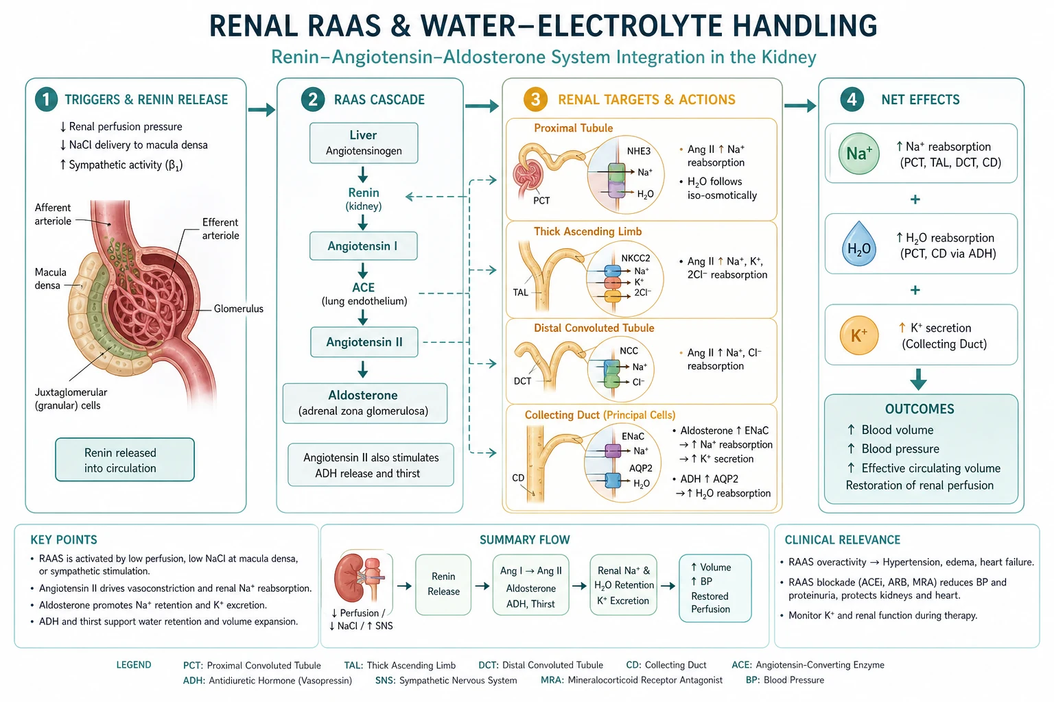
One-liner: Low renal perfusion or sodium delivery → renin → angiotensin II (efferent constriction, aldosterone, thirst/ADH) → distal Na reabsorption with K/H secretion; free water is controlled by ADH via AQP2 on a medullary gradient built by the loop. [1]
Sensors and the juxtaglomerular apparatus
Three classical renin triggers (state all three in a viva): [1]
- Reduced renal perfusion pressure (afferent arteriolar baroreceptor / JGA).
- Reduced NaCl delivery to the macula densa (tubuloglomerular feedback arm).
- Sympathetic β1 stimulation of JG cells (and α-adrenergic effects on vessels). [1]
Juxtaglomerular apparatus anatomy (exam sketch): afferent arteriole JG cells (renin granules) + macula densa of thick ascending limb + extraglomerular mesangium. Local prostaglandins (PGE2, PGI2) support afferent dilation when volume is low — this is why NSAIDs are dangerous in that state. [1]
RAAS cascade — exact sequence

| Step | Where | Product / action |
|---|---|---|
| Angiotensinogen | Liver (constitutive) | Substrate |
| Renin | JG cells | Rate-limiting; cleaves to Ang I |
| ACE | Pulmonary endothelium (and elsewhere) | Ang I → Ang II; also bradykinin degradation |
| Angiotensin II | Plasma / tissues | AT1 receptor effects (below) |
| Aldosterone | Zona glomerulosa | Distal Na↑, K↓, H↓ |
Angiotensin II — must-list actions: [1]
- Potent arteriolar vasoconstriction (raises SVR and BP).
- Preferential efferent glomerular constriction → supports glomerular hydrostatic pressure and GFR when renal perfusion is low.
- Stimulates aldosterone release.
- Stimulates ADH and thirst.
- Direct proximal Na reabsorption.
- Facilitates sympathetic transmission. [1]
Exam drug map: ACE inhibitors block Ang II generation and raise bradykinin (cough, angioedema). ARBs block AT1. Both reduce efferent tone → GFR can fall when afferent pressure is already low. Direct renin inhibitors exist but are less exam-dominant in ANZ.[6]
Aldosterone — principal cell physiology
Site: late distal tubule / cortical collecting duct principal cells. [1]
Mechanism: mineralocorticoid receptor → genomic ↑ of ENaC (apical Na entry) and basolateral Na/K-ATPase → Na reabsorbed; lumen-negative potential drives K secretion (ROMK) and favours H secretion (α-intercalated cells). [1]
Clinical tetrad of hyperaldosteronism: hypertension + expanded volume + hypokalaemia + metabolic alkalosis. [1]
Spironolactone / eplerenone: block receptor → hyperkalaemia risk, especially with ACEi/ARB, CKD, or K supplements. [1]
Cortisol caveat: 11β-HSD2 normally inactivates cortisol in the mineralocorticoid receptor cell; when overwhelmed (apparent mineralocorticoid excess, liquorice) cortisol acts like aldosterone. [1]
Water balance — ADH, thirst, osmoreceptors
Plasma osmolality ≈ 2×[Na] + glucose/18 + urea/2.8 (conventional teaching units) — Na dominates. [1]
Osmotic trigger: ~1–2% rise in Posm → hypothalamic osmoreceptors → supraoptic/paraventricular nuclei → posterior pituitary ADH (AVP). [1]
Non-osmotic triggers (high yield): large volume loss / hypotension (baroreceptors), nausea, pain, angiotensin II, some drugs. [1]
ADH renal effect: V2 receptors on collecting duct principal cells → cAMP → aquaporin-2 insertion → free water reabsorption. Without ADH, duct is water-impermeable → dilute urine (diabetes insipidus pattern). [1]
Thirst runs in parallel — defence against hypernatraemia if access to water exists.[2][4]
| Disorder | Problem | Urine | Plasma |
|---|---|---|---|
| Central DI | No ADH | Large volume dilute | Rising Na/osmolality |
| Nephrogenic DI | Kidney ignores ADH | Same | Same |
| SIADH | Inappropriate ADH | Inappropriately concentrated | Hyponatraemia, euvolemic |
Anaesthetic DI hooks: pituitary surgery (central), lithium (nephrogenic), severe hypernatraemia management with controlled free water. [1]
Potassium handling — viva board
- Freely filtered; ~65–70% reabsorbed proximally; further in loop; regulated excretion is distal (flow- and aldosterone-dependent).
- Hyperkalaemia stimulates aldosterone → kaliuresis (feedback).
- Distal Na delivery and flow matter: loop diuretics increase distal delivery → K loss; low flow states reduce kaliuresis.
- Insulin, β2-agonists, alkalosis shift K into cells (not true total-body loss); acidosis, succinylcholine, tissue injury shift K out.
- Perioperative hyperkalaemia triage: ECG changes, stop K, protect heart (calcium), shift (insulin–glucose, salbutamol), remove (resins, dialysis). [1]
Countercurrent multiplier — describe the graph in words
Draw script: long loop of Henle descending into medulla; thick ascending limb (TAL) labelled “NKCC2, water-impermeable”; medullary interstitium gradient ~300 cortex → up to ~1200 mOsm/kg at papilla; collecting duct running through gradient with AQP2 “on/off”; vasa recta as countercurrent exchanger preserving the gradient. [1]
Multiplier principle: [1]
- TAL actively reabsorbs Na–K–2Cl (NKCC2 — loop diuretic target) without water → tubular fluid dilutes; interstitium concentrates.
- Descending limb equilibrates with hypertonic medulla (water out).
- Multiplication along the long axis builds a vertical gradient.
- Urea recycling contributes to inner medullary tonicity (exam bonus).
- Vasa recta remove reabsorbed solute/water without washing out the gradient (exchanger). [1]
ADH then allows collecting-duct water to follow the gradient → concentrated urine. Loop diuretics abolish the gradient → cannot concentrate; also increase Na and water excretion. [1]
Natriuretic peptides
ANP (atrial stretch) and BNP (ventricular wall stress): promote natriuresis/diuresis, vasodilation, suppress renin and aldosterone — the physiologic counterweight to volume overload. BNP/NT-proBNP are clinical markers of heart failure strain, not pure “volume meters.”[5]
Acid–base renal limb (brief, linked)
Proximal: reclaim HCO3 via NHE3 + carbonic anhydrase. Distal: generate new HCO3 while excreting titratable acid and NH4+. Links to aldosterone (H secretion) and hypokalaemic alkalosis of hyperaldosteronism. Full Stewart/buffer detail lives in acid–base leaves. [1]
Electrolyte perioperative decision board
| Problem | Physiology link | Anaesthetic action |
|---|---|---|
| ACEi/ARB morning dose + dry | Blunted Ang II support of GFR/BP | Expect induction hypotension; careful fluids/vasopressors; hold per local protocol for major cases |
| Spironolactone + ACEi | Dual RAAS block | Check K; ECG if high |
| SGLT2i | Osmotic diuresis, euglycaemic ketoacidosis risk | Hold perioperatively per guidance; watch volume |
| Hyponatraemia | Often ADH-related or free water excess | Slow correction if chronic; avoid ODS |
| Hypernatraemia | Free water deficit | Replace free water carefully; treat DI if present |
| Loop diuretic chronic use | Flat medullary gradient, low K/Mg | Replace K/Mg; volume status assessment |
Equations and numbers board
- GFR ≈ 125 mL/min (young adult); falls with age.
- Filtration fraction ≈ 0.2.
- Medullary max osmolality ≈ 1200 mOsm/kg (teaching figure).
- Osmotic ADH threshold: ~1–2% Posm change.
- Plasma osmolality teaching formula: 2[Na] + glucose/18 + urea/2.8 (conventional units).
- Effective circulating volume defence hierarchy: baroreflex (seconds) → RAAS/ADH (minutes–hours) → renal Na handling (hours–days). [1]
Volume defence (RAAS)
- Renin triggers ×3
- Ang II: SVR + efferent GFR support
- Aldosterone: Na in, K/H out
- Slow-ish endocrine
Water defence (ADH)
- Osmotic 1–2% + non-osmotic
- AQP2 collecting duct
- Needs medullary gradient
- DI vs SIADH poles
Graph / SAQ viva scripts
Draw RAAS cascade with ACE in the lung and list four Ang II effects. [1]
Draw nephron and mark NKCC2 (loop), NCC (thiazide), ENaC (aldosterone), AQP2 (ADH). [1]
Explain why loop diuretics cause hypokalaemia: increased distal Na delivery and flow → increased distal K secretion; plus secondary aldosteronism if volume falls. [1]
SIADH vs hypovolaemic hyponatraemia: volume status, urine Na, and whether ADH is “appropriate.” [1]
Extended viva dialogue
Examiner: What stimulates renin release? [1]
Candidate: Reduced afferent arteriolar pressure, reduced NaCl at the macula densa, and sympathetic β1 stimulation of juxtaglomerular cells. [1]
Examiner: How does aldosterone affect acid–base? [1]
Candidate: It promotes distal hydrogen ion secretion (and potassium loss). Excess produces hypokalaemic metabolic alkalosis; deficiency contributes to hyperkalaemic metabolic acidosis (type 4 RTA pattern). [1]
Examiner: Why can ACE inhibitors cause a rise in creatinine? [1]
Candidate: In states dependent on efferent constriction to maintain glomerular pressure — bilateral renal artery stenosis physiology, severe underfilling — removing angiotensin II dilates the efferent arteriole and GFR falls. A modest creatinine rise can be expected; a large fall needs review of volume and dual blockade. [1]
Clinical synthesis: On the list, RAAS is not abstract endocrinology — it is why the dry patient on ramipril and ibuprofen becomes oliguric and hypotensive at induction. [1]
Worked SAQ model answers
SAQ: Outline the renin–angiotensin–aldosterone cascade and its anaesthetic relevance (10 marks)
Renin is released from juxtaglomerular cells when renal perfusion pressure falls, macula densa NaCl delivery falls, or sympathetic β1 tone rises. Renin cleaves hepatic angiotensinogen to angiotensin I; pulmonary endothelial ACE converts this to angiotensin II. Angiotensin II causes arteriolar vasoconstriction, preferential efferent glomerular constriction that supports GFR in underfilling, aldosterone release, ADH and thirst stimulation, and proximal sodium reabsorption. Aldosterone acts on collecting-duct principal cells to increase ENaC and Na/K-ATPase activity, retaining sodium and secreting potassium and hydrogen. [1]
Anaesthetic relevance: ACE inhibitors and ARBs blunt angiotensin II support of blood pressure and GFR, predisposing to induction hypotension and functional creatinine rise when combined with hypovolaemia or NSAIDs. Aldosterone antagonists raise potassium. Surgical stress and hypovolaemia activate RAAS, promoting sodium and water retention postoperatively. [1]
SAQ: Compare osmotic and non-osmotic control of ADH (5 marks)
Osmoreceptors detect a 1–2% rise in plasma osmolality and trigger ADH release, inserting aquaporin-2 in the collecting duct. Non-osmotic stimuli include substantial volume loss or hypotension via baroreceptors, nausea, pain and angiotensin II. Non-osmotic ADH explains hyponatraemia when free water is given while effective arterial underfilling or postoperative stress keeps ADH high despite low osmolality. [1]
References
- [1]Xiong LI, et al. Sexual dimorphic pattern of renal transporters and channels in spontaneously hypertensive rats Am J Physiol Cell Physiol, 2026.PMID 42290570
- [2]Drummond JB. Pathophysiology and aetiologies of hypernatremia Best Pract Res Clin Endocrinol Metab, 2026.PMID 41176477
- [3]Gilbert SJ. Sodium and Water Disorders Adv Kidney Dis Health, 2025.PMID 40175029
- [4]D'Acierno M, et al. The biology of water homeostasis Nephrol Dial Transplant, 2025.PMID 39435642
- [5]Wang Z, et al. Cardiovascular protection by SGLT2 inhibitors: an integrative review of mechanistic networks, clinical evidence, and safety considerations Front Cardiovasc Med, 2026.PMID 42344351
- [6]Adin D, et al. Influence of serum chloride concentrations on the renin-angiotensin-aldosterone system in dogs with congestive heart failure Am J Physiol Renal Physiol, 2026.PMID 42324236