Anaes · Applied physiology — thermoregulation and heat balance
Thermoregulation
Also known as Body temperature regulation · Perioperative hypothermia · Heat loss under anaesthesia · Forced-air warming · Shivering · Redistribution hypothermia · Non-shivering thermogenesis · Brown adipose tissue · Malignant hyperthermia
Thermoregulation keeps the human core temperature within a narrow band around 37 degrees C, and anaesthesia dismantles the control system so effectively that inadvertent perioperative hypothermia becomes one of the commonest and most preventable complications in the operating theatre. The framework rests on six exam-critical ideas. First, the hypothalamus is the body's thermostat: it integrates central (preoptic) and peripheral (skin) thermoreceptor input and, through a threshold system, drives heat production (basal metabolism, shivering, and brown-fat non-shivering thermogenesis) against heat loss (radiation about 40 to 60 per cent at rest, convection, conduction, and evaporation). Second, general and neuraxial anaesthesia widen the inter-threshold range by 2 to 4 degrees C, abolishing the vasoconstriction and shivering defences so the patient becomes poikilothermic. Third, the resulting heat loss has three phases: redistribution (core-to-peripheral, a fall of about 1 to 1.5 degrees C in the first hour and not true heat loss), a linear decline of about 0.5 to 1 degree C per hour for 2 to 3 hours, then a plateau when vasoconstriction returns. Fourth, mild hypothermia (below 36 degrees C) is harmful: it increases morbid cardiac events (Frank, relative risk 2.2), triples surgical site infection (Kurz, 19 versus 6 per cent), increases blood loss and transfusion (Schmied), and prolongs drug action (Heier, vecuronium duration doubled). Fifth, malignant hyperthermia is a ryanodine-receptor channelopathy triggered by suxamethonium and volatiles, presenting with unexplained rising end-tidal carbon dioxide and treated with dantrolene. Sixth, prevention by prewarming, forced-air warming, fluid warming, and a warm theatre is the standard of care. Anchored on Sessler's reviews (Lancet 2016, Anesthesiology 2008 and 2013, Journal of Clinical Anesthesia 2024), Cannon on brown adipose tissue, and the four landmark randomised trials of normothermia.
On this page & tools
Your progress
Saved locally on this device.
8 MCQs with explanations
Target exams
Red flags
Why this matters to the anaesthetist
More than half of all anaesthetised patients become hypothermic unless they are actively warmed, and a fall in core temperature of even 1 to 2 degrees C has measurable, harmful consequences. Mild perioperative hypothermia — defined as a core temperature below 36 degrees C — increases the rate of morbid cardiac events, surgical site infection, bleeding and transfusion, and prolongs the action of anaesthetic drugs; it also makes the patient shiver, doubling or tripling oxygen demand at exactly the moment when respiratory and cardiovascular reserve is blunted. The remarkable thing is that almost all of this is preventable. Understanding normal thermoregulation, how anaesthesia dismantles it, and how to restore it is therefore one of the highest-yield topics in the applied-physiology examination and one of the clearest examples of physiology translating directly into safer practice [1][3].
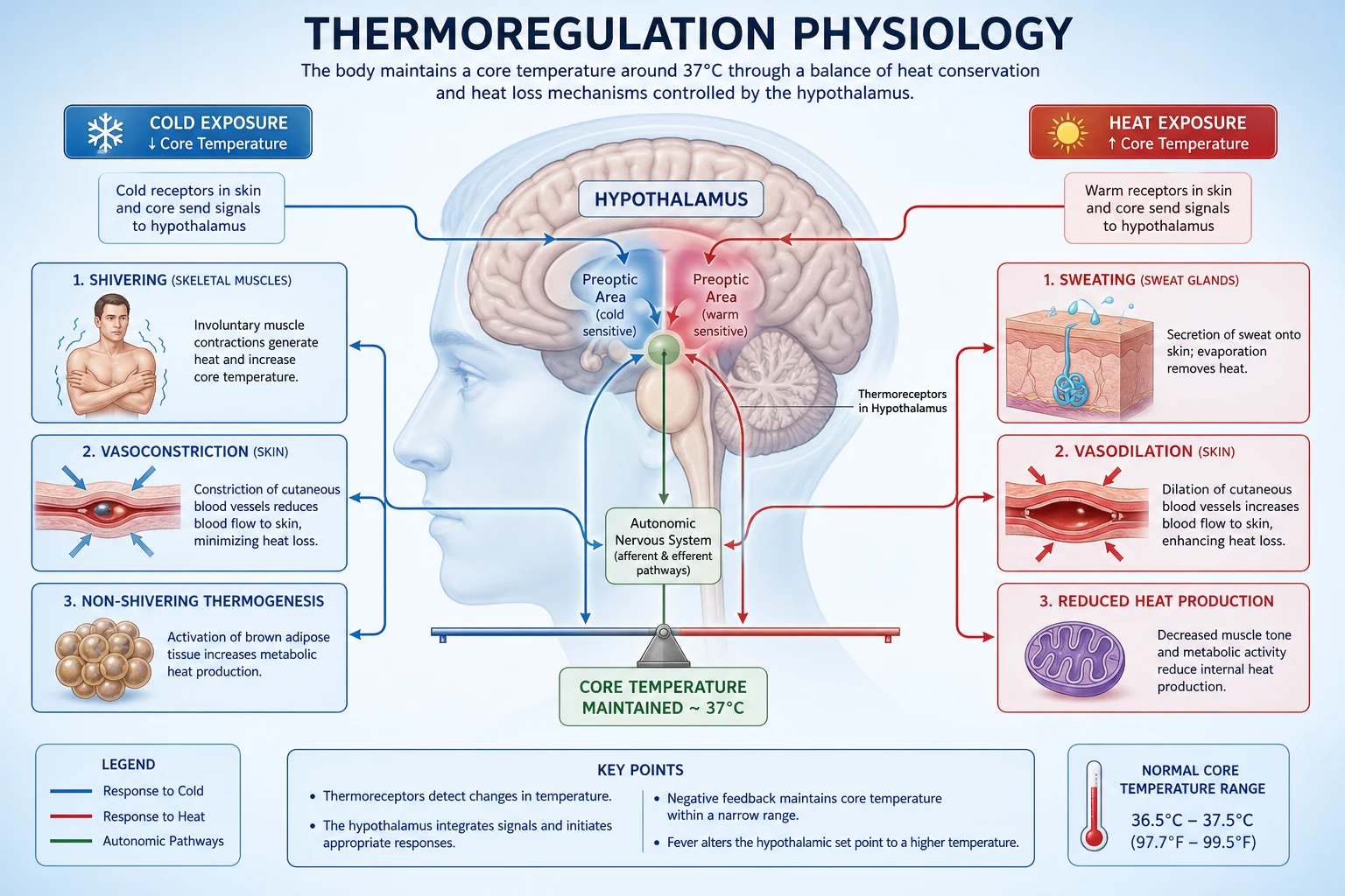
Normal body temperature and the thermoneutral zone
Core temperature is tightly regulated at about 37 degrees C when measured orally (range 36.5 to 37.5 degrees C). Rectal and core (pulmonary artery, oesophageal, nasopharyngeal) readings run about 0.3 to 0.5 degrees C higher; axillary and skin readings run lower and are peripheral rather than core measurements. The "normal" of 37 degrees C is itself a moving target, because temperature shows a characteristic diurnal (circadian) variation of about 0.5 to 1 degree C: the nadir occurs in the early morning (around 04:00 to 06:00) and the peak in the early evening (around 18:00 to 20:00). Menstruating women add a further monthly variation of about 0.3 to 0.5 degrees C, with a rise after ovulation driven by progesterone. [1]
Fever, hyperthermia and the set point — a distinction that changes management
A high body temperature is not a single entity, and the anaesthetist must distinguish three patterns because each demands different management. [1]
Fever is a regulated upward shift of the hypothalamic set point by endogenous pyrogens (interleukin-1, interleukin-6, tumour necrosis factor, and the prostaglandin E2 they induce in the preoptic area). The hypothalamus now "believes" the normal temperature is, say, 39 degrees C, and it defends that new set point just as vigorously as it defends 37 — the patient vasoconstricts, shivers, and feels cold (a rigor) until the new set point is reached. Antipyretics work in fever because they block prostaglandin E2 and lower the set point back toward normal. Fever is therefore an upward-reset thermostat, and the thermoregulatory machinery is intact and working hard. [1]
Hyperthermia (including heat illness and malignant hyperthermia) is an UNregulated rise in body temperature in which the set point is normal but heat production or environmental heat overwhelms the body's capacity to lose heat. The thermoregulatory responses are maximal (the patient is vasodilated and sweating) but cannot keep up. Antipyretics do NOT help, because the set point is already normal; the treatment is to remove the heat source or the heat-generating process and to cool actively. [1]
Hypothermia, the perioperative concern of this topic, is a fall of core temperature below the set point that the (anaesthesia-impaired) system cannot prevent. The practical point for the anaesthetist: a febrile patient's set point is reset upward and mild perioperative cooling may simply drag them back toward 37 (often acceptable); a hyperthermic patient needs active cooling and treatment of the cause; and the routine intraoperative fall toward 34 to 35 degrees C is unwanted hypothermia that must be prevented by warming [1][2].
Heat production
Heat is an unavoidable by-product of metabolism: every chemical reaction and every contraction of muscle liberates energy, and all but the small fraction captured as useful work appears eventually as heat. There are four sources the examiner expects you to name and rank. [1]
Basal metabolic rate (BMR) is the heat produced at complete rest, in a post-absorptive state, in the thermoneutral zone. At rest the body produces about 70 to 100 watts (about 80 kilojoules per square metre per hour, or roughly 1700 kilocalories per day). The organs contributing most at rest are the liver and the viscera (about 20 per cent each), the brain (about 20 per cent), the heart (about 10 per cent), and the kidneys (about 7 per cent) — the metabolically active core organs that never stop working. Skeletal muscle at rest contributes surprisingly little (about 20 per cent), but this changes dramatically with activity. BMR is set largely by thyroid hormone (T3 up-regulates the basal rate of oxidative metabolism and the sodium-potassium ATPase) and is modulated by catecholamines; it falls with hypothyroidism, starvation and ageing. [1]
Voluntary muscle activity is the most controllable source of additional heat. Walking, lifting, even the muscle tone of wakefulness, all raise heat production several-fold above BMR. This is the behavioural response to cold — the awake person moves, curls up, and puts on clothes — and it is the first defence that general anaesthesia removes. [1]
Shivering is the principal involuntary thermogenic response to cold in the adult. It consists of asynchronous, involuntary, oscillating contractions of skeletal muscle, coordinated by the posterior hypothalamus through shivering centres in the brainstem (dorsomedial posterior hypothalamus). Because the contractions are asynchronous and reciprocal (agonist against antagonist), they produce little net movement but generate large amounts of heat. Shivering raises metabolic heat production four to five times resting BMR and can transiently reach very high rates, but it is metabolically expensive (it consumes oxygen and substrate and produces carbon dioxide) and it is fatiguing, which is why it cannot be sustained indefinitely and why it is so poorly tolerated in the elderly and in cardiac disease [2].
Non-shivering thermogenesis (NST) is heat production without muscle contraction, and it is mediated almost entirely by brown adipose tissue. Brown fat is rich in mitochondria (which give it its colour), in sympathetic nerve endings, and in a unique mitochondrial protein called uncoupling protein 1 (UCP1, thermogenin). When the sympathetic nervous system (driven by the hypothalamus) releases norepinephrine onto brown adipocytes, lipolysis releases free fatty acids that are oxidised; UCP1 then acts as a proton channel in the inner mitochondrial membrane, allowing protons to re-enter the mitochondrial matrix and dissipate the proton-motive force as HEAT rather than driving ATP synthesis. The oxidation is therefore "uncoupled" from phosphorylation, and the energy is released as heat directly [5].
Endocrine contributions. The thyroid sets the basal metabolic rate (the long-term thermostat of heat production); catecholamines (epinephrine and norepinephrine) provide rapid, short-term calorigenic stimulation; and in prolonged cold, the adrenal cortex and anterior pituitary increase cortisol and growth hormone. These hormonal mechanisms are slow (minutes to hours for catecholamines, days to weeks for thyroid up-regulation) compared with the neural mechanisms, but thyroid status is the single biggest determinant of a person's resting heat output. [1]
Heat loss
Heat is lost from the body by four physical mechanisms. Their relative contributions at rest in a temperate environment are the classic examination percentages, and the candidate must be able to state them and explain how anaesthesia and surgery alter each. [1]
Radiation (about 40 to 60 per cent). This is the transfer of heat as infrared electromagnetic radiation between the body (warm) and the cooler surrounding surfaces (walls, ceiling, equipment). It is the dominant mode of heat loss at rest in a normally dressed person in a temperate room. The magnitude depends on the temperature difference between the skin and the surrounding surfaces and on the body surface area exposed. Two consequences follow. First, the patient loses heat whenever any exposed skin is warmer than the surrounding surfaces — which is almost always true in a cool operating theatre. Second, warming the ambient surfaces (raising theatre temperature, using radiant warmers) directly reduces radiative loss, whereas warming the air does so only indirectly (by warming the surfaces). [1]
Convection (about 25 to 30 per cent). Air next to the skin is warmed, becomes less dense, rises, and is replaced by cooler air; in a draught, cooler air is continuously brought into contact with the skin (the "wind-chill" effect). Convection loss depends on air movement and the skin-to-air temperature gradient. It is the rationale for the warm-air blanket (forced-air warming), which replaces the cool moving air at the skin with a continuously renewed layer of warm air, and for covering the patient with drapes (which trap a still layer of air). Intraoperatively, convection is amplified by laminar airflow theatres and by the high fresh-gas flows of some breathing systems. [1]
Evaporation (about 10 to 15 per cent at rest, much more intraoperatively). The conversion of water from liquid to vapour consumes the latent heat of vaporisation (about 2.4 kilojoules per millilitre), and that heat is taken from the body surface. Two sites contribute: the skin (insensible water loss plus sweat — the latter a powerful active heat-loss mechanism, each litre of sweat removing about 2.4 megajoules of heat) and the respiratory tract (warming and humidifying inspired gas). In the theatre, evaporation rises dramatically with an open body cavity (bowel exposed, peritoneal surfaces wet), with cold dry inspired gas (the rationale for a heat-and-moisture exchanger or a circle system with soda lime), and with surgical skin preparation (alcohol-based prep evaporating from a large skin area). Evaporative loss is the reason large open-cavity and burns surgery are particularly challenging thermally. [1]
Conduction (about 3 to 5 per cent at rest). This is direct transfer of heat from the body to a cooler solid surface in contact with it — the operating table, a cold mattress, wet drapes, a cold intravenous fluid bag placed on the patient. Though small as a resting percentage, it becomes large when the patient lies on an uninsulated table (a large contact area) or when cold fluids are infused directly into the circulation (conduction plus the heat needed to warm the fluid to body temperature). Conduction is the rationale for a warming mattress, an insulating pad between patient and table, and a fluid warmer for large-volume infusion. [1]
The four mechanisms — R C E C
The control system — the hypothalamus as thermostat
Thermoregulation is a classical negative-feedback control system, and the candidate should describe it in the language of any control loop: a controlled variable (core temperature), a reference set point (about 37 degrees C), sensors (thermoreceptors), a controller (the hypothalamus), effectors (the thermogenic and heat-loss responses), and a feedback path. [1]
The hypothalamus is the integrator. The preoptic/anterior hypothalamus contains the central thermosensitive neurons and is the region that mediates heat LOSS — it coordinates sweating and cutaneous vasodilation when core temperature rises. The posterior hypothalamus integrates afferent input and mediates heat PRODUCTION and conservation — it drives vasoconstriction, shivering, and non-shivering thermogenesis when core temperature falls. The older mnemonic — "anterior is for heat loss, posterior is for production" — captures this division and is worth remembering [2].
Afferents (the sensors). There are two populations of thermoreceptors, both firing continuously and changing their firing rate with temperature. Central thermoreceptors lie in the preoptic area of the anterior hypothalamus, the brainstem, and the spinal cord; they sense core (blood) temperature and are the dominant input to the control system. Peripheral thermoreceptors lie in the skin (and to a lesser degree in the mucous membranes and viscera) and convey both the skin temperature and the rate of change of skin temperature. Cold receptors outnumber warm receptors in the skin by roughly 10 to 1, which is why the system is more sensitive to a fall in temperature than to a rise. The afferent signal to the hypothalamus is a weighted average of core and skin temperatures, with core given roughly ten times the weight of skin. [1]
The threshold concept. Thermoregulatory responses are not graded continuously from the set point but are triggered at defined threshold temperatures. There is a narrow inter-threshold range (about 0.2 degrees C in the awake person) within which NO active response occurs — the body is in the thermoneutral zone and relies on basal vasomotor tone. Above the sweating threshold the body sweats and vasodilates; below the vasoconstriction threshold it vasoconstricts, and below the shivering threshold it shivers. This gain-and-threshold model is central to understanding anaesthesia, because anaesthetics work not by abolishing the responses but by shifting their thresholds [1][3].
Gain and maximum intensity. Two further properties define each response once its threshold is crossed. The gain is the slope of the response — how aggressively the effector ramps up per degree of deviation. The maximum intensity is the ceiling of the response. In the awake adult, vasoconstriction begins first (highest threshold, about 36.5 degrees C), then non-shivering thermogenesis, then shivering (lowest threshold, about 35.5 degrees C). Sweating and vasodilation share the upper thresholds near 37.5 degrees C. The system is therefore arranged in tiers, each with its own threshold, gain, and ceiling. [1]
Efferents (the effectors). In response to heat, the body: (1) sweats (eccrine sweat glands, cholinergic sympathetic innervation, up to 1 to 2 litres per hour in the heat); (2) cutaneously vasodilates, opening arteriovenous anastomoses in the skin (especially fingers, toes, ears, nose) to bring warm blood to the surface; and (3) behaviourally seeks cool. In response to cold, the body: (1) cutaneously vasoconstricts, diverting warm blood to the core; (2) shivers if vasoconstriction is insufficient; (3) mounts non-shivering thermogenesis (brown fat, especially in neonates); and (4) increases the endocrine calorigenic drive (catecholamines, thyroid). Behavioural responses (moving, clothing, shelter) are the most powerful of all in the awake person — and the first to be lost under anaesthesia. [1]
How anaesthesia defeats the control system — threshold shifts
The single most important physiological fact for the anaesthetist is that general and regional anaesthesia do not abolish thermoregulation outright; they increase the separation between the thresholds. The inter-threshold range, normally about 0.2 degrees C, widens to about 2 to 4 degrees C under general anaesthesia. The vasoconstriction and shivering thresholds are each lowered by roughly 2 to 3 degrees C, so the patient mounts no cold-defence response until the core temperature has fallen markedly. Over this widened permissive range the patient is effectively poikilothermic — core temperature drifts passively towards the ambient temperature, just as a cold-blooded animal's would [1][3].
A worked example. Imagine an awake patient at 37 degrees C. The sweating and vasodilation thresholds sit at about 37.5 degrees C, the vasoconstriction threshold at about 36.7 degrees C, and the shivering threshold at about 35.5 degrees C — a narrow inter-threshold range of about 0.2 degrees C around 37, with each response ready to engage within a fraction of a degree. Now induce anaesthesia: the thresholds all slide downward, the vasoconstriction threshold to about 34.5 degrees C and the shivering threshold to about 34 degrees C, while the gain and maximum intensity of each response are also reduced. Between 34.5 and about 37.5 degrees C the patient mounts no thermoregulatory defence at all — core simply follows the environment. This is why an unwarmed anaesthetised patient reliably drifts down to the mid-35s and then, if the anaesthetic is long enough, finally hits the lowered vasoconstriction threshold and plateaus. [1]
Regional (neuraxial) anaesthesia and thermoregulation
Spinal and epidural anaesthesia produce hypothermia by two mechanisms, and the fall can be just as severe as under general anaesthesia while being far less noticed — because the patient is awake and "comfortable." First, the sympathetic and somatic block abolishes vasoconstriction and shivering in the blocked dermatomes (often the legs, a large thermal compartment), so the lower body behaves like an open radiator, losing heat without defence. Second, the vasodilated, warm legs send a falsely reassuring afferent signal: the hypothalamus perceives the legs as warm and infers whole-body warmth, so it does not engage the (intact) upper-body defences until the core has fallen substantially [1].
Two practical consequences follow. Epidural and spinal anaesthesia can drop the core temperature by 1 to 2 degrees C over the first hour in an unwarmed patient — comparable to general anaesthesia — so active warming is just as important. And the vasoconstriction that would normally terminate phase 3 occurs later and less effectively, because the largest thermal compartment (the legs) cannot vasoconstrict. Obstetric regional anaesthesia is the classic setting: the parturient under spinal for caesarean section cools, the neonate may be born hypothermic, and shivering (which is common and distressing in awake obstetric patients) is treated with warming and, if needed, a small dose of pethidine or an alpha-2 agonist. [1]
The three phases of perioperative heat loss
The classic description of the time-course of intraoperative core temperature, worked out largely by Sessler and colleagues, divides the fall into three phases. This is among the most frequently examined pieces of thermoregulation and the candidate should be able to draw the temperature-versus-time curve [1][4].

Phase 1 — redistribution (the first hour, the steepest fall). The first and largest component of the intraoperative temperature drop is NOT a loss of heat from the body at all. Under anaesthesia the tonic vasoconstriction that normally keeps warm blood in the core is abolished; the arteriovenous anastomoses in the periphery open, and the warm core equilibrates with the cool periphery. The total body heat content is essentially unchanged, but heat has moved from the core (where it is measured) to the periphery (where it is not), so the measured core temperature falls by about 1 to 1.5 degrees C in the first hour. This is why the drop is steepest immediately after induction and why it is so hard to treat: you cannot replace heat that has not actually been lost. It can only be prevented. [1]
Phase 2 — linear decline (hours 1 to 3). After redistribution has largely completed, core temperature falls more slowly and linearly as actual heat loss to the environment exceeds metabolic heat production. The rate is about 0.5 to 1 degree C per hour and depends on the theatre temperature, the size of any open cavity, the dryness and temperature of inspired gas, the temperature of infused fluids, and whether the patient is actively warmed. This is the phase at which active warming (forced-air, fluid warming) is most effective, because the heat being lost can be replaced. [1]
Phase 3 — plateau (after 3 to 4 hours). In a long anaesthetic, core temperature eventually stabilises when vasoconstriction returns (the threshold for vasoconstriction, although lowered by anaesthesia, is eventually reached). Once vasoconstriction returns, peripheral blood flow falls, heat is again retained in the core, and heat loss once again matches heat production. The plateau therefore signals that the body's (anaesthesia-impaired) thermoregulatory defence has finally engaged. The plateau occurs at a lower temperature in the unwarmed patient and may never occur at all if the patient loses heat faster than the impaired defence can compensate (as in an open abdomen with cold fluids and a cold theatre). [1]
The consequences of perioperative hypothermia
A fall in core temperature of even 1 to 2 degrees C has measurable and clinically important consequences. The classic teaching point is that mild hypothermia is not a benign curiosity but a cause of excess morbidity, established by a remarkable series of randomised trials in the 1990s. [1]

Cardiac events. This is the most feared consequence and the leading cause of excess perioperative mortality attributable to hypothermia. Hypothermia increases sympathetic tone and noradrenaline levels, causes vasoconstriction and hypertension (raising myocardial oxygen demand), and — below about 34.5 degrees C — provokes arrhythmias (the classic sequence is first premature ventricular contractions, then atrial fibrillation, then ventricular fibrillation as temperature falls further). The landmark randomised trial by Frank and colleagues (JAMA 1997) in 300 high-risk patients (documented coronary disease or risk factors) undergoing non-cardiac surgery found that maintaining normothermia (mean 36.7 degrees C) versus allowing hypothermia (mean 35.4 degrees C) reduced morbid cardiac events (unstable angina, ischaemia, cardiac arrest, myocardial infarction) from 6.3 to 1.4 per cent (relative risk 2.2 for hypothermia, a 55 per cent reduction), and reduced ventricular tachycardia from 7.9 to 2.4 per cent [6]. The practical message: in the patient with cardiac risk factors, perioperative normothermia is a cardioprotective intervention as important as beta-blockade.
Coagulopathy and increased blood loss. Hypothermia impairs both platelet function and the coagulation enzyme cascade (the clotting factors are temperature-sensitive enzymes whose kinetics slow as temperature falls — a 1 to 2 degree fall roughly halves the activity of the clotting factors). It also causes a sequestration of platelets in the liver and spleen. Importantly, this is a functional coagulopathy that is NOT reliably reflected in the standard coagulation tests (which are run at 37 degrees C and so "correct" the hypothermia in the laboratory). Schmied and colleagues (Lancet 1996) showed in patients undergoing total hip arthroplasty that mild hypothermia (a core temperature of about 35 degrees C versus 36.6 degrees C) increased estimated blood loss by about 500 millilitres and significantly increased allogeneic transfusion requirements [8]. The practical message: in major surgery with large expected blood loss, hypothermia is a contributor to bleeding and transfusion that the laboratory screen will not warn you about.
Surgical site infection. Hypothermia causes thermoregulatory vasoconstriction, which reduces subcutaneous oxygen tension; tissue oxygen is the substrate for the oxidative (respiratory) burst by which neutrophils kill bacteria, and it is also needed for collagen deposition (hydroxylation of proline and lysine) and wound healing. Hypothermia directly impairs immune cell function. Kurz, Sessler and Lenhardt (NEJM 1996) randomised 200 patients having colorectal surgery to normothermia (36.6 degrees C) or hypothermia (34.7 degrees C) and found surgical wound infection fell from 19 per cent to 6 per cent, and hospital stay was shortened by about 2.6 days [7]. The practical message: in clean-contaminated and contaminated surgery, normothermia is an infection-control measure as effective as antibiotic prophylaxis and sterile technique.
Prolonged drug action. Hypothermia slows the hepatic metabolism and renal excretion of many drugs and reduces the clearance of volatile agents (though it also reduces their minimum alveolar concentration, by about 5 per cent per degree). The cleanest demonstration is the effect on neuromuscular blockers: Heier and colleagues (Anesthesiology 1991) showed that a 2 degree C fall in core temperature roughly doubled the duration of action of vecuronium and prolonged its spontaneous recovery [9]. Propofol, midazolam and the opioid infusions are similarly prolonged. The practical message: a hypothermic patient will wake and breathe later, and the neuromuscular blockade will outlast expectation — a recipe for residual paralysis and airway compromise in recovery.
Shivering. Shivering is the most visible consequence and, paradoxically, one of the most dangerous. The involuntary asynchronous muscle contractions raise whole-body oxygen consumption by 200 to 500 per cent and carbon dioxide production in parallel, placing a large load on the cardiorespiratory system. In the patient with cardiac or respiratory disease this can precipitate myocardial ischaemia, hypoxaemia and respiratory failure. Shivering is also distressing, it interferes with monitoring (movement artefact on ECG and pulse oximetry), and it raises intraocular and intracranial pressure. Treatment is active warming plus, if needed, a shivering-suppressant: pethidine (meperidine) is the most effective (it acts at N-methyl-D-aspartate and alpha-2 receptors and lowers the shivering threshold more than equianalgesic opioids), clonidine and dexmedetomidine (alpha-2 agonists) are useful, and tramadol has a role [3].
Patient discomfort and prolonged recovery. Subjectively, patients rate feeling cold as more distressing than pain in the early recovery period. A cold patient stays in the recovery room longer, has a longer hospital stay, and (via the mechanisms above) has a higher complication rate. Warming is a patient-experience and a throughput issue, not only a safety issue. [1]
Malignant hyperthermia and heat illness
Malignant hyperthermia (MH) is the dramatic, opposite end of the thermoregulation spectrum — not failure of the thermostat but uncontrolled heat production by muscle. It is an inherited calcium-release channelopathy of the skeletal-muscle sarcoplasmic reticulum, most commonly a mutation of the ryanodine receptor type 1 (RYR1) gene (less commonly the dihydropyridine receptor). Inheritance is autosomal dominant with variable penetrance. In a susceptible individual, the triggering agents — suxamethonium and all the volatile anaesthetic agents (halothane historically; sevoflurane, isoflurane, desflurane) — cause the ryanodine receptor to open pathologically, releasing uncontrolled amounts of calcium into the myoplasm. The sustained muscle contraction and the frantic activity of the calcium pumps (which hydrolyse ATP to pump the calcium back) generate enormous heat [3].
The clinical picture is driven by hypermetabolism: the earliest and most reliable sign is an unexplained, rapidly rising end-tidal carbon dioxide despite adequate or increased ventilation (because muscle is producing carbon dioxide faster than it can be removed). Masseter muscle rigidity (jaw clenching after suxamethonium), generalised muscle rigidity, tachycardia, a rising temperature (which is often a LATE sign and must never be waited for), hyperkalaemia (from muscle breakdown), acidosis (metabolic and respiratory), myoglobinuria and a rise in creatine kinase follow. [1]
Management is a true emergency and proceeds in parallel: (1) call for help and get the dantrolene; (2) stop all triggers immediately — discontinue the volatile agent, flush the circuit, switch to total intravenous anaesthesia with propofol, and use air and oxygen (a "clean" machine is ideal, though modern washout is faster than in the halothane era); (3) give intravenous dantrolene at 2.5 milligrams per kilogram, repeated every few minutes to a maximum of about 10 milligrams per kilogram until the metabolic signs subside — dantrolene is the specific ryanodine-receptor antagonist that blocks further calcium release; (4) cool the patient actively (cold intravenous fluids, surface cooling, gastric and bladder lavage) but stop cooling at about 38 degrees C to avoid overshoot; (5) treat the hyperkalaemia (insulin and dextrose, calcium chloride for ECG changes) and the acidosis (hyperventilate, consider bicarbonate); (6) monitor for and treat arrhythmias, maintain urine output with fluids and mannitol (which is in the old dantrolene formulation) to protect against myoglobinuric renal failure; and (7) arrange intensive care, because the syndrome can recur (recrudescence) over 24 to 48 hours and there is a risk of disseminated intravascular coagulation. The patient and first-degree relatives must be referred for RYR1 testing and a caffeine-halothane contracture test on muscle biopsy [3].
[1]Heat illness (environmental hyperthermia) is relevant to anaesthesia in the context of the febrile or heat-stressed patient and postoperative pyrexia, and is distinguished from MH by its setting and its lack of relation to triggering agents. Classic heat stroke occurs in the elderly and the very young in a hot environment (impaired dissipation); exertional heat stroke occurs in the fit and young exercising in heat (overwhelming heat production). The core temperature is high (above 40 degrees C), the skin is hot but may be dry (if sweating is exhausted) or sweating, and the defining feature is central nervous system dysfunction (confusion, seizures, coma). Management is rapid cooling (evaporative, ice-water immersion, cold intravenous fluids) and supportive care. Postoperative fever, by contrast, is most often inflammatory or infective in origin and the temperature seldom reaches the dangerous range in the first 24 hours; the anaesthetist must distinguish benign early pyrexia (a common inflammatory response) from a developing sepsis or, rarely, MH. [1]
Special populations — paediatric and elderly differences
Neonates and infants are at the highest risk of perioperative hypothermia and deserve particular attention. The reasons are physiological and structural: a large surface-area-to-mass ratio (roughly three times that of an adult) means heat loss is proportionally greater; a thin subcutaneous fat layer and immature skin provide poor insulation; the glycogen and brown-fat stores are limited, especially in the premature or growth-restricted infant; and the ability to shiver is poorly developed, so the neonate depends almost entirely on brown-fat non-shivering thermogenesis [5]. The consequences of cold stress in a neonate are rapid and severe: increased oxygen consumption, apnoea, hypoglycaemia (exhausted glycogen), metabolic acidosis, pulmonary vasoconstriction (which can reopen the ductus arteriosus and cause right-to-left shunting and hypoxaemia), and bradycardia. Prevention is intensive and begins before induction: a warm theatre (at least 24 to 26 degrees C for neonates), a forced-air warmer or radiant warmer, warmed skin preparation, a hat (the head is a large fraction of the surface area), wrapped limbs, and warmed inspired gases and fluids.
The elderly are also high-risk, for different reasons. Thermoregulatory responses are blunted: the vasoconstriction threshold falls, the maximum vasoconstrictor response is reduced, and shivering is less vigorous and less well sustained (less muscle mass, weaker central drive). The perception of cold is impaired, so the behavioural defence is lost. The metabolic reserve is lower (lower BMR, often less subcutaneous fat), and comorbidity (cardiac, respiratory, vascular) means the consequences of hypothermia — especially the cardiac and shivering effects — are less well tolerated. The practical message: monitor temperature in every elderly patient, warm actively, and do not assume that a quiet, not-shivering elderly patient is normothermic — they may simply lack the response [3].
Warming measures — prevention is the standard of care
The goal is to keep core temperature at or above 36 degrees C (most guidelines define normothermia as 36.0 degrees C or above and hypothermia as below 36.0, with 35.0 to 35.9 mild, 34.0 to 34.9 moderate, and below 34 severe). Prevention is far more effective than treatment, because once heat is lost (phase 2) or redistributed (phase 1) it is hard to recover. The measures, in approximate order of effectiveness and importance [1][3][10][11]:

Prewarming (10 to 30 minutes before induction). Because phase 1 redistribution is driven by the core-to-peripheral temperature gradient, the most logical and effective prevention is to abolish that gradient before anaesthesia removes vasoconstrictor tone. Warming the periphery for 10 to 30 minutes before induction raises peripheral tissue temperature towards core temperature; when anaesthesia then opens the peripheral vessels, little or no heat redistributes, and the phase 1 fall is markedly reduced or abolished. Prewarming is the single most effective way to prevent the steepest, hardest-to-treat part of the temperature fall [10].
Forced-air warming (the Bair Hugger and similar). This is the most effective single intraoperative warming method for an anaesthetised patient. A powered blower pushes temperature-controlled warm air through a porous disposable blanket draped over the patient; the warm air emerges over the skin surface, replacing the cool still air and dramatically reducing convective loss while adding heat. It is effective from the moment of induction, is practical for most surgery, and is the mainstay of perioperative warming. The one caution is in major orthopaedic implant surgery, where theoretical concern has been raised about forced-air warming disrupting the laminar airflow and increasing airborne contamination near the implant (this is debated; resistive warming and fluid warming are alternatives) [3].
Fluid warming. One unit of red cells or one litre of crystalloid at room temperature (about 20 degrees C) removes about the heat equivalent of a 0.25 degree C fall in core temperature in an adult; infusing cold fluid fast therefore cools the patient directly. Whenever large volumes are infused (major haemorrhage, large crystalloid resuscitation, massive transfusion), a fluid warmer is essential. It is not a substitute for surface warming but a necessary complement when the blood-volume route of heat loss is significant. [1]
Insulation and a head covering. Simple passive insulation reduces heat loss cheaply and effectively: a plastic wrap or a blanket over exposed areas, an insulating pad between the patient and the cold operating table (reducing conductive loss), and — especially in children — a hat or head wrap, because the head is a large fraction of body surface area in the small child and a significant route of loss even in adults. Plastic limb wrapping (a "space blanket" or plastic bag) is particularly useful for the legs under regional anaesthesia. [1]
Ambient theatre temperature. Raising the theatre temperature reduces all modes of loss (radiation, convection, conduction) and is the single most important passive measure, especially at induction (before draping) and in children. The cost is staff comfort and infection-control concerns at very high temperatures; a practical target is at least 21 degrees C for adults and 24 to 26 degrees C for neonates. The conflict between a warm theatre (good for the patient) and a cool theatre (preferred by the gowned surgical team in heavy gowns and lights) is perennial and is resolved by warming the patient actively and accepting a moderately cool theatre once draping is complete. [1]
The warming ladder — P F F I A
Rewarming in recovery — rate, after-drop and rewarming shock
Once a patient is hypothermic, rewarming must be done with an understanding of its kinetics and hazards. A forced-air warmer raises core temperature by about 0.5 to 1 degree C per hour in a typical adult; faster than this is hard to achieve because the patient's own metabolic heat production is depressed by the residual anaesthetic and by the hypothermia itself. Two specific hazards deserve mention. [1]
The after-drop. As the periphery is actively warmed and perfusion improves, cold peripheral blood returns to the core and can transiently LOWER the core temperature by a further 0.2 to 0.5 degrees C before it climbs. This is the "after-drop," and it is the reason to keep warming (and monitoring) through the early recovery period rather than declaring victory when the blanket goes on. [1]
Rewarming shock. If warming is too aggressive, the sudden peripheral vasodilation can cause a sharp fall in venous return and blood pressure ("rewarming shock"), unmasking hypovolaemia that was masked by cold-induced vasoconstriction. The defence is to warm at a moderate rate, to ensure the patient is adequately volume-resuscitated, and to anticipate the vasodilation — sometimes a brief period of reduced warming or vasopressor support is needed as the patient crosses from vasoconstricted to vasodilated. [1]
Temperature measurement sites
Temperature must be measured in every anaesthetic of more than about 30 minutes, and the choice of site is itself an examination topic. Sites are classified as core (best representing the temperature of the vital organs), intermediate (close to core with a small lag), and peripheral (skin, which is not core). [1]
The pulmonary artery catheter thermistor gives the truest core (mixed) temperature but is invasive and reserved for the sickest patients. The oesophageal probe (placed in the lower third, behind the heart) and the nasopharyngeal probe (near the sphenoid, close to the hypothalamus and the great vessels) are the practical intraoperative core measurements and track core temperature closely during rapid change, including the phase 1 redistribution [3][11]. The tympanic membrane temperature is an attractive surrogate for core (the tympanum shares its blood supply with the hypothalamus via the internal carotid), but the infrared probe is operator-dependent, reads falsely low with poor technique or earwax, and carries a small risk of tympanic perforation; a contact tympanic probe is more accurate but less used. The bladder temperature (via a specialised urinary catheter thermistor) follows core well but lags during low urine output, and is therefore less reliable in the shocked or dehydrated patient. The rectal temperature lags core (it sits in a slow-flow sump), is falsely high if the probe sits in stool, and is not recommended during rapid change. Skin and axillary temperatures are peripheral and may differ from core by 2 degrees C or more, so they are unsuitable for monitoring core during anaesthesia, though they are useful for following trends [12].
Continuous comparison studies confirm that the pulmonary-artery, oesophageal, nasopharyngeal and bladder measurements agree closely with true core (within about 0.2 degrees C), while tympanic infrared and skin measurements are less accurate, especially during rapid change — which is exactly when the perioperative patient needs an accurate number [12].
The practical rule: in a general anaesthetic of more than 30 minutes, monitor a core or near-core site (oesophageal or nasopharyngeal for head, neck and body surgery, bladder for abdominal surgery, tympanic as a surrogate for short cases); start warming early (ideally prewarm); aim to keep core temperature at or above 36.0 degrees C; and treat any drop as a signal to intensify warming [3].
A practical perioperative temperature-management protocol
The examination and the ward round both reward a simple, defensible protocol. Before induction: measure the patient's temperature; if below 36 degrees C, start warming; prewarm the periphery for 10 to 30 minutes where possible, especially for long or major cases; raise the ambient theatre temperature toward 21 degrees C (24 to 26 for neonates); prepare warmed fluids and a heat-and-moisture exchanger. At induction: apply a forced-air warming blanket over the largest available area; insulate the head and any exposed limbs. Intraoperatively: monitor a core site (oesophageal or nasopharyngeal, or bladder) continuously; fluid-warm any infusion; keep the cavity closed and the drapes on as much as possible; check the temperature every 15 to 30 minutes and intensify warming if it falls below 36 degrees C. In recovery: continue forced-air warming; treat shivering with warmth and pethidine or an alpha-2 agonist; reverse neuromuscular blockade fully (remembering that hypothermia prolongs it); monitor for the after-drop and rewarming shock; and only discharge from recovery once the core temperature is at or above 36 degrees C and stable. Document the temperature at each stage — inadvertent perioperative hypothermia is now a tracked quality indicator in many health systems [3][11].
Key facts to take to the examination
- Core temperature is about 37 degrees C (oral), diurnal variation about 0.5 to 1 degree C, thermoneutral zone about 28 degrees C naked.
- Fever raises the set point (antipyretics work); hyperthermia overwhelms a normal set point (cool actively, antipyretics do not work).
- Heat production: BMR (liver, brain, heart at rest), shivering (skeletal muscle, 4 to 5 times BMR), non-shivering thermogenesis (brown fat, UCP1, norepinephrine — the neonate's main mechanism), endocrine (thyroid, catecholamines).
- Heat loss at rest: radiation 40 to 60 per cent (largest), convection 25 to 30 per cent, evaporation 10 to 15 per cent, conduction 3 to 5 per cent.
- Control: hypothalamus — anterior/preoptic mediates heat loss, posterior mediates heat production; afferents central and peripheral; efferents sweating, vasodilation/vasoconstriction, shivering, NST, behavioural, endocrine; threshold and gain model.
- Anaesthesia widens the inter-threshold range from 0.2 to 2 to 4 degrees C — the patient is poikilothermic.
- Three phases: redistribution (first hour, 1 to 1.5 degrees C, not true loss), linear (0.5 to 1 degree C per hour), plateau (vasoconstriction returns).
- Consequences of hypothermia below 36 degrees C: cardiac (Frank, RR 2.2), wound infection (Kurz, 19 versus 6 per cent), blood loss (Schmied), prolonged drug action (Heier, vecuronium doubled), shivering (oxygen demand 200 to 500 per cent).
- Malignant hyperthermia: RYR1 channelopathy, suxamethonium and volatiles trigger, earliest sign is rising end-tidal carbon dioxide, treat with dantrolene 2.5 mg/kg and stop triggers.
- Warming: prewarming prevents phase 1, forced-air warming is the mainstay, fluid warming for large volumes, ambient at least 21 degrees C (24 to 26 for neonates); rewarm at 0.5 to 1 degree C per hour, beware after-drop and rewarming shock.
- Measure a core site (oesophageal, nasopharyngeal, bladder) in every anaesthetic over 30 minutes; target at or above 36.0 degrees C. [1]
This is applied physiology with a direct line to outcome: the patient who is warmed is the patient who bleeds less, gets fewer infections, has fewer cardiac events, wakes sooner, and is more comfortable. Few topics in the examination repay mastery so clearly. [1]
References
- [1]Sessler DI. Perioperative thermoregulation and heat balance Lancet, 2016.PMID 26775126
- [2]Sessler DI. The thermoregulation story Anesthesiology, 2013.PMID 23221865
- [3]Sessler DI. Temperature monitoring and perioperative thermoregulation Anesthesiology, 2008.PMID 18648241
- [4]Sessler DI. How three linked clinical observations led to an understanding of perioperative heat balance: A personal reflection on the scientific process J Clin Anesth, 2024.PMID 38733707
- [5]Cannon B, Nedergaard J. Brown adipose tissue: function and physiological significance Physiol Rev, 2004.PMID 14715917
- [6]Frank SM, Fleisher LA, Breslow MJ, et al. Perioperative maintenance of normothermia reduces the incidence of morbid cardiac events. A randomized clinical trial JAMA, 1997.PMID 9087467
- [7]Kurz A, Sessler DI, Lenhardt R. Perioperative normothermia to reduce the incidence of surgical-wound infection and shorten hospitalization. Study of Wound Infection and Temperature Group N Engl J Med, 1996.PMID 8606715
- [8]Schmied H, Kurz A, Sessler DI, et al. Mild hypothermia increases blood loss and transfusion requirements during total hip arthroplasty Lancet, 1996.PMID 8569362
- [9]Heier T, Caldwell JE, Sessler DI, et al. Mild intraoperative hypothermia increases duration of action and spontaneous recovery of vecuronium blockade during nitrous oxide-isoflurane anesthesia in humans Anesthesiology, 1991.PMID 1673591
- [10]Heuer L. [Pre-warming - how can perioperative hypothermia be avoided?] Anasthesiol Intensivmed Notfallmed Schmerzther, 2003.PMID 12975737
- [11]Insler SR, Sessler DI. Perioperative thermoregulation and temperature monitoring Anesthesiol Clin, 2006.PMID 17342966
- [12]Ehlers UE, Ulmer J, Keller M, et al. Comparison of continuous temperature measurement methods in the intensive care unit: standard bladder catheter measurements versus non-invasive transcutaneous sensors J Clin Monit Comput, 2025.PMID 39066870