Anaes · Applied cardiovascular & respiratory physiology
Ventilation, perfusion & dead space
Also known as Ventilation-perfusion ratio · V/Q mismatch · Shunt · Dead space · Hypoxaemia · Alveolar-arterial gradient
Gas exchange requires that ventilation and perfusion are matched, and most clinical hypoxaemia is a disorder of that matching. The framework rests on five exam-critical ideas: the ventilation-perfusion (V/Q) ratio is normally about 0.8 to 1.0, and both ventilation and perfusion increase from apex to base, but perfusion more, so the apex is high V/Q and the base low V/Q; ventilation-perfusion mismatch spans a spectrum from shunt (V/Q of zero, perfusion without ventilation) through normal to dead space (V/Q very high, ventilation without perfusion); true shunt is hypoxaemia that is NOT corrected by 100 percent oxygen, whereas low-V/Q mismatch IS corrected by oxygen — the test that distinguishes them; dead space is ventilated lung that does not exchange gas, raised by pulmonary embolism; and the five causes of hypoxaemia are low inspired oxygen, hypoventilation, diffusion impairment, shunt, and ventilation-perfusion mismatch — separated by the alveolar-arterial oxygen gradient. Built on the acute-hypoxaemic-respiratory-failure imaging review (Coppola 2026), the Eisenmenger-syndrome review (the classic pulmonary shunt, Baino 2026), the patent-foramen-ovale platypnea study (right-to-left shunt hypoxaemia, Ramidi 2026), the pulmonary-embolism perfusion-imaging study (Evans 2026), the portopulmonary-gas-exchange study (Lacoste-Palasset 2026), and the partial-pressure-of-oxygen review (Hoecker 2026).
On this page & tools
Your progress
Saved locally on this device.
8 MCQs with explanations
Target exams
Red flags
Why this matters to the anaesthetist
Hypoxaemia under anaesthesia is almost always a V/Q story: atelectasis (low V/Q and shunt), tube malposition, pulmonary embolism (high V/Q dead space), low cardiac output amplifying venous admixture, or HPV failure during one-lung ventilation. Primary candidates must define V/Q, draw the spectrum from shunt to dead space, write the shunt and Bohr equations, list the five causes of hypoxaemia, and explain why 100% oxygen distinguishes true shunt from low V/Q. [1]
The V/Q ratio
For the whole lung at rest: alveolar ventilation VA ≈ 4 L/min, cardiac output Qt ≈ 5 L/min, so overall V/Q ≈ 0.8. Individual units range from 0 (perfused but unventilated = shunt) to infinity (ventilated but unperfused = dead space). Ideal units sit near 1. Arterial blood is the flow-weighted mixture of end-capillary contents from all units; expired gas is the ventilation-weighted mixture of alveolar gases. [1]
Regional distribution — West zones and gravity
In the upright lung: [1]
- Blood flow increases from apex to base (gravity, recruitment/distension of vessels).
- Ventilation also increases from apex to base, but less steeply.
- Therefore apex: high V/Q (relatively over-ventilated), base: low V/Q (relatively over-perfused). [1]
West zones (exam classic): [1]
| Zone | Condition | Physiology |
|---|---|---|
| Zone 1 | PA > Pa > Pv | No flow — alveolar dead space (pathological if extensive) |
| Zone 2 | Pa > PA > Pv | Intermittent flow (waterfall) |
| Zone 3 | Pa > Pv > PA | Continuous flow — most of normal lung |
| Zone 4 | Base, low volumes | Extra-alveolar vessel compression → reduced flow |
Under anaesthesia in the supine patient the vertical gradient shrinks but still exists (dependent lung). Lateral position and OLV exaggerate inequalities. PEEP can convert zone 3 toward zone 1/2 if alveolar pressure rises above pulmonary arterial pressure — raising alveolar dead space. [1]
The V/Q spectrum and gas exchange
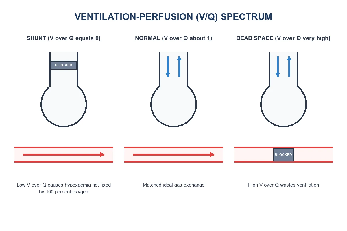
Low V/Q units: alveolar gas approaches mixed venous gas — low PO2, high PCO2. They contribute disproportionately to arterial hypoxaemia because the O2 dissociation curve is flat at high saturation: well-ventilated units cannot fully compensate by carrying extra O2 once Hb is nearly full. CO2 is different — the CO2 content curve is steeper/near-linear, so high-V/Q units can excrete extra CO2 and largely compensate for low-V/Q units' CO2 retention. Result: V/Q mismatch causes hypoxaemia more readily than hypercapnia; hypercapnia appears when total VA is inadequate or dead space is extreme. [1]
High V/Q units: waste ventilation (like dead space), raise the work of breathing for a given CO2 elimination, increase PaCO2–EtCO2 gradient. [1]
Shunt — true shunt and venous admixture
True shunt (Qs): blood bypasses ventilated alveoli entirely (V/Q = 0). Causes: anatomical (bronchial veins, Thebesian veins — normal 2–5%), intracardiac R→L, pulmonary AVMs, consolidated/collapsed lung. [1]
Venous admixture: the calculated shunt that would produce the observed CaO2 if all admixture were true shunt — includes true shunt plus contribution of low-V/Q units. [1]
Shunt equation: Qs/Qt = (Cc′O2 − CaO2) / (Cc′O2 − CvO2) [1]
Cc′O2 is ideal end-capillary content from PAO2 via alveolar gas equation. On FiO2 1.0, nitrogen is gone and the calculated shunt approximates true shunt more closely. [1]
Critical property: hypoxaemia from true shunt is not abolished by 100% oxygen (though PaO2 may rise somewhat as dissolved O2 in non-shunt blood increases). Hypoxaemia from low V/Q usually is largely correctable with oxygen because PAO2 in low-V/Q units rises. [1]
Dead space — anatomic, alveolar, physiologic
Anatomic dead space (VD anat): conducting airways ~2 mL/kg (~150 mL). Fowler method (N2 washout) measures it. [1]
Alveolar dead space (VD alv): ventilated alveoli with no (or inadequate) perfusion — PE, low CO, zone 1 from high PEEP, emphysema. [1]
Physiologic dead space (VD phys) = VD anat + VD alv [1]
Bohr equation: VD/VT = (PaCO2 − PECO2) / PaCO2 [1]
Enghoff modification uses PaCO2 as a surrogate for ideal alveolar PCO2. Normal VD/VT ≈ 0.2–0.35. Raised VD/VT → need higher VE to keep PaCO2 normal; EtCO2 underestimates PaCO2 (widened a–ET gradient). [1]
West & Wagner concepts: high V/Q units contribute to alveolar dead-space-like behaviour. [1]
The five causes of hypoxaemia (must list)
- Low inspired PO2 (altitude, hypoxic mixture) — A–a normal
- Hypoventilation — A–a normal; high PaCO2
- Diffusion impairment — A–a raised; uncommon sole cause at rest
- Shunt — A–a raised; poor response to O2
- V/Q mismatch — A–a raised; usually responds to O2 [1]
(Some lists add low mixed venous O2 as an amplifier rather than a primary cause.) [1]
Hypoxic pulmonary vasoconstriction as V/Q defender
HPV diverts blood from hypoxic alveoli, narrowing the scatter of V/Q ratios and protecting PaO2. Inhibitors (volatiles, vasodilators) widen scatter and worsen venous admixture — link to the oxygen-cascade/HPV topic. [1]
Shunt versus low V/Q — the oxygen test
| True shunt | Low V/Q mismatch | |
|---|---|---|
| 100% O2 | PaO2 remains relatively low | PaO2 usually rises substantially |
| A–a on high FiO2 | Large | Smaller once units oxygenated |
| Examples | Atelectasis complete, R→L cardiac | Bronchospasm, mild pneumonia, COPD |
Atelectasis under anaesthesia often behaves as shunt (compression, absorption atelectasis with high FiO2). [1]
Equations toolkit
- PAO2 = FiO2(Patm − 47) − PaCO2/R
- A–a = PAO2 − PaO2
- Qs/Qt = (Cc′O2 − CaO2)/(Cc′O2 − CvO2)
- VD/VT = (PaCO2 − PECO2)/PaCO2
- VA = VE × (1 − VD/VT)
- PaCO2 ∝ V̇CO2 / VA [1]
Anaesthetic relevance
- GA + supine + high FiO2: FRC falls, dependent atelectasis → shunt-like fractions; PEEP and recruitment help.
- PE: sudden rise in VD/VT, fall in EtCO2, rise in a–ET CO2 gradient, hypoxaemia, RV strain.
- Low cardiac output: lower PvO2 → any given shunt fraction produces lower PaO2; also more zone 1 if PA high.
- OLV: intentional extreme V/Q inequality; HPV and positional blood flow determine PaO2.
- COPD: high VD/VT and low V/Q regions coexist; oxygen careful titration. [1]
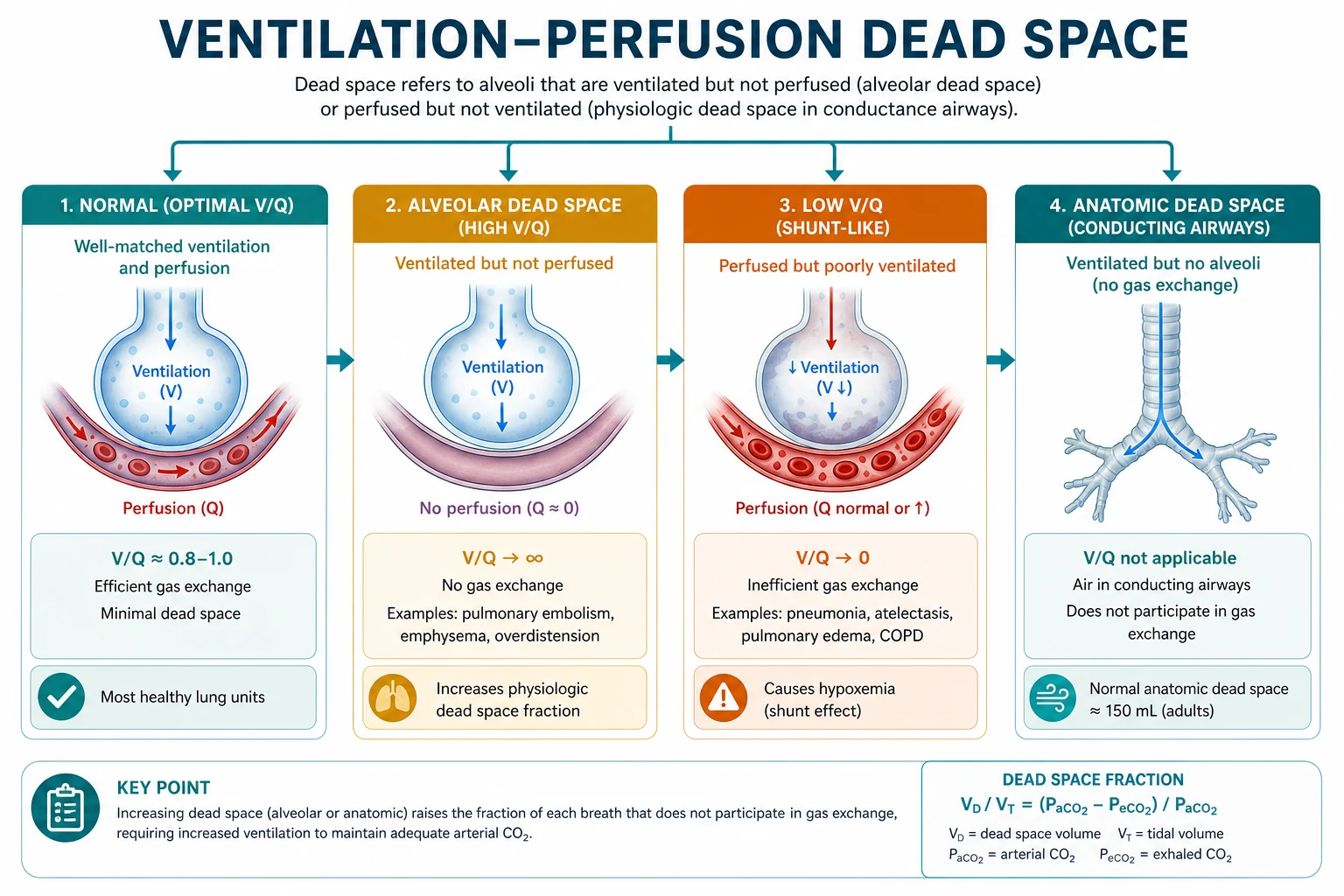
Shunt (low extreme)
- V/Q = 0
- Blood bypasses gas exchange
- Hypoxaemia poorly fixed by 100% O2
- Qs/Qt equation quantifies
Dead space (high extreme)
- V/Q → ∞
- Ventilation wasted
- Raises VD/VT and a–ET CO2 gap
- Bohr equation quantifies
Graph descriptions for the viva
- O2–CO2 diagram (Rahn–Fenn): plot PACO2 vs PAO2 for units from shunt point (mixed venous gas) to inspired gas point; ideal alveolar point on the R line.
- Multiple inert gas elimination technique (MIGET) concept: distribution of perfusion and ventilation against V/Q ratio — bimodal distributions in ARDS/COPD.
- Iso-shunt diagrams: PaO2 vs FiO2 for different shunt fractions — shows limited benefit of high FiO2 once Qs/Qt is large. [1]
Viva traps
- Overall V/Q is 0.8 not 1 — remember VA 4 vs Q 5.
- Anatomic shunt of 2–5% is normal — zero calculated shunt is not expected.
- Dead space fraction uses PaCO2 and mixed expired CO2 — not EtCO2 alone in the classic Bohr form (though EtCO2 approximates end-tidal alveolar gas for trends).
- Zone 1 is not normal in healthy spontaneous breathing at rest — if present extensively, dead space is pathological.
- Hypercapnia is not required for the diagnosis of V/Q mismatch. [1]
Additional depth — absorption atelectasis
When FiO2 is high, nitrogen's splinting effect in poorly ventilated units disappears; ongoing gas uptake collapses the unit → conversion of low V/Q to shunt. This is why 100% oxygen for long periods promotes atelectasis and why a balance exists between preoxygenation safety and absorption collapse — recruitment and modest PEEP mitigate. [1]
Additional depth — position and blood flow
Upright → base-predominant flow. Supine → caudal-dependent. Prone in ARDS → more uniform dorsal expansion and improved matching (clinical ARDS application of V/Q physiology). Lateral decubitus for thoracic surgery: dependent lung receives more blood but is compressed; HPV and surgical manipulation alter distribution minute to minute. [1]
SAQ: five causes of hypoxaemia with A–a gradient
- Low inspired oxygen — normal A–a
- Hypoventilation — normal A–a, high PaCO2
- Diffusion limitation — raised A–a
- Shunt — raised A–a, poor O2 response
- V/Q mismatch — raised A–a, usually good O2 response [1]
Dead space versus shunt clinical contrast table in prose
Shunt makes blood that never saw alveolar gas; dead space makes gas that never saw blood. Shunt hurts oxygenation more than CO2 elimination; dead space hurts CO2 elimination efficiency and widens a–ET CO2 gradients. Both can coexist in ARDS: flooded units shunt while overdistended units act as dead space, especially with high PEEP. [1]
MIGET conceptual paragraph
The multiple inert gas elimination technique infuses dissolved inert gases of different solubilities and measures arterial and expired retention/excretion to recover a distribution of ventilation and perfusion against V/Q. Normal lung shows a unimodal distribution near V/Q 1; disease produces low and high V/Q modes. You will not perform MIGET in theatre, but describing the concept scores marks for depth. [1]
West zone manipulation by the ventilator
High alveolar pressure from PEEP or dynamic hyperinflation can create West zone 1 conditions at non-dependent regions: ventilation without perfusion, raised physiologic dead space, falling EtCO2, rising PaCO2, and wasted work. Reducing overdistension can improve CO2 elimination without increasing set minute ventilation — applied V/Q thinking. [1]
Primary exam expansion
Mathematical identities you must write under pressure
VA = f × (VT − VD) PaCO2 = K × V̇CO2 / VA Qs/Qt = (Cc′O2 − CaO2)/(Cc′O2 − CvO2) VD/VT = (PaCO2 − PECO2)/PaCO2 A−a gradient = [FiO2(Patm − 47) − PaCO2/R] − PaO2 [1]
Why 100% oxygen cannot fully correct true shunt
Blood traversing true shunt has content ≈ CvO2. Non-shunt blood can carry at most ~1.34×Hb + 0.003×PAO2. Dissolved oxygen adds only ~2 mL/dL even at PAO2 ~660 mmHg — not enough to offset a large admixture of venous blood. Hence SpO2 remains depressed when Qs/Qt is large. [1]
Atelectasis types under anaesthesia
Compression atelectasis (dependent lung, reduced FRC), absorption atelectasis (high FiO2 in closed units), and surfactant impairment. All create low V/Q or shunt. Recruitment manoeuvres reopen units; PEEP keeps them open; both reshape the V/Q distribution toward normal. [1]
Pulmonary embolism as a dead-space prototype
Sudden rise in alveolar dead space: EtCO2 falls, PaCO2 may rise or stay same if VE increases, a–ET gradient widens, VD/VT rises, hypoxaemia from mechanisms above, RV afterload rises. Capnography is a continuous dead-space monitor in this setting. [1]
COPD V/Q pattern
Broadened distribution with both low and high V/Q regions; high VD/VT; chronic HPV and vascular remodelling; oxygen carefully titrated because removing hypoxic stimulus can increase blood flow to low V/Q regions (worse venous admixture) and reduce ventilation slightly in retainers. [1]
Prone positioning physiology
In ARDS, dorsal lung is often poorly ventilated but still perfused (shunt). Prone positioning improves dorsal expansion, homogenises transpulmonary pressures, and improves matching — an applied V/Q intervention proven to affect outcomes in severe ARDS when done properly. [1]
Exam graph: iso-shunt diagram
Describe PaO2 on the y-axis versus FiO2 on the x-axis with curves for Qs/Qt of 0%, 10%, 20%, 30%, 50%. As shunt fraction rises, the curve flattens: increasing FiO2 buys less and less PaO2. This picture explains refractory hypoxaemia. [1]
Positioning and one-lung anaesthesia V/Q
Lateral decubitus: dependent lung better perfused; non-dependent better ventilated if both lungs open — relative mismatch. When non-dependent lung is collapsed for surgery, its ventilation goes to zero while residual perfusion becomes shunt until HPV and surgical ligation reduce flow. [1]
Extended viva dialogue
Examiner: Define the ventilation–perfusion ratio and give the normal whole-lung value. [1]
Candidate: V/Q is alveolar ventilation divided by blood flow for a lung unit or the lung as a whole. Whole-lung alveolar ventilation is about 4 L/min and cardiac output about 5 L/min, so overall V/Q is about 0.8. Individual units range from zero (shunt) to infinity (dead space). [1]
Examiner: Why does V/Q mismatch cause hypoxaemia more than hypercapnia? [1]
Candidate: The oxygen dissociation curve is flat at high saturation, so high-V/Q units cannot carry much extra oxygen to compensate for desaturated blood from low-V/Q units. The carbon dioxide content curve is steeper and more linear, so high-V/Q units can eliminate extra CO2 and largely compensate. Therefore arterial hypoxaemia appears first; hypercapnia implies inadequate total alveolar ventilation or extreme dead space. [1]
Examiner: Write the shunt and Bohr equations and state when 100% oxygen distinguishes shunt from low V/Q. [1]
Candidate: Qs/Qt equals (Cc-prime-O2 minus CaO2) divided by (Cc-prime-O2 minus CvO2). VD/VT equals (PaCO2 minus mixed expired PCO2) divided by PaCO2. True shunt hypoxaemia is not abolished by pure oxygen because shunted blood never contacts alveolar gas; low-V/Q hypoxaemia usually improves substantially as alveolar PO2 rises in poorly ventilated units. [1]
Examiner: Describe West zones and how PEEP can create dead space. [1]
Candidate: Zone 1 has alveolar pressure greater than arterial and venous pressures so vessels collapse and act as alveolar dead space. Zone 2 has intermittent flow, zone 3 continuous flow. High PEEP raises alveolar pressure and can expand zone-1-like conditions, raising physiologic dead space, widening the arterial-to-end-tidal CO2 gradient, and lowering cardiac output via venous return effects. [1]
Clinical synthesis: Intraoperative desaturation algorithms are V/Q algorithms: tube position (mainstem creates massive low V/Q/shunt), recruitment, FiO2, cardiac output (mixed venous oxygen), and embolism (dead space). Speak in those terms and you sound like a consultant. [1]
Red flags
- True shunt hypoxaemia is not abolished by 100% oxygen; low-V/Q usually is improved — the distinguishing test.
- A–a gradient separates hypoventilation/low PIO2 (normal A–a) from shunt/mismatch/diffusion (raised A–a).
- Apex high V/Q, base low V/Q in the upright lung.
- PE is the classic acute alveolar dead-space state — wasted ventilation, raised a–ET gradient.
- HPV defends matching and is impaired by volatiles and many vasodilators. [1]
References
- [1]Coppola S, et al. Lung Imaging in Acute Hypoxemic Respiratory Failure: From Physics to Bedside Applications J Clin Med, 2026.PMID 42279206
- [2]Baino B, et al. Eisenmenger Syndrome: The Pulmonology Perspective Chest, 2026.PMID 42341977
- [3]Ramidi SR, et al. Refractory Hypoxemia Secondary to Patent Foramen Ovale Presenting With Platypnea-Orthodeoxia Syndrome Cureus, 2026.PMID 42333279
- [4]Evans B, et al. Quantitative perfusion imaging from non-contrast micro-ct for pulmonary embolism evaluation in preclinical models Phys Med Biol, 2026.PMID 42335950
- [5]Lacoste-Palasset T, et al. Impact of Pulmonary Arterial Hypertension Therapies on Gas Exchange in Portopulmonary Hypertension Chest, 2026.PMID 42066896
- [6]Hoecker RN. Partial Pressure of Oxygen 2026.PMID 29630271