Anaes · Regional anaesthesia
Interscalene brachial plexus block
Also known as Interscalene block · ISB · Shoulder block · Interscalene brachial plexus block
Exam-exhaustive interscalene block: indications for shoulder surgery, C5–C7 coverage with ulnar spare, near-universal phrenic palsy, pneumothorax and LAST risks, low-volume and superior-trunk alternatives, and ultrasound technique for ANZCA Final and equivalents.
On this page & tools
Your progress
Saved locally on this device.
Target exams
Red flags
Why this is examined / the one-line answer
Interscalene block (ISB) is the signature upper-limb regional topic because indication, coverage gap, and phrenic cost are clean viva hooks. Examiners want sonoanatomy, why not for hand surgery, phrenic almost always, volume–phrenic relationship, and LAST/pneumothorax prevention.[1][4]
One-liner: I use ultrasound ISB for shoulder surgery with the lowest effective volume, warn about phrenic palsy and ulnar spare, and choose superior trunk or other alternatives when respiratory reserve is poor. [1]
Indications and coverage
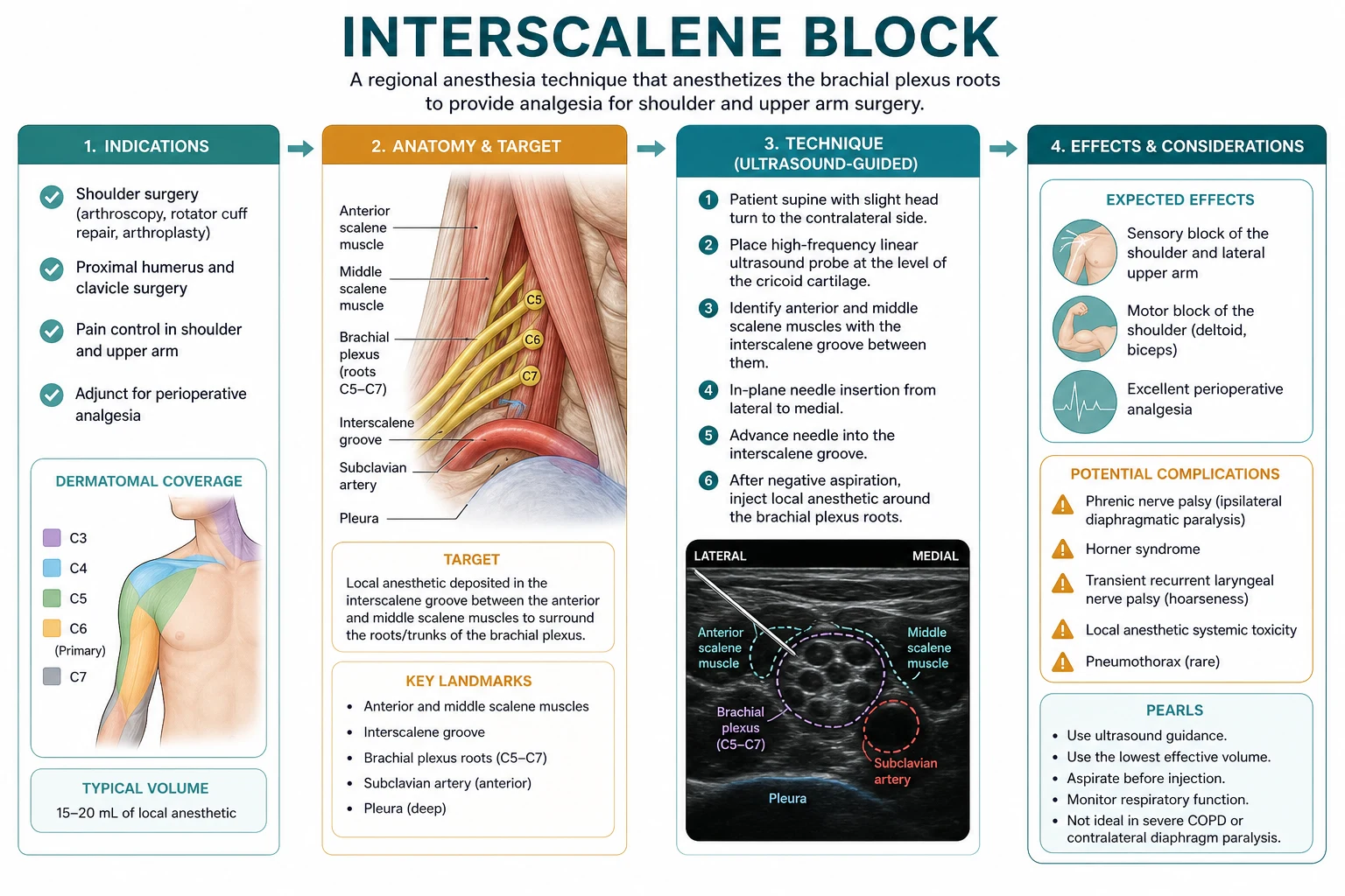
Indicated: shoulder and proximal humerus surgery (arthroscopy, arthroplasty, fracture fixation) — dense C5–C7 root/trunk analgesia. [1]
Not indicated as sole block: hand/forearm surgery needing ulnar (C8–T1) cover — inferior trunk often spared → choose supraclavicular/infraclavicular/axillary as appropriate. [1]
Expected side effects to consent: ipsilateral hemidiaphragmatic paresis, Horner syndrome, recurrent laryngeal nerve block (hoarseness), motor block of arm, block failure, LAST, pneumothorax (uncommon with US), nerve injury. [1]
Anatomy and sonoanatomy
- Brachial plexus roots/trunks exit between anterior and middle scalene muscles.
- Probe: high-frequency linear, transverse in interscalene groove at cricoid level / posterior to SCM.
- “Traffic light” root appearance common teaching image.
- Phrenic nerve lies on anterior scalene — short distance explains near-universal phrenic involvement with traditional volumes.
- Nearby: carotid/internal jugular medially, vertebral vessels, pleura caudally, neuraxis medially if needle goes wrong. [1]
Ultrasound technique (exam sequence)
- Consent: phrenic, Horner, hoarseness, failure, LAST, pneumothorax, nerve injury.
- Full monitoring; IV access; lipid available.[4]
- Position: semi-sitting or supine, head turned slightly contralateral.
- Identify SCM, scalenes, roots; confirm with slight proximal/distal scan.
- In-plane lateral-to-medial (common) with continuous tip visualisation.
- Aspirate, inject small aliquots, watch perineural spread — not intramuscular, not intravascular.
- Volumes: historical 20–30+ mL; modern ultrasound often 5–15 mL of ropivacaine/levobupivacaine 0.25–0.5% depending on goal — lower volume reduces (does not abolish) phrenic effect.[1][3]
Riazi et al.: 5 mL vs 20 mL — lower volume preserved analgesia quality with better respiratory function in classic ultrasound-era evidence.[1]
Phrenic nerve palsy — the defining feature
- Incidence of ipsilateral hemidiaphragmatic paresis approaches universal with traditional volumes.
- Usually tolerated in healthy patients; dyspnoea can be significant in low reserve.
- Contraindications / strong cautions: contralateral phrenic palsy, severe COPD/ILD with poor reserve, some neuromuscular disease, bilateral ISB (never casually).
- Low-volume strategies and superior trunk block reduce hemidiaphragmatic paralysis rates versus classic ISB while aiming to keep shoulder analgesia.[2][3]
Complications table
| Complication | Mechanism | Mitigation |
|---|---|---|
| Phrenic palsy | LA to C3–C5 / phrenic on anterior scalene | Low volume; alternatives; avoid in low reserve |
| LAST | IV injection (vertebral/carotid) | Aspirate, fractionate, lipid, US tip control |
| Neuraxial / epidural / spinal spread | Medial injection | Needle path, low volume, monitor |
| Pneumothorax | Caudal pleural puncture | US, shallow path, avoid deep caudal drive |
| Horner / RLN block | Sympathetic / nearby nerves | Counsel; usually transient |
| Nerve injury | Intraneural injection / trauma | Low pressure, stop if severe pain/resistance |
Continuous interscalene catheter
Useful after arthroplasty/major open shoulder for multi-day analgesia. Same phrenic concerns (may fluctuate with infusion), secure carefully on mobile neck, watch for migration and LAST from infusion errors, physiotherapy fall risk with dense motor block. [1]
Compare: which upper trunk approach?
SAQ answer scaffold
A 68-year-old with severe COPD needs arthroscopic rotator cuff repair. Discuss regional options. [1]
- ISB pros/cons (3): excellent shoulder analgesia but phrenic cost.
- Risk in COPD (3): may not tolerate hemidiaphragm loss.
- Low-volume ISB vs superior trunk / extrascalene options (3).[2][3]
- GA + multimodal / other regional (2).
- Consent and rescue dyspnoea plan (2).
Viva stem bank and model phrases
Stem 1: “Why not ISB for carpal tunnel?”
Model: “Ulnar and inferior trunk sparing — wrong coverage; choose a more distal plexus block.” [1]
Stem 2: “Patient short of breath 20 minutes after ISB.”
Model: “Ipsilateral phrenic block with hemidiaphragm paresis until I exclude pneumothorax and other causes — oxygen, sit up, assess, ultrasound diaphragm/CXR as needed.” [1]
Stem 3: “Safe volume?”
Model: “Lowest effective; ultrasound-era evidence supports single-digit to low-teens millilitres often suffice — twenty millilitres is not mandatory and worsens phrenic effect.”[1]
Stem 4: “LAST prevention?”
Model: “Ultrasound tip control, aspiration, fractionated injection, dose limits, lipid emulsion immediately available.”[4]
Stem 5: “Contralateral phrenic palsy.”
Model: “I avoid ISB — risk of bilateral diaphragm dysfunction is unacceptable.” [1]
Stem 6: “Superior trunk versus ISB?”
Model: “Superior trunk aims for shoulder analgesia with lower rates of hemidiaphragmatic paralysis than classic interscalene — still counsel residual phrenic risk.”[2]
Stem 7: “Horner syndrome after block — cancel surgery?”
Model: “Usually expected collateral effect; reassure if isolated and patient stable — does not by itself mean catastrophe.” [1]
Common traps
- Using ISB for hand surgery
- Large volumes “to be sure”
- Ignoring COPD/contralateral phrenic issues
- No lipid available
- Medial needle wandering
- Missing pneumothorax differential for dyspnoea
- Bilateral ISB thinking [1]
ISB consent — PHORN
Intraoperative and PACU management after ISB
Expect dense motor block of shoulder abductors and elbow flexors; protect the arm in a sling. Supplement with multimodal systemic analgesia for bone pain and posterior port sites that may be incompletely covered. If GA is combined, reduce opioid accordingly but do not assume zero requirement. In PACU, assess for dyspnoea, Horner, hoarseness, and adequacy of analgesia before phase-2 recovery. For day-case shoulder arthroscopy, ensure escort, sling teaching, and written block-duration advice — falls and stove burns on a numb arm are preventable disasters. [1]
Crisis pivots
Acute dyspnoea after block
Sit up, oxygen, examine chest, ultrasound diaphragm if skilled, exclude pneumothorax and anaphylaxis/LAST. Most pure phrenic palsies improve with time and reassurance in healthy lungs; low-reserve patients may need overnight observation or ventilatory support rarely.
Seizure during injection
Stop injection, call for help, ABC, benzodiazepine, lipid emulsion per ASRA, do not give more local anaesthetic.[4]
Complete spinal / high neuraxial after medial injection
Support airway and blood pressure; this is why medial trajectories and large volumes are dangerous.
Patchy block in theatre
Do not chase toxic top-ups; convert to GA or supplement carefully within dose limits; fix the plan for next time (volume, location, timing).
Dosing worked example
70 kg adult for shoulder arthroscopy analgesia: ropivacaine 0.5% 10 mL (50 mg) under ultrasound at superior trunk/interscalene with visualised spread may suffice for many analgesic goals; compare with historical 20–30 mL that multiplies phrenic and LAST mass without guaranteed better pain scores.[1] Always recalculate if surgeon also infiltrates.
Teaching sequence for first solo ISB
- Watch side marked and consent phrenic explicitly.
- Position and landmark scan until roots unmistakable.
- Needle path drawn mentally lateral-to-medial avoiding medial vessels.
- Tip on screen continuously — no blind millimetres.
- 1 mL test then spread then continue to planned low volume.
- Stop if paraesthesia severe or arterial blood.
- Reassess breathing before leaving the bay. [1]
Supervisors should be ready to convert to superior trunk or GA if images are poor or the patient cannot tolerate dyspnoea risk. [1]
Examiner mental map
- Indication = shoulder.
- Sonoanatomy between scalenes.
- Ulnar spare.
- Phrenic almost always / low volume / alternatives.
- LAST and pneumothorax prevention.
- Dyspnoea differential and when to avoid. [1]
That is a complete interscalene viva in six moves. [1]
References
- [1]Riazi S, Carmichael N, Awad I, et al. Effect of local anaesthetic volume (20 vs 5 ml) on the efficacy and respiratory consequences of ultrasound-guided interscalene brachial plexus block Br J Anaesth, 2008.PMID 18682410
- [2]da Cunha DF, et al. Incidence of hemidiaphragmatic paralysis in superior trunk versus interscalene block upper limb surgeries: a systematic review, meta-analysis, and trial sequential analysis Anesth Pain Med (Seoul), 2026.PMID 42130052
- [3]Verbeke AL, et al. The diaphragm-sparing effect of interscalene block with a low-volume of ropivacaine 0.1% vs. 0.5%: A double-blind, controlled, randomised trial Eur J Anaesthesiol Intensive Care, 2026.PMID 42244823
- [4]Neal JM, et al. The Third American Society of Regional Anesthesia and Pain Medicine Practice Advisory on Local Anesthetic Systemic Toxicity: Executive Summary 2017 Reg Anesth Pain Med, 2018.PMID 29356773