Anaes · Regional anaesthesia
Lower limb regional anaesthesia: plexus and peripheral blocks
Also known as lower limb regional blocks
Exam-exhaustive lower-limb regional anaesthesia for ANZCA Final: femoral and fascia iliaca, adductor canal, sciatic and popliteal, obturator, IPACK, ankle block; ultrasound landmarks, dosing principles, LAST rescue, compartment-syndrome surveillance, and ERAS motor-sparing strategy.
On this page & tools
Your progress
Saved locally on this device.
8 MCQs with explanations
Target exams
Red flags
Why this is examined / the one-line answer
Lower-limb regional anaesthesia is a high-yield Final topic because it forces the candidate to think like a consultant: match nerve territory to the incision, protect mobilisation goals after arthroplasty, avoid converting a useful analgesic block into a falls risk or a missed compartment syndrome, and still respect local anaesthetic mass limits and anticoagulation timing. Examiners do not reward a laundry list of block names; they reward a territory-first decision matrix with ultrasound landmarks, realistic volumes, complications specific to each approach, and a clear plan when the block is incomplete.
[2]The one-line viva opener is: I choose lower-limb blocks by surgical territory — femoral or fascia iliaca for anterior thigh and hip fracture analgesia, adductor canal for motor-sparing knee analgesia, sciatic or popliteal for posterior leg and foot, and IPACK for posterior knee capsule — combine only after adding local anaesthetic doses, use ultrasound, agree compartment surveillance with orthopaedics when risk is high, and keep 20 percent lipid emulsion with a 1.5 mL/kg bolus plan ready. Everything below is the anatomy, technique detail, ERAS context, and crisis pivots that justify that sentence.
[1]This topic sits next to lumbosacral plexus anatomy, ultrasound peripheral nerve block safety, neuraxial alternatives for hip and knee arthroplasty, and the LAST and ASRA anticoagulation frameworks that govern all large-volume lower-limb work.[1][2]
Preoperative assessment and risk stratification
Surgical map first
Start with the operation, not the block menu. Hip fracture (neck of femur) analgesia pathways prioritise fascia iliaca or femoral catheter or single-shot as part of multimodal care while the patient awaits theatre and postoperatively. Elective total hip arthroplasty may use periarticular infiltration, PENG (pericapsular nerve group) or fascia iliaca variants depending on institutional ERAS. Total knee arthroplasty (TKA) in modern ERAS almost never needs a dense high-volume femoral block that paralyses the quadriceps for 24 hours — adductor canal (saphenous-dominant) plus local infiltration analgesia plus or minus IPACK is the usual consultant answer. Below-knee amputation or foot and ankle surgery needs sciatic (often popliteal) plus saphenous/femoral territory for the medial strip. Anterior cruciate ligament reconstruction and high tibial osteotomy sit between knee and compartment-risk thinking.
[1]Patient factors that change the plan
Document baseline neurology (diabetic polyneuropathy, sciatica, previous lumbar surgery), anticoagulation and antiplatelet timing for deep blocks and any planned neuraxial, infection at needle sites, allergy to amide local anaesthetics, body habitus and ability to position for ultrasound, cognitive status and consent capacity, and risk of acute compartment syndrome — tibial plateau or shaft fracture, crush injury, reperfusion after vascular repair, severe soft-tissue swelling. If compartment risk is real, dense prolonged motor and sensory block without a written orthopaedic surveillance plan is indefensible.
[3]VTE, cement, and systemic risk
Major lower-limb orthopaedic lists often combine regional analgesia with general or spinal anaesthesia, cemented implants (BCIS risk in frail hip fracture), and aggressive VTE prophylaxis that constrains neuraxial and deep block timing.[2][3] Preoperative haemoglobin, cardiorespiratory reserve, and falls risk after femoral-range motor block all belong in the same briefing.
Applied anatomy of the lower limb nerve supply
Lumbar plexus contributions
The lumbar plexus (L1–L4, often with L2–L4 dominant for the major lower-limb nerves) gives the iliohypogastric, ilioinguinal, genitofemoral, lateral femoral cutaneous (LFCN), obturator, and femoral nerves. For the anaesthetist:
[2]- Femoral nerve (L2–L4): exits under the inguinal ligament lateral to the femoral artery, within the femoral triangle, deep to fascia iliaca. Motor to quadriceps (knee extension), sensory to anterior thigh and, via saphenous continuation, medial leg to the medial malleolus.
- Obturator nerve (L2–L4): exits the obturator foramen; anterior and posterior divisions. Motor to adductors; sensory contribution to medial thigh and often a variable articular contribution to the knee. Missed obturator territory is a classic incomplete medial knee block after femoral-only techniques.
- LFCN (L2–L3): pure sensory to the lateral thigh; relevant for tourniquet discomfort and some lateral hip approaches; commonly covered with supra-inguinal fascia iliaca spread.
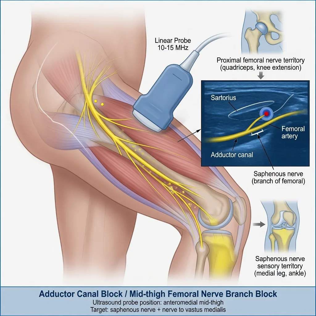
- Saphenous nerve: terminal sensory branch of femoral; targeted in the adductor canal (mid-thigh, under sartorius, lateral to femoral artery) for motor-sparing knee analgesia.
Sacral plexus and sciatic
The sacral plexus via the sciatic nerve (L4–S3) supplies the posterior thigh (tibial portion hamstrings), and all of the leg and foot except the saphenous strip. In the popliteal fossa the sciatic divides into tibial and common peroneal (fibular) nerves — the classic popliteal ultrasound target. Sciatic block at any level produces foot drop until it wears off; counsel and protect the foot.
[2]Knee joint innervation — why TKA is multi-block
Anterior knee capsule: femoral (including articular branches), obturator contributions, and local infiltration. Posterior capsule: genicular and articular branches related to the sciatic/tibial complex and the IPACK (infiltration between popliteal artery and capsule of the knee) plane. A femoral block alone does not cover posterior knee pain — examiners love this trap.
[1]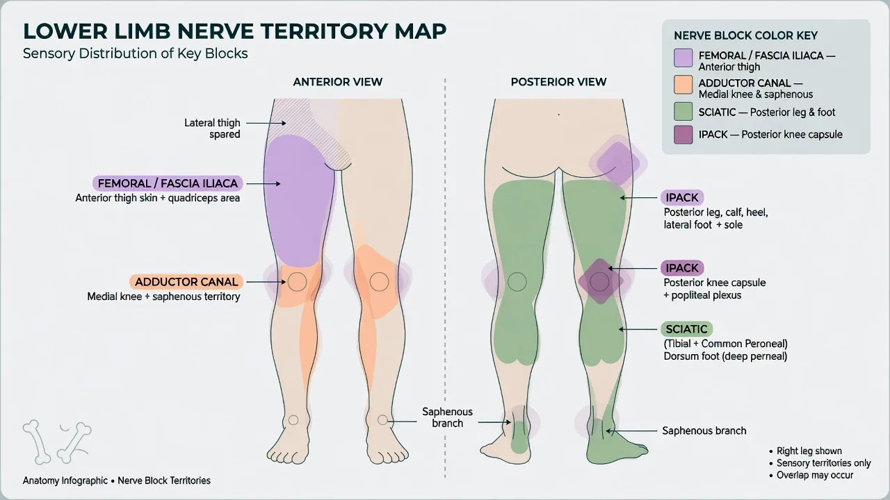
Anaesthetic goals
- Match block(s) to surgical and analgesic territory — not habit.
- Preserve mobilisation after arthroplasty when ERAS demands early weight-bearing.
- Minimise falls risk from dense quadriceps weakness.
- Respect total local anaesthetic mass across combined blocks and infiltration.
- Preserve compartment syndrome surveillance when the injury pattern demands it.
- Prevent and treat LAST with lipid readiness and fractionated injection technique.[1]
- Obey ASRA-compatible timing for neuraxial and deep plexus/peripheral techniques when anticoagulants are in play.[2]
- Document baseline neurology and expected duration of sensory and motor block.
Technique options and decision matrix
Ultrasound principles common to all approaches
Prefer high-frequency linear probes for femoral, adductor canal, and popliteal sciatic; curvilinear may help deep fascia iliaca in large patients. Sterile technique, continuous needle-tip visualisation, aspiration before each aliquot, fractionated 3–5 mL boluses, stop for high resistance or severe paraesthesia with injection, and never exceed maximum dose calculated on lean body weight. Typical exam-friendly concentration bands: ropivacaine 0.2–0.5 percent or levobupivacaine/bupivacaine 0.25–0.5 percent for surgical anaesthesia or dense analgesia; dilute 0.1–0.2 percent for continuous catheters and motor-sparing infusions. Adjuncts (dexamethasone IV or perineural per local policy, clonidine, dexmedetomidine) prolong duration in some studies — consent and local formulary apply.
[1]Femoral nerve block
Patient supine, probe transverse at the inguinal crease: femoral artery medial, nerve typically lateral and slightly deeper, hyperechoic triangular or oval under fascia iliaca. In-plane lateral-to-medial or out-of-plane with tip control. Volume classically 15–20 mL for single-shot analgesia (institutional range 10–30 mL). Expect quadriceps weakness — falls precautions mandatory.
[2]Fascia iliaca block (infra-inguinal and supra-inguinal)
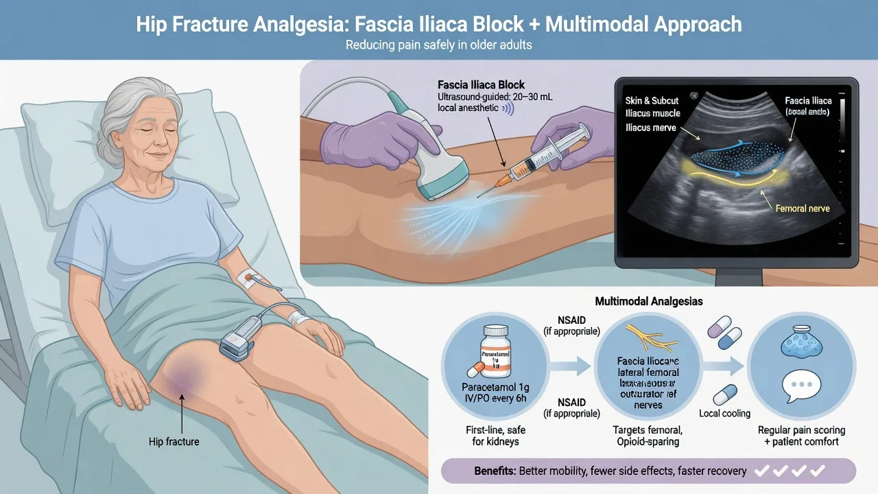
Fascia iliaca block deposits local anaesthetic under the fascia iliaca plane aiming to bathe femoral ± LFCN ± partial obturator via fascial spread. Supra-inguinal approaches under ultrasound (deep to internal oblique / iliacus plane cephalad to the inguinal ligament) are increasingly used in hip fracture pathways for more reliable proximal spread. Volumes often 30–40 mL of dilute solution for fracture analgesia — this is a large volume technique: do the arithmetic if combining with other blocks. Ideal as multimodal analgesia for neck of femur fracture while awaiting theatre and for postoperative pain; not a sole surgical anaesthetic for hip arthroplasty.
[1]Adductor canal (saphenous) block
Mid-thigh, probe transverse: femoral artery deep to sartorius; saphenous nerve near the artery within the adductor canal (Hunter). Target perivascular spread lateral to the artery under the vastoadductor membrane / canal roof. Volume often 10–20 mL. Relative motor sparing of vastus medialis and quadriceps compared with classical femoral block is the ERAS rationale for TKA — not absolute motor sparing if large volumes spill proximally toward the femoral nerve.
[2]Sciatic nerve — popliteal approach
Patient prone, lateral, or supine with leg elevated. At the popliteal crease or slightly proximal, identify popliteal artery; tibial nerve usually superficial/lateral to artery; common peroneal more lateral — track proximally to the sciatic bifurcation “figure-of-eight” or common sheath. Inject 15–25 mL around the sciatic at or just proximal to bifurcation for ankle/foot surgery or below-knee amputation (with saphenous coverage). Foot drop until offset — protect heel and educate patient.
[2]IPACK
Ultrasound between the popliteal artery and the posterior femoral cortex / knee capsule in the popliteal fossa region, depositing local anaesthetic to bathe posterior capsule articular branches without intending dense tibial/common peroneal motor block. Typical volumes 10–20 mL of dilute solution as part of TKA multimodal regimens with adductor canal block and periarticular infiltration.
[1]Ankle block
Five nerves: tibial, deep peroneal, superficial peroneal, sural, saphenous. Landmark or ultrasound-assisted. Excellent ambulatory forefoot anaesthesia; avoid in infection or when compartment risk exists more proximally.
[2]Neuraxial as the single-shot alternative
Spinal (or CSE/epidural) remains excellent surgical anaesthesia for many hip and knee arthroplasties when ASRA timing allows, often combined with motor-sparing peripheral techniques for postoperative analgesia rather than dense bilateral motor blocks.[2] For bilateral procedures or paediatric lower limb work, caudal or lumbar epidural/spinal strategies often dominate.
Monitoring and equipment
Standard monitoring during block placement (ECG, SpO2, NIBP). Resuscitation trolley, airway equipment, and 20 percent lipid emulsion immediately available with a posted ASRA LAST checklist: stop injecting, call for help, 100 percent oxygen, benzodiazepines for seizures, lipid 1.5 mL/kg lean body weight bolus over 2–3 minutes then infusion approximately 0.25 mL/kg/min, reduced adrenaline dosing in LAST arrest relative to standard ACLS, avoid vasopressin, calcium channel blockers, and pure beta-blockade as first-line LAST therapy.[1] Ultrasound machine with sterile cover, appropriate needle (typically 50–100 mm insulated or block needle), optional nerve stimulator as adjunct (quadriceps twitch for femoral; plantar flexion/dorsiflexion for sciatic), and clear labelling of injectate syringes.
Intraoperative management
Combining blocks — the arithmetic
Example TKA ERAS package: adductor canal 15 mL ropivacaine 0.2–0.5 percent + IPACK 15 mL dilute + surgeon periarticular infiltration (often with further local anaesthetic, NSAID, adrenaline). You must add all amide mass and stay under toxic limits (classic teaching ceilings such as levobupivacaine/bupivacaine approximately 2 mg/kg and ropivacaine approximately 3 mg/kg are starting points — institutional maxima and lean body weight rules apply; never stack full femoral plus full sciatic high-volume doses plus infiltration without arithmetic).
[1]Testing and rescue
Before incision under pure regional anaesthesia, test relevant sensory territories and motor function. Incomplete medial knee: consider obturator rescue or infiltration. Incomplete posterior knee: IPACK or sciatic-level rescue if still within dose budget. Convert to general anaesthesia early rather than repeated top-ups that breach dose limits.
[2]Hip fracture pathway nuance
Fascia iliaca or femoral analgesia reduces opioid load in frail patients and can facilitate positioning for spinal anaesthesia. Cemented hemiarthroplasty still carries BCIS risk — regional analgesia does not remove the need for euvolaemia, communication at cementation, and vasopressor readiness.[3]
Crisis pivots — what changes the plan
Local anaesthetic systemic toxicity
Stop injection, ABC with 100 percent oxygen, control seizures, start lipid per ASRA dosing, prepare for prolonged CPR if cardiovascular collapse.[1] Lower-limb fascial plane blocks with large volumes are not “safe by geography.”
Acute compartment syndrome under block
Pain out of proportion, pain on passive stretch, rising analgesic requirement, paraesthesia, and later pulselessness are teaching signs — but a dense sensory block can blunt the pain signal. Pivot: use dilute solutions, single-shot short-acting agents when risk is high, avoid dense multi-nerve blocks for tibial fractures without orthopaedic agreement, schedule regular compartment checks (pressure monitoring when indicated), and escalate early for fasciotomy rather than attributing all pain to “block wearing off unevenly.”
[3]Incomplete block and LAST from topping up
Do not chase completeness with another 20 mL of concentrated local anaesthetic. Convert technique or deepen general anaesthesia.
[1]Falls and nerve injury
Post-femoral block falls, pressure injury on a numb heel after sciatic block, and needle trauma or intraneural injection with persistent deficit all require documentation, neurology follow-up, and honest consent discussions preoperatively.
[2]Lower limb territory map
Postoperative / PACU plan
Document sensory and motor levels, protect the insensate limb, fall precautions after any femoral-range weakness, counsel expected duration, and plan breakthrough multimodal analgesia (paracetamol, NSAID if not contraindicated, carefully titrated opioid, ice, elevation). Continuous catheters need infusion protocols, daily review, infection surveillance, and coordinated removal with VTE prophylaxis timing.[2] After TKA, physiotherapy goals and block density must be co-designed — a dense motor block that prevents straight-leg raise on day zero may undermine ERAS.
Special populations and comorbidities
Frail hip fracture: fascia iliaca / femoral as part of national pathways; minimise opioids and delirium; plan spinal versus general with anticoagulation and aortic stenosis nuance. Diabetic neuropathy: document baseline sensory loss so residual deficit is not blamed solely on the block. Paediatrics: weight-based dosing strict; caudal or lumbar neuraxial often preferred for bilateral or young children; ultrasound peripheral blocks excellent for older children with ambulatory foot surgery. Obesity: deeper targets, curvilinear probe, longer needles, higher risk of incomplete blocks and dose miscalculation if total body weight is used naively. Trauma with crush or reperfusion: compartment risk dominates — choose short, dilute, or no dense block. Anticoagulated patients: deep plexus and neuraxial need ASRA table discipline; superficial ankle blocks and some distal techniques have different risk profiles but are not risk-free.[2]
SAQ answer scaffold
Stem: A 68-year-old patient is listed for primary TKA on an ERAS pathway. They have diet-controlled diabetes and take aspirin 100 mg daily. Outline your regional analgesic strategy.
[2]- Goals (2 marks): motor-sparing analgesia, early mobilisation, multimodal package, falls prevention.
- Anatomy (3 marks): anterior knee (femoral/saphenous ± obturator), posterior capsule (IPACK/sciatic-related), why femoral alone fails posteriorly.
- Technique choice (4 marks): adductor canal + IPACK + periarticular infiltration; optional spinal for surgical anaesthesia if timing allows; ultrasound details and approximate volumes; LA mass arithmetic.
- Complications (3 marks): LAST with lipid 1.5 mL/kg; nerve injury; residual motor weakness/falls; ASRA principles if anticoagulants escalate.[1][2]
- Compartment caveat (2 marks): not the primary issue in elective TKA, but state the principle for high-risk fractures.
Alternative stem: Neck of femur fracture — fascia iliaca technique, volumes, role before positioning for spinal, and what regional does not prevent (BCIS at cementation).[3]
Viva stem bank and model phrases
Stem 1: “Which block for TKA ERAS?”
Model: “I favour adductor canal plus IPACK and local infiltration rather than a high-volume femoral block that delays mobilisation and increases falls risk.”
Stem 2: “Femoral block done — patient still has posterior knee pain.”
Model: “Expected — femoral territory does not cover the posterior capsule. I would use IPACK or sciatic-level coverage within remaining local anaesthetic dose, or rely on periarticular infiltration and systemic multimodal analgesia.”
Stem 3: “Tibial fracture — can I do a dense sciatic and femoral block?”
Model: “Only with an explicit orthopaedic surveillance plan. Dense sensory block can mask compartment syndrome. I prefer dilute or short-acting techniques, high index of suspicion, and early fasciotomy discussion.”
Stem 4: “What is your LAST dose of lipid?”
Model: “Twenty percent lipid emulsion, 1.5 mL per kilogram lean body weight bolus, then infusion, per ASRA — I know where the kit is before I start the block.”[1]
Stem 5: “Hip fracture patient for cemented hemiarthroplasty — regional plan?”
Model: “Fascia iliaca or femoral for analgesia and positioning, spinal if ASRA timing allows, prepare for BCIS at cementation with oxygen, euvolaemia, and vasopressors — the block does not treat embolisation physiology.”[3]
Stem 6: “List the five nerves of an ankle block.”
Model: “Tibial, deep peroneal, superficial peroneal, sural, and saphenous.”
Common traps
- Expecting femoral block alone to cover posterior knee pain after TKA
- Missing obturator contribution to medial knee
- Exceeding local anaesthetic maximum with multiple fascial plane blocks plus infiltration
- Dense multi-nerve blocks in high compartment-risk tibial trauma without surveillance agreement
- Forgetting sciatic territory for lateral ankle and most of the foot
- Ignoring foot drop protection after sciatic/popliteal block
- Using total body weight for toxic dose calculations in obesity
- Placing deep blocks without checking the ASRA last-dose interval for anticoagulants
ANZCA Final candidates are expected to integrate lower-limb blocks with ERAS arthroplasty pathways, hip fracture national standards, ASRA anticoagulation timing (or local ANZ equivalents based on the same principles), and ASRA LAST rescue numbers — not merely name the femoral nerve.
References
- [1]Neal JM et al. The Third American Society of Regional Anesthesia and Pain Medicine Practice Advisory on Local Anesthetic Systemic Toxicity: Executive Summary 2017 Reg Anesth Pain Med, 2018.PMID 29356773
- [2]Horlocker TT et al. Regional Anesthesia in the Patient Receiving Antithrombotic or Thrombolytic Therapy: American Society of Regional Anesthesia and Pain Medicine Evidence-Based Guidelines (Fourth Edition) Reg Anesth Pain Med, 2018.PMID 29561531
- [3]Donaldson AJ et al. Bone cement implantation syndrome Br J Anaesth, 2009.PMID 19059919