Anaes · Regional anaesthesia
Ultrasound-guided peripheral nerve blocks
Also known as US-guided regional anaesthesia · Peripheral nerve block ultrasound · In-plane PNB · Regional anaesthesia safety
Exam-exhaustive ultrasound PNB: probe and needle skills, upper and lower limb block selection by surgery, fascial plane blocks, ultrasound vs nerve stimulator evidence, intraneural injection and LAST prevention, and ASRA anticoagulation principles for peripheral blocks.
On this page & tools
Your progress
Saved locally on this device.
Target exams
Red flags
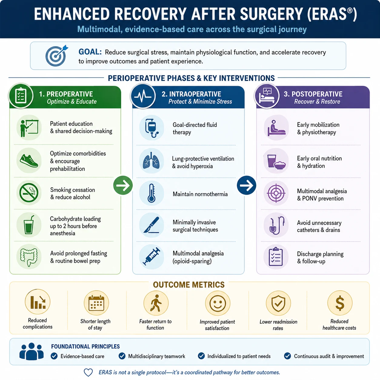
Why this is examined / the one-line answer
Ultrasound-guided peripheral nerve blockade is core Final skills territory: block selection, sono-skills, safety (LAST, nerve injury, anticoagulation), and honest comparison with landmark/stimulator techniques. Examiners reward structured workflow more than brand-name needles.[1][4]
One-liner: I match block to surgery, use in-plane ultrasound with visible tip and perineural spread, inject incrementally at low pressure, and prevent LAST with dose discipline and lipid readiness. [1]
Core ultrasound skills
| Skill | Exam point |
|---|---|
| Probe selection | High-frequency linear for superficial; curvilinear for deep |
| Ergonomics | Screen, probe, needle aligned; sit if needed |
| Axis | Short-axis nerve view common; long-axis for confirmation |
| Needle approach | In-plane preferred for continuous tip view; out-of-plane only with discipline |
| Artefacts | Anisotropy, reverberation, mirror — do not inject on faith |
| Hydrodissection | Small fluid opens plane and confirms tip |
Goal of injection: perineural spread surrounding the target — not intraneural (fascicular) and not intravascular. [1]
Safety workflow (every block)
- Indication, consent, contraindications (infection at site, patient refusal, uncorrected coagulopathy for deep blocks).
- Monitoring, IV access, resuscitation drugs, lipid emulsion present for non-trivial doses.[4]
- Stop before max dose; include surgical infiltrate in the total.
- Asepsis.
- Visualise tip always before advancing.
- Aspirate; inject 1–3 mL test aliquots; watch spread; low pressure.
- Stop for severe pain, high resistance, or intravascular pattern.
- Document drug, dose, time, laterality, and complications discussed.
Neurologic complication advisories emphasise patient selection, technique discipline, and follow-up of deficits.[1]
Upper limb — choose by surgery
| Surgery | Prefer | Avoid thinking |
|---|---|---|
| Shoulder | Interscalene / superior trunk | Using axillary alone |
| Arm / elbow / hand (single block) | Supraclavicular or infraclavicular | ISB for hand (ulnar spare) |
| Forearm / hand | Axillary or distal nerves | Missing musculocutaneous |
| Wrist/hand awake | Wrist blocks / distal US | Unnecessary phrenic risk |
Anatomy-driven contraindications: interscalene ≈ phrenic; supraclavicular ≈ pleura. [1]
Lower limb — choose by surgery
| Surgery | Prefer | Notes |
|---|---|---|
| Hip / femur neck | Fascia iliaca / femoral ± obturator; consider PENG concepts | Not complete hip capsule alone always |
| Knee | Femoral/adductor canal + sciatic as needed | Adductor canal more motor-sparing for quads |
| Foot / ankle | Popliteal sciatic + saphenous | Two nerves often |
| Below-knee amputation | Sciatic + femoral/saphenous | Cover both territories |
Fascial plane blocks (concept level)
TAP, QL, ESP and chest-wall planes are volume-dependent techniques for trunk analgesia — match to incision, respect LAST maths, limited visceral cover.[2] Large volumes → LAST awareness; anticoagulation risk intermediate for some deep planes.[3][4]
Complications
Intraneural injection
Suspect: high pressure, severe paraesthesia, nerve swelling on US. Stop; withdraw; do not continue forcing. Most subepineural deposits are not catastrophic, but intrafascicular injury is the fear — prevention beats heroics.[1]
LAST
Early: tinnitus, metallic taste, agitation, seizures. Late: cardiovascular collapse. Management: stop LA, ABC, benzodiazepines for seizures, 20% lipid emulsion per ASRA, modified ACLS (avoid certain drugs per checklist), prolonged CPR if needed.[4]
Others
Infection, haematoma, allergic reaction, block failure, residual motor block falls, pneumothorax (supra/interscalene), phrenic (interscalene/supraclavicular). [1]
Anticoagulation and PNB
Not identical to neuraxial rules, but not risk-free. Deep, non-compressible sites (deep lumbar plexus, some paravertebral/QL, deep neck) carry higher haematoma consequence → apply ASRA interventional guidance and local policy; superficial compressible blocks are more permissive but still judged case-by-case.[3]
Ultrasound vs landmark/stimulator — viva lines
| US advantages | Residual role of stimulator |
|---|---|
| See anatomy, vessels, needle, spread | Teaching adjunct; confirmation |
| Lower LA volume possible for some blocks | When US images poor |
| Fewer vascular punctures in skilled hands | Combined dual guidance sometimes |
| Still not zero nerve injury | Operator skill remains decisive |
US does not make blocks risk-free — it makes careful operators safer and careless operators differently dangerous. [1]
SAQ answer scaffold
Describe your approach to performing an ultrasound-guided peripheral nerve block safely. [1]
- Selection and consent (2).
- Preparation including lipid (2).[4]
- US workflow and in-plane tip control (3).
- Injection discipline and stop rules (3).[1]
- Post-block monitoring and failure plan (2).
- Anticoagulation risk stratification if relevant (2).[3]
Viva stem bank and model phrases
Stem 1: “In-plane or out-of-plane?”
Model: “I prefer in-plane so the entire needle path and tip are visible before I inject.” [1]
Stem 2: “How do you prevent LAST?”
Model: “Minimum effective dose, aspiration, fractionated injection, ultrasound, max-dose maths including infiltrate, and lipid immediately available.”[4]
Stem 3: “Patient screams and high pressure on syringe.”
Model: “I stop — possible intraneural injection — withdraw, reassess, do not force.” [1]
Stem 4: “Is ultrasound mandatory?”
Model: “It is the modern standard for most PNBs where available; landmark/stimulator remain fallback and teaching tools, not excuses for never learning US.” [1]
Stem 5: “Apixaban and popliteal block?”
Model: “I risk-stratify with ASRA-style guidance; superficial distal blocks differ from deep plexus — document shared decision.”[3]
Stem 6: “Wrong block commonest failure?”
Model: “Mismatch of block to surgical incision — anatomy selection before needle skill.” [1]
Stem 7: “Chest wall planes?”
Model: “Fascial plane techniques are useful adjuncts when matched to incision with volume and LAST discipline.”[2]
Common traps
- Emptying 30 mL without watching spread
- Advancing without tip view
- Ignoring surgeon’s infiltrate in mg totals
- Treating all PNB as anticoagulation-irrelevant
- No lipid in the room
- Wrong level for the operation
- Declaring success by needle proximity without spread [1]
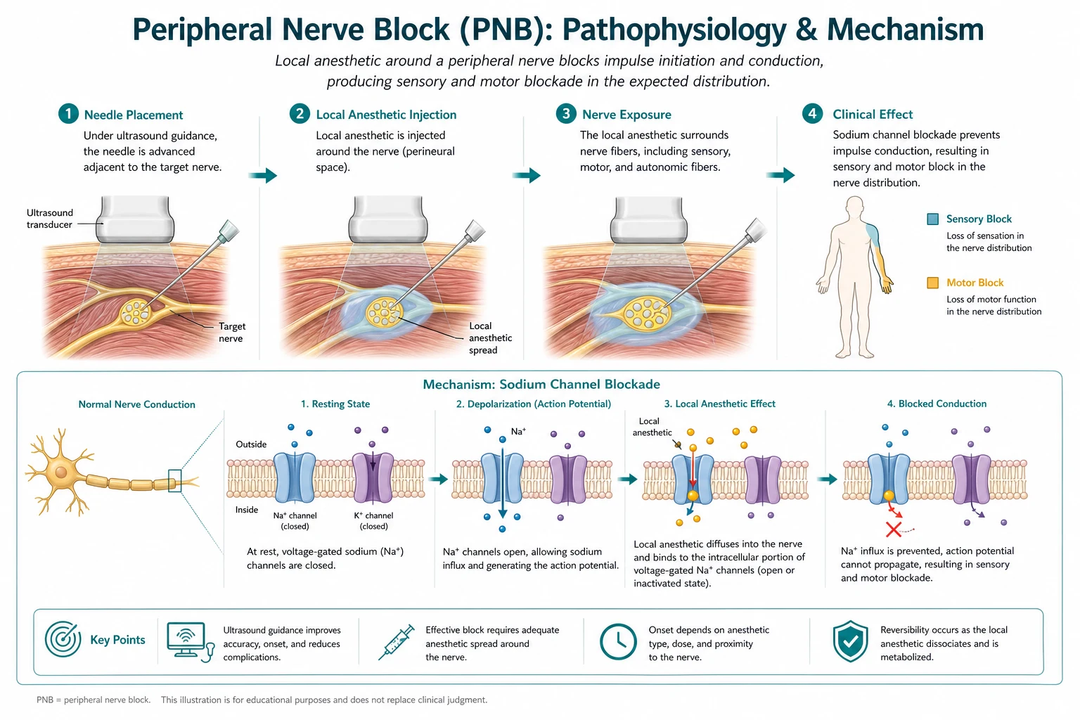
US block safety — SEE LA
Equipment checklist and infection control
Ultrasound machine with appropriate preset, sterile probe cover and gel for invasive blocks, short-bevel block needle of suitable length, extension tubing optional for stability, calibrated syringes, skin prep, sterile gloves/gown as per depth and catheter plans, marker for side check. Single-shot clean technique still requires rigorous skin antisepsis. Catheters need full sterile barriers. Never share multidose LA vials across patients casually — infection outbreaks have occurred in regional practice. [1]
Worked selection cases
Case 1 — ORIF distal radius: axillary or infraclavicular/supraclavicular depending on tourniquet and surgeon preference; avoid ISB. [1]
Case 2 — total knee arthroplasty ERAS: spinal or GA plus adductor canal ± IPACK/local infiltration; dense femoral may impair early physio. [1]
Case 3 — rib fractures multi-level: ESP or PVB with dose awareness; multimodal systemic co-analgesia; anticoagulation status first.[2]
Case 4 — AV fistula creation: supraclavicular or axillary; counsel phrenic if supra chosen in COPD. [1]
Dual guidance and nerve stimulator
Ultrasound plus low-current nerve stimulation can add confidence for trainees, but motor response does not guarantee extraneural position and absence of response does not guarantee safety. Opening injection pressure monitoring (where available) is an adjunct against high-pressure intraneural injection — still secondary to visualisation and stop-rules.[1]
Postoperative follow-up of neurological symptoms
Persistent sensory or motor deficit beyond expected block duration needs urgent review: document distribution, compare to surgical neuropraxia risk, consider neurology/imaging, and report via local quality systems. Most deficits resolve, but delayed recognition of compressive haematoma at deep sites is the nightmare scenario paralleling neuraxial haematoma thinking.[1][3]
Examiner mental map
- US workflow and in-plane discipline.
- Block selection by surgery.
- LAST prevention and treatment.
- Intraneural stop rules.
- Anticoagulation risk tiers.
- Fascial planes as volume adjuncts. [1]
Discipline plus anatomy passes; gadget worship without safety fails. [1]
References
- [1]Neal JM, Brull R, Horn JL, et al. Efficacy of ultrasound and nerve stimulation guidance in peripheral nerve block: A systematic review and meta-analysis IUBMB Life, 2017.PMID 28714206
- [2]Chin KJ, Versyck B, Pawa A. American Society of Regional Anesthesia and Pain Medicine Local Anesthetic Systemic Toxicity checklist: 2020 version Reg Anesth Pain Med, 2021.PMID 33148630
- [3]Narouze S, Benzon HT, Provenzano D, et al. Interventional Spine and Pain Procedures in Patients on Antiplatelet and Anticoagulant Medications (Second Edition): Guidelines From the American Society of Regional Anesthesia and Pain Medicine, the European Society of Regional Anaesthesia and Pain Therapy, the American Academy of Pain Medicine, the International Neuromodulation Society, the North American Neuromodulation Society, and the World Institute of Pain Reg Anesth Pain Med, 2018.PMID 29278603
- [4]Neal JM, Barrington MJ, Fettiplace MR, et al. The Third American Society of Regional Anesthesia and Pain Medicine Practice Advisory on Local Anesthetic Systemic Toxicity: Executive Summary 2017 Reg Anesth Pain Med, 2018.PMID 29356773