Anaes · Regional anaesthesia
Upper limb regional anaesthesia: brachial plexus approaches
Also known as upper limb regional blocks
Exam-exhaustive upper-limb brachial plexus anaesthesia: interscalene, supraclavicular, infraclavicular, axillary; ultrasound landmarks, phrenic and pneumothorax risk, LAST rescue with lipid 1.5 mL/kg, incomplete-block rescue, and ANZCA Final viva stems.
On this page & tools
Your progress
Saved locally on this device.
8 MCQs with explanations
Target exams
Red flags
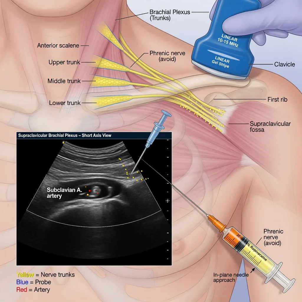
Why this is examined / the one-line answer
Upper-limb regional anaesthesia is examiner gold because it compresses three skills into one viva: gross and sonoanatomy of the brachial plexus, a decision matrix that matches approach to surgical site and patient respiratory reserve, and crisis management of local anaesthetic systemic toxicity and approach-specific complications (phrenic paresis, pneumothorax, intravascular injection, incomplete distal coverage). The candidate who only recites “interscalene for shoulder” without naming phrenic block, ulnar sparing, and lipid dosing fails the discriminating part of the station.
[1]The one-line answer: I match plexus level to territory — interscalene for shoulder accepting ipsilateral phrenic paresis, supraclavicular for dense arm and forearm anaesthesia with pneumothorax risk, infraclavicular for below-elbow surgery and catheters, axillary for distal surgery with deliberate musculocutaneous coverage — always under ultrasound with fractionated injection, ASRA dose limits, and lipid 1.5 mL/kg ready for LAST.
[2]Preoperative assessment and risk stratification
Surgical territory
Map the incision and tourniquet: shoulder and proximal humerus (interscalene or superior trunk variants); mid-humerus to hand (supraclavicular often excellent); elbow, forearm, and hand with preference for catheter (infraclavicular); hand and forearm with arm abducted and ultrasound-friendly axilla (axillary). Distal rescue blocks (median, ulnar, radial at forearm or wrist) salvage incomplete proximal blocks without restacking a full plexus volume.
[2]Respiratory and systemic risk
Severe COPD, interstitial lung disease, contralateral phrenic or diaphragmatic impairment, morbid obesity with limited reserve, and bilateral interscalene plans (almost never justified) all push you away from classical interscalene volume near the roots. Obstructive sleep apnoea and day-case shoulder surgery need a plan for rebound pain when the block wears off and for residual phrenic effect in recovery.
[1]Anticoagulation, infection, neuropathy
Deep approaches (infraclavicular, some interscalene and supraclavicular variants depending on path) require ASRA-compatible risk assessment with antithrombotics.[2] Infection at the needle site is a hard stop. Document pre-existing neuropathy (ulnar neuritis, cervical radiculopathy) so residual deficit is not automatically attributed to the block. Consent must include failure, conversion to general anaesthesia, LAST, nerve injury, pneumothorax (supraclavicular), and hemidiaphragmatic paresis (interscalene).
Dose planning before the needle moves
Calculate maximum local anaesthetic mass on lean body weight before drawing up. Typical single-shot plexus volumes 15–25 mL of ropivacaine 0.5 percent or equivalent under ultrasound — lower volumes are increasingly used with precise deposition. Adjuncts (dexamethasone, alpha-2 agonists) per evidence and consent.
[1]Applied anatomy — roots to terminal nerves
The ladder examiners expect
- Roots C5–T1 (variable C4/T2 contributions) exit intervertebral foramina and form the plexus between anterior and middle scalene muscles (interscalene groove).
- Trunks: upper (C5–C6), middle (C7), lower (C8–T1) — interscalene targets roots/trunks for shoulder; lower trunk/ulnar often missed (ulnar sparing).
- Divisions form behind the clavicle — supraclavicular “spinal of the arm” at the clustered divisions lateral to the subclavian artery, above the first rib (pleura nearby).
- Cords (lateral, posterior, medial) named relative to the axillary artery under pectoralis minor — infraclavicular target.
- Terminal nerves around the axillary artery in the axilla: median, ulnar, radial, and musculocutaneous (often already left the sheath into coracobrachialis — must be blocked separately in axillary approaches).
Sensory and motor testing after block should follow this map: shoulder abduction (axillary nerve / superior trunk), elbow flexion (musculocutaneous), wrist extension (radial), thumb opposition (median), little finger sensation (ulnar).
[2]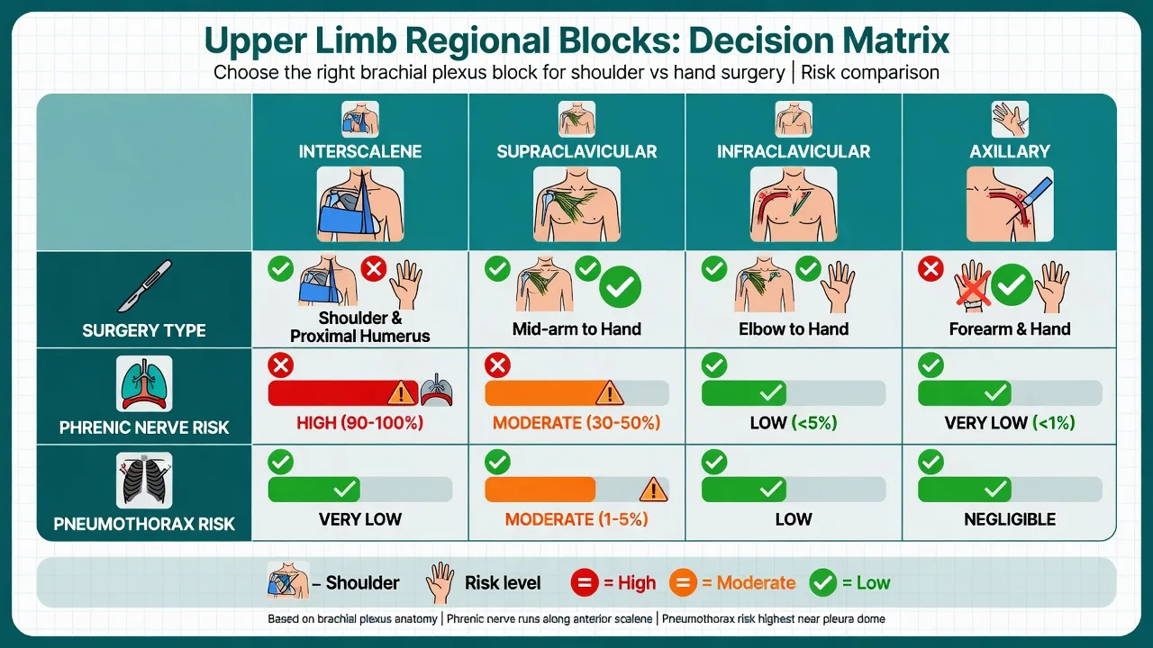
Anaesthetic goals
- Surgical anaesthesia or dense analgesia matched to territory.
- Accept only those collateral effects the patient’s physiology can tolerate (especially phrenic block).
- Avoid intravascular and intraneural injection.
- Stay under toxic local anaesthetic mass, including any distal rescue.
- Plan early general anaesthesia conversion if the block is incomplete — do not “top up to toxicity.”
- Protect the insensate limb and plan post-block analgesia for rebound pain.
Technique options and decision matrix
Interscalene
Ultrasound at the level of the cricoid / C6: identify carotid, internal jugular, sternocleidomastoid, anterior and middle scalene, and the stacked nerve roots (classically “stop-light” appearance). In-plane lateral-to-medial common. Deposit local anaesthetic around superior and middle trunk elements for shoulder surgery. Phrenic nerve lies on anterior scalene — even low volumes often produce ipsilateral hemidiaphragmatic paresis; low-volume and extrafascial/superior trunk variants aim to reduce but not eliminate this. Horner syndrome (cervical sympathetic), recurrent laryngeal nerve block (hoarseness), and epidural/intrathecal injection (catastrophic if unrecognised) are classic teaching complications. Do not choose interscalene for isolated hand surgery — ulnar sparing and unnecessary phrenic risk.
[1]Supraclavicular
Probe above clavicle, subclavian artery in short axis, brachial plexus superolateral as a “bunch of grapes,” first rib and pleura deep. Corner pocket deposition near the lower trunk improves ulnar coverage compared with interscalene. Pneumothorax is the signature risk — ultrasound visualisation of rib and pleura reduces but never zeros the incidence. Intravascular injection into subclavian or transverse cervical vessels is another LAST pathway.
[2]Infraclavicular (including costoclavicular variants)
Probe below clavicle, identify axillary artery under pectoralis major/minor; cords arranged around the artery (classic “U” distribution varies). Single injection posterior to the artery often produces reliable distal anaesthesia. Excellent for catheters because the site is stable and clean under the clavicle. Pneumothorax risk is lower than supraclavicular but vascular puncture risk is real.
[2]Axillary
Arm abducted, probe high axilla: axillary artery with median (often superficial), ulnar, and radial nerves around it; musculocutaneous tracked into coracobrachialis laterally. Multiple injections or circumferential spread required. Low pneumothorax and phrenic risk — preferred when respiratory disease forbids interscalene and surgery is distal. Incomplete block if musculocutaneous is forgotten — patient feels the lateral forearm incision.
[2]Distal rescue and supplementary techniques
Selective median/ulnar/radial blocks at the mid-forearm or wrist, intercostobrachial coverage for medial upper arm tourniquet pain (T2, not brachial plexus), and superficial cervical plexus for cape distribution after shoulder surgery complete the toolkit.
[2]Awake versus sedated versus GA with block
Awake or lightly sedated block allows neurological monitoring during injection; deep sedation or general anaesthesia with block is common for children and anxious adults but removes early warning of intraneural injection — ultrasound discipline must be stricter. Shoulder arthroscopy beach-chair position adds cerebral perfusion and airway access considerations when general anaesthesia is combined.
[3]Monitoring and equipment
ECG, SpO2, NIBP during performance; intravenous access before injection; ultrasound with sterile cover; block needle of appropriate length (25–100 mm depending on approach); optional nerve stimulator (deltoid twitch for interscalene; hand twitches for distal approaches) as adjunct, not a licence to inject without tip visualisation. Lipid emulsion 20 percent and LAST cognitive aid immediately available.[1] Airway equipment ready — rare total spinal or severe LAST needs DAS-aligned airway rescue thinking.[3]
Intraoperative management
Test the block systematically before incision. Supplement deficient territories with distal blocks or field infiltration within remaining dose budget. For beach-chair shoulder surgery under general anaesthesia with interscalene: careful blood pressure management relative to sitting position cerebral perfusion, secure airway, and eye protection. Document sensory and motor levels. If converting to general anaesthesia mid-case, treat residual partial block as a contribution to postoperative analgesia, not as a reason to omit multimodal systemic drugs entirely.
[1]Crisis pivots — LAST and approach-specific disasters
LAST
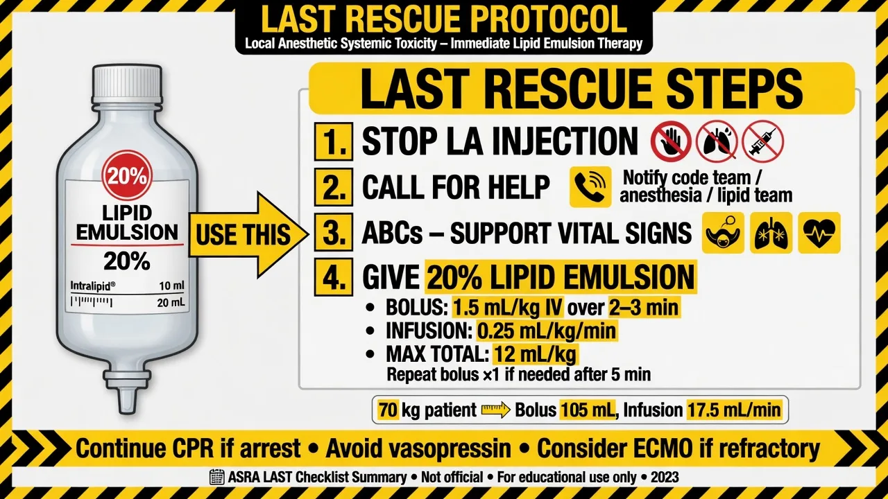
Stop local anaesthetic, call for help, airway with 100 percent oxygen, treat seizures with benzodiazepines (propofol carefully if haemodynamically stable), start 20 percent lipid emulsion 1.5 mL/kg lean body weight bolus then infusion, prepare for prolonged CPR, use reduced adrenaline doses in arrest compared with standard ACLS, avoid vasopressin and calcium channel blockers as primary LAST therapy.[1]
Phrenic paresis with respiratory distress
Sit up if safe, oxygen, physiotherapy, non-invasive support if needed, exclude pneumothorax (especially after supraclavicular), and rarely ventilatory support until block offsets. Prevention is better: avoid classical interscalene in severe respiratory disease.
[2]Pneumothorax
Suspicion after supraclavicular: pleuritic pain, desaturation, reduced air entry — chest radiograph or ultrasound, oxygen, chest drain if indicated.
[2]High spinal / epidural spread from interscalene
Hypotension, bradycardia, ascending block, apnoea — airway, cardiovascular support, explanation of unanticipated neuraxial anaesthesia.
[3]Incomplete block mid-surgery
Do not inject large additional concentrated volumes. Convert to general anaesthesia or carefully planned distal rescue.
[2]LAST first steps
Postoperative / PACU plan
Document block density, protect the arm in a sling, warn about delayed LAST (can present after PACU arrival), counsel on expected duration and night-time rebound pain after shoulder surgery (multimodal pack: paracetamol, NSAID if suitable, breakthrough opioid, ice). Continuous catheters need infusion protocols, disconnection rules, and infection surveillance. Neurologic deficit beyond expected duration needs urgent review.
[2]Special populations and comorbidities
Severe respiratory disease: avoid classical interscalene; prefer axillary/infraclavicular for distal surgery or GA with multimodal systemic analgesia for shoulder if regional phrenic risk is unacceptable. Anticoagulated patients: apply ASRA principles for deep blocks.[2] Paediatrics: strict weight-based dosing, ultrasound, often under general anaesthesia. Day surgery: reliable escort, written advice, phone follow-up pathway for dyspnoea or prolonged deficit. Rheumatoid arthritis and stiff necks: positioning and airway planning if GA conversion likely; cervical spine caution.
Local anaesthetic dosing arithmetic for upper limb
Ultrasound has reduced required volumes, but examiners still expect you to defend a number. A typical single-shot brachial plexus volume under ultrasound is 15–25 mL of ropivacaine 0.5 percent (75–125 mg) or equipotent levobupivacaine/bupivacaine at lower concentrations for denser motor block when needed. For continuous catheters, dilute ropivacaine 0.1–0.2 percent infusions (for example 5–10 mL/h with optional PCA boluses per protocol) prolong analgesia while limiting motor density. Always calculate on lean body weight, add any distal rescue and surgical infiltration, and stop before toxic ceilings (classic teaching anchors: bupivacaine/levobupivacaine near 2 mg/kg; ropivacaine near 3 mg/kg — institutional maxima and patient factors override slogans). [1]
Adjuncts commonly discussed in vivas: dexamethasone (IV 4–8 mg or perineural per local evidence and consent) prolongs block duration in many trials; clonidine or dexmedetomidine perineural prolongs block but may increase hypotension and sedation; adrenaline 1:200,000–1:400,000 as a vascular marker and to reduce systemic absorption in some approaches — avoid large adrenaline doses near end-artery debates is more a digital block issue than plexus. Never use adrenaline-containing solutions as a substitute for aspiration and fractionation. [1]
Incomplete block map — how to rescue without LAST
- Interscalene with ulnar sparing: expected for pure hand surgery — wrong primary choice; for shoulder, ulnar sparing is usually acceptable.
- Supraclavicular with ulnar miss: inject more toward the corner pocket near the lower trunk if dose remains; or distal ulnar rescue at elbow/wrist.
- Axillary missing musculocutaneous: track into coracobrachialis and block separately; lateral forearm incision will declare your miss.
- Radial sparing after axillary: radial often lies deep to the artery — ensure circumferential or targeted deep injection.
- Tourniquet pain on medial upper arm: intercostobrachial (T2) field block on the medial arm, not more plexus volume. [1]
Beach-chair shoulder surgery specifics
When interscalene is combined with general anaesthesia in the beach-chair position, cerebral perfusion pressure falls with the hydrostatic gradient from heart to brain. Maintain blood pressure close to baseline rather than allowing “permissive” relative hypotension simply because the field is bloodless. Secure the airway meticulously; accidental extubation in beach-chair is awkward. PONV risk after shoulder arthroscopy is significant — dexamethasone plus ondansetron is a common dual therapy. Rebound pain at night after single-shot interscalene is predictable — prescribe a multimodal pack and counsel the patient before discharge. [1]
Human factors and consent language
Consent phrases that score: temporary hemidiaphragm weakness and shortness of breath; small risk of pneumothorax needing a drain; nerve injury usually temporary; local anaesthetic toxicity treated with a fat emulsion we keep immediately available; block failure and conversion to general anaesthesia. Mark the side, stop before wrong-side block (never events), and perform a pre-block timeout including allergy, maximum dose, and lipid location. [1]
Nerve stimulator endpoints (still examinable)
Although ultrasound is first-line, stimulator endpoints remain viva currency: deltoid or biceps twitch at interscalene (upper trunk); forearm or hand twitches at supraclavicular/infraclavicular; specific median (finger flexion), ulnar (thumb adduction/finger flexion), radial (wrist/finger extension), and musculocutaneous (biceps) responses at axillary level. Acceptable current thresholds historically near 0.2–0.5 mA for proximity without intraneural injection — inject with low pressure, stop if high resistance or severe pain. Stimulator without ultrasound is inferior for safety in modern practice but may be all that exists in resource-limited settings — know both. [1]
Continuous catheter troubleshooting
Catheter dislodgement, leakage, incomplete secondary block, and infection are common. Fixation with tunnelling for longer use, clear labelling of local anaesthetic infusions (never confuse with IV lines), bacterial filter, and daily review. Rebound pain after intentional catheter removal needs a planned oral multimodal bridge started before cessation. For outpatient catheters, reliable contact pathways and criteria to return (fever, progressive neurological deficit, dyspnoea after interscalene catheter) are mandatory. [1]
Comparison with general anaesthesia alone for upper limb surgery
Regional reduces opioid use, improves early analgesia, and may reduce PONV and recovery time for ambulatory hand surgery. GA alone remains appropriate for bilateral procedures, infection at block sites, patient refusal, coagulopathy for deep blocks, and when surgical duration and positioning make an incomplete block intolerable. Combined techniques (GA plus block) are common for shoulder arthroscopy — the block is for postoperative analgesia more than sole anaesthesia. [1]
Paediatric upper limb blocks
Weight-based maximum doses are strict; ultrasound guidance is strongly preferred; blocks are usually performed under general anaesthesia after induction. Parental counselling about motor block and protection of the numb limb matters. Caudal or systemic multimodal analgesia may be better for very young children with minor procedures. [1]
SAQ answer scaffold
Stem: A 55-year-old with COPD (FEV1 40 percent predicted) needs open reduction of a distal radius fracture. Discuss regional options.
[2]- Territory (2 marks): distal radius — infraclavicular or axillary or supraclavicular; interscalene unnecessary and harmful.
- Why not interscalene (3 marks): near-universal phrenic paresis; COPD reserve inadequate.
- Preferred technique (4 marks): ultrasound axillary or infraclavicular details; musculocutaneous coverage; volumes and LA choice; tourniquet and intercostobrachial if needed.
- LAST (3 marks): prevention and lipid 1.5 mL/kg pathway.[1]
- Failure plan (2 marks): GA conversion; DAS-ready airway equipment.[3]
Shoulder arthroscopy stem: interscalene indications, phrenic risk, low-volume variants, rebound pain, beach-chair cerebral perfusion if GA combined.
[2]Viva stem bank and model phrases
Stem 1: “Block for shoulder arthroscopy?”
Model: “Ultrasound interscalene if respiratory reserve is adequate, accepting temporary ipsilateral phrenic paresis; lipid 1.5 mL/kg is my LAST opener; I plan multimodal analgesia for rebound pain.”
Stem 2: “Why is the little finger still sensitive after interscalene?”
Model: “Ulnar sparing — lower trunk often incompletely blocked at interscalene level. Interscalene is the wrong primary block for hand surgery.”
Stem 3: “Patient desaturates after supraclavicular block.”
Model: “Differential includes pneumothorax, phrenic effect if spread cephalad, sedation, and LAST. I examine, oxygenate, use lung ultrasound or chest radiograph, and treat on findings.”
Stem 4: “Where is the musculocutaneous nerve in the axilla?”
Model: “It usually leaves the plexus early into coracobrachialis lateral to the artery — I track and block it separately or the lateral forearm will feel the knife.”
Stem 5: “Give me the lipid dose.”
Model: “Twenty percent lipid, 1.5 mL per kilogram lean body weight bolus, then infusion per ASRA.”[1]
Stem 6: “Deep block on apixaban — principles?”
Model: “I use the current ASRA drug-specific timing table for last dose, renal function, and dose intensity — I do not invent hours from memory.”[2]
Common traps
- Interscalene for hand surgery (ulnar miss and unnecessary phrenic block)
- Ignoring phrenic paresis in severe COPD
- Combining multiple large-volume blocks without adding LA doses
- Missing musculocutaneous at axillary level
- Not knowing where the lipid emulsion is kept
- Injecting without seeing the needle tip near the pleura at supraclavicular level
- Assuming ultrasound makes pneumothorax impossible
- Failing to consent for conversion to general anaesthesia
ANZCA Final viva stations frequently pair brachial plexus choice with either LAST numbers or a respiratory comorbidity that forbids classical interscalene — lead with the decision matrix, then the exact rescue dose.
References
- [1]Neal JM et al. The Third American Society of Regional Anesthesia and Pain Medicine Practice Advisory on Local Anesthetic Systemic Toxicity: Executive Summary 2017 Reg Anesth Pain Med, 2018.PMID 29356773
- [2]Horlocker TT et al. Regional Anesthesia in the Patient Receiving Antithrombotic or Thrombolytic Therapy: American Society of Regional Anesthesia and Pain Medicine Evidence-Based Guidelines (Fourth Edition) Reg Anesth Pain Med, 2018.PMID 29561531
- [3]Frerk C et al. Difficult Airway Society 2015 guidelines for management of unanticipated difficult intubation in adults Br J Anaesth, 2015.PMID 26556848