Anaes · Cardiac anaesthesia
Anaesthesia for aortic dissection and major aortic surgery
Also known as Type A aortic dissection anaesthesia · DHCA arch repair · Major aortic surgery
Exam-exhaustive anaesthesia for Stanford Type A emergency and major aortic surgery: shear-stress haemodynamic control, TOE priorities, CPB cannulation and malperfusion, DHCA with cerebral protection, descending aortic spinal strategies, coagulopathy and restrictive transfusion context for ANZCA Final and equivalents.
On this page & tools
Your progress
Saved locally on this device.
10 MCQs with explanations
Target exams
Red flags
Why this is examined / the one-line answer
Type A aortic dissection and major open aortic surgery sit at the top of the cardiac anaesthesia viva hierarchy for ANZCA Final and parallel fellowship exams. The examiner is testing whether you can control shear stress without delaying definitive surgery, use TOE as a decision tool, anticipate cannulation and malperfusion traps, describe deep hypothermic circulatory arrest (DHCA) with cerebral protection, and manage catastrophic coagulopathy after long bypass and hypothermia.
[1]One-line opener: Type A is a surgical emergency — beta-block first then control pressure, TOE for flap and complications, prepare for CPB with possible DHCA and selective cerebral perfusion, and treat malperfusion and bleeding as parallel crises. Do not treat Type A medically “overnight” in the ICU while hoping the flap behaves.
[2]Classification and urgency
Stanford Type A involves the ascending aorta (with or without arch and descending extension) → emergency open surgery in almost all operable candidates. Stanford Type B begins distal to the left subclavian artery → medical blood-pressure control and often TEVAR if complicated (rupture, malperfusion, refractory pain/hypertension). DeBakey I–III describes longitudinal extent and remains examinable vocabulary. Anaesthetic planning follows: root involvement (aortic valve/coronary ostia), arch involvement (cerebral strategy), descending extent (spinal cord), and malperfusion syndromes (coronary, cerebral, spinal, visceral, renal, limb).
[1]Time matters: mortality rises hourly for untreated Type A. Anaesthesia must not create avoidable delay with non-essential investigations once the diagnosis and operative plan are clear, but CT angiography (when the patient is stable enough for transfer imaging) defines extent and malperfusion anatomy that TOE cannot fully replace.
[2]Preoperative assessment, transfer, and haemodynamic goals
Immediate priorities in ED/ICU and during transfer
Large-bore IV access, arterial monitoring strategy, cross-matched blood and products, cell salvage readiness, TOE-capable theatre, cardiac surgical and perfusion teams activated. Control pain and anxiety that drive hypertension. Keep the patient calm; avoid hypertensive laryngoscopy surprises later.
[1]Shear-stress control: aortic wall stress relates to pressure and the rate of pressure rise (dP/dt), which tracks heart rate and contractility. Classic teaching: heart-rate control first with beta-blockade when not contraindicated (e.g. esmolol infusion titrated; alternative agents if asthma/severe LV failure complicate pure beta-blockade), then vasodilators to systolic pressure targets agreed with the team (often systolic approximately 100–120 mmHg as tolerated, never at the expense of coronary or cerebral perfusion in a shocked patient). Giving a pure vasodilator before rate control can increase dP/dt as stroke volume rises into a lower afterload — examiners listen for that order.
[2]Assessment that changes anaesthetic technique
Look for tamponade (muffled sounds, raised JVP, hypotension, electrical alternans), new AI murmur, stroke or lateralising neurology, anuria or abdominal pain (visceral/renal malperfusion), pulse deficits, and ECG ischaemia (coronary ostial involvement). Review CT for true versus false lumen, entry tear, and branch compromise. Note which arms have equal pressures — do not rely on a single arterial line in an involved limb.
[2]Consent and logistics
High mortality and stroke risk discussion with family when possible. Massive transfusion potential. Possible prolonged ICU, renal replacement, and spinal deficit in extensive repairs. Prepare antifibrinolytics, viscoelastic testing pathway, and ICU ventilator/bed.
[1]Applied anatomy, physiology, and pharmacology
Dissection physiology
Blood enters the media creating a false lumen that may compress the true lumen and branch vessels (static or dynamic obstruction). Retrograde extension threatens the coronary ostia and aortic valve commissures (acute AI). Free rupture into the pericardium produces tamponade; into the pleura produces haemothorax. Inflammatory and ischaemic cascades after long CPB and hypothermia drive vasoplegia and coagulopathy.
[2]CPB numbers every candidate must state
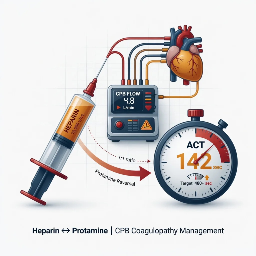
Full heparinisation typically 300–400 IU/kg aiming for ACT ≥400–480 s before full bypass (institutional targets vary slightly; state a range and your hospital’s gate). Typical adult full-flow targets around 2.2–2.5 L/min/m² with MAP often managed in a 50–80 mmHg band unless a higher pressure is chosen for spinal or cerebral reasons. Protamine for neutralisation is classically on the order of 1 mg per 100 IU of heparin given, titrated to ACT and bleeding, with vigilance for adverse reactions.[3]
Cerebral protection physiology
During open arch repair, forward cerebral flow may stop unless selective antegrade cerebral perfusion (SACP) is provided. DHCA reduces cerebral metabolic rate; safe durations are finite and temperature-dependent — shorter is always better. Alpha-stat blood-gas management is commonly used in adults; pH-stat appears in some congenital and DHCA programmes — know that your unit has a protocol and that the viva expects awareness of both names. Cooling and rewarming gradients should be controlled to reduce cerebral injury risk. Near-infrared spectroscopy (NIRS) is often used as a trend monitor for hemispheric asymmetry during SACP.
[2]Pharmacology of induction and maintenance
High-dose opioid techniques, propofol or etomidate carefully titrated, and volatile or TIVA maintenance are all used. Blunt laryngoscopy (opioid, beta-blockade, topical/IV lignocaine as adjuncts) to avoid hypertensive rupture/extension. Have vasopressors ready — induction can unmask tamponade or vasoplegia. Antifibrinolytics (tranexamic acid per cardiac protocol) reduce fibrinolysis on CPB. Inotropes and vasopressors for separation as guided by TOE.
[1]Anaesthetic goals
- Do not delay definitive surgery for Type A once the decision to operate is made.
- Control heart rate then blood pressure to limit shear.
- Maintain coronary and cerebral perfusion — avoid overshoot hypotension.
- TOE-guided structural diagnosis and CPB management.[2]
- Safe cannulation with malperfusion vigilance.
- Cerebral protection plan before the arch is open.
- Spinal protection plan for extensive descending work.
- Anticipate coagulopathy — products, cell salvage, viscoelastic guidance, careful protamine.[1][3]
Technique options and decision matrix
Monitoring strategy
Right radial arterial line is classic when left subclavian or arch work may affect left-sided readings; many teams place right radial plus femoral arterial monitoring to compare proximal and distal pressures and to detect malperfusion. Five-electrode ECG with ST analysis, large-bore venous access, central venous access, TOE, urinary catheter with temperature, nasopharyngeal and bladder or other core temperatures for DHCA, NIRS, and processed EEG in some units. Pulmonary artery catheters are selective, not mandatory.
[2]Induction with tamponade risk
If pericardial blood is present, induce in theatre with surgeon scrubbed and prep/drape ready for immediate sternotomy. Gentle induction, maintain preload and coronary perfusion pressure, avoid pure venodilating high-dose propofol boluses without support. Extreme pure tamponade physiology may need drainage as sternotomy starts.
[2]TOE priorities in Type A
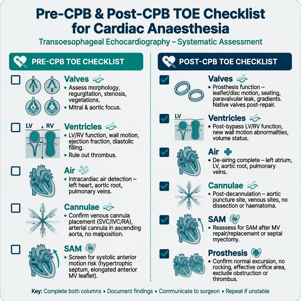
Confirm intimal flap where visible, aortic regurgitation mechanism (leaflet prolapse versus root dilation), pericardial effusion and signs of tamponade physiology, biventricular function, regional wall motion (coronary involvement), and guide cannulation and de-airing. TOE cannot image the entire aorta — distal arch and abdominal aorta need CT correlation when available. Follow a structured ASE/SCA examination mindset rather than two random views.[2]
CPB cannulation options
New malperfusion after starting CPB (falling NIRS, rising lactate, asymmetric pressures, bowel or limb signals, unexplained acidosis) is a cannulation/true-lumen emergency — say it aloud and change strategy with the surgeon immediately (alternate arterial site, true-lumen cannulation, fenestration strategies as indicated).
[2]Arch repair and DHCA
Cool to institutional target for the planned arrest duration; ice to head often used; pharmacological cerebral protection adjuncts vary and are secondary to temperature and SACP. SACP via axillary or direct ostial perfusion maintains some cerebral flow during open arch. Document DHCA and SACP times. Rewarm slowly; watch for vasoplegia and coagulopathy as temperature rises.
[2]Monitoring and equipment (checklist culture)
Perfusion checklist: heparin given and ACT at target before full flow; vents and suckers; cardioplegia plan if root/coronary work; temperature probes; NIRS baseline; blood products in room; cell saver; vasoactive infusions primed; defibrillator pads on; TOE in place before sternotomy when anatomy allows. Protamine only when surgical bleeding control is ready and ACT strategy is clear.[3]
Intraoperative management
- Communicate clamp, arrest, and reperfusion times every phase.
- Manage AI and LV distension on CPB with venting strategies.
- Separate from CPB using a structured approach: rhythm, rate, residual air, ventricular function, valve competence, afterload, coronary perfusion — TOE is central.
- Protamine: slow administration, watch for pulmonary hypertension and systemic hypotension of adverse reactions; support and stop infusion if severe reaction occurs.[3]
- Transfusion: use viscoelastic testing and clinical bleeding; in stable cardiac surgical contexts, restrictive red-cell strategies as in TRICS III reduce transfusion without increasing mortality when carefully applied — do not withhold blood from an exsanguinating open aorta.[1]
Descending aorta / spinal protection
For extensive descending or thoracoabdominal repair (open or hybrid): CSF drainage in selected protocols (pressure targets institutional, infection and haematoma risks), permissive higher distal perfusion pressures when a distal perfusion technique is used, moderate hypothermia, intercostal reimplantation strategies, and neuromonitoring (motor evoked potentials) where available. Avoid hypotension after unclamping. Emergence with neurological exam planning when feasible. (Link conceptually to dedicated thoracoabdominal topics.)
[2]Coagulopathy and transfusion
Deep hypothermia, long CPB, extensive suture lines, and consumptive coagulopathy produce complex bleeding. Algorithm: temperature normalise, surgical haemostasis, protamine adequacy, platelets, fibrinogen/cryoprecipitate or concentrates, plasma as indicated, antifibrinolytics, calcium, correct severe acidaemia, factor concentrates per protocol, and early re-exploration if surgical bleeding is likely. Cell salvage reduces allogeneic exposure. Restrictive thresholds apply when bleeding is controlled and oxygen delivery is adequate.[1][3]
Crisis pivots — what changes the plan
- Tamponade on induction — surgeon-ready sternotomy, support perfusion pressure.
- Malperfusion after cannulation — change arterial inflow strategy immediately.
- Inability to wean from CPB — RRRAC-type structure plus TOE: residual issues, rate/rhythm, residual air, acute ischaemia, contractility, afterload/preload, technical surgical problems.
- Protamine reaction — stop protamine, support RV and systemic pressure, pulmonary vasodilators as indicated, reheparinise and return to bypass in extreme cases.
- Stroke signals — NIRS asymmetry, delayed awakening: optimise pressure, haemoglobin, glucose, temperature; imaging when stable.
- Spinal deficit postoperatively — raise MAP, CSF drain management if present, urgent surgical review of intercostal supply strategy.
- Massive bleeding — major haemorrhage protocol, damage-control packing, correct coagulum.
Postoperative / ICU / PACU plan
Sedation and controlled ventilation until warm, dry, and haemodynamically stable; early neurological assessment when safe. Blood pressure targets that protect anastomoses without hypoperfusion. Watch lactate clearance, urine output, distal pulses, spinal function, drain losses, and TOE or TTE for tamponade if instability returns. Glycaemic control, VTE prophylaxis when bleeding allows, and family communication about stroke risk and recovery trajectory.
[3]Special populations and comorbidities
Pregnancy: rare catastrophic Type A — maternal cardiac surgery with fetal considerations, multidisciplinary. Connective tissue disease (Marfan, Loeys–Dietz): younger patients, fragile tissues, often root replacement strategies. Prior cardiac surgery: redo sternotomy risks. Severe AI and low EF: difficult separation physiology. Renal malperfusion: early RRT planning. Type B complicated: often endovascular first-line in hybrid theatres with different anaesthetic emphases (spinal protection still relevant).
[1]SAQ answer scaffold
A 15-mark SAQ: A 58-year-old man with sudden tearing chest pain has a CT showing Type A dissection involving the arch. BP is 190/100 mmHg, heart rate 110/min.
[2]- Immediate management (3 marks): rate then pressure control; pain relief; activate theatre; avoid delay.
- Monitoring and induction (3 marks): dual arterial strategy concept; TOE; surgeon-ready induction.
- CPB and cerebral protection (5 marks): heparin/ACT; cannulation options; DHCA + SACP; alpha-stat awareness; malperfusion response.
- Bleeding strategy (4 marks): hypothermia coagulopathy; products; viscoelastic; TRICS III context when stable; protamine caution.[1][3]
Viva stem bank and model phrases
Stem 1: “Why beta-block before GTN?”
Model: “I reduce dP/dt first; pure vasodilation with reflex tachycardia can increase shear.”
Stem 2: “Femoral CPB starts and the right radial pressure collapses.”
Model: “I suspect malperfusion or monitoring/true-lumen problems — I announce it and change cannulation strategy with the surgeon immediately.”
Stem 3: “What does TOE add?”
Model: “Flap where visible, AI mechanism, tamponade, ventricular function, cannulation and air — but not a full aorta map.”[2]
Stem 4: “Safe DHCA duration?”
Model: “No unlimited safe time; I minimise arrest duration, use hypothermia and selective antegrade cerebral perfusion per protocol, and document times.”
Stem 5: “Opening sentence for Type A?”
Model: “Beta-block first, control pressure, TOE, prepare for DHCA.”
Common traps
- Treating Type A medically overnight without a surgical plan
- Single arterial line on an involved limb
- No cerebral protection plan for open arch
- Delayed blood product logistics
- Ignoring malperfusion after femoral cannulation
- Aggressive pure vasodilation before heart-rate control
- Withholding blood in exsanguination because of “restrictive transfusion” slogans
- Protamine bolused into a closed pulmonary circulation without vigilance

Detailed haemodynamic control algorithm (exam depth)
In the emergency department or ICU transfer bay, place the patient on continuous ECG and arterial monitoring as soon as possible. Control pain with carefully titrated opioid so that catecholamine-driven hypertension falls without abolishing the airway drive in a semi-conscious patient. Start a short-acting beta-blocker infusion (esmolol is the classic teaching agent because it is titratable and stops quickly if shock appears). Target heart rate commonly in the low 60s when tolerated, then add a vasodilator such as GTN or clevidipine to reach the agreed systolic band. If pure vasodilatation is started first, reflex tachycardia may increase dP/dt and worsen shear — this sequencing error is a classic viva fail. Contraindications to beta-blockade (severe asthma, acute decompensated heart failure, marked bradycardia, high-grade AV block) force alternative negative inotropy/chronotropy strategies (for example careful calcium-channel blockade in selected protocols) with senior input. Never chase a cosmetic blood-pressure number in a patient with coronary ostial compromise and rising lactate; coronary and cerebral perfusion trump neat chart targets.
[2]During transfer to theatre, avoid unnecessary stimulation, keep infusions running, and reassess pulses and neurology on arrival. In theatre, place the second arterial line early if arch or subclavian involvement is known, and do not induce until the surgeon and perfusionist declare readiness when tamponade or rupture risk is high.
[2]Cannulation malperfusion scenarios in more detail
Dynamic obstruction of a branch vessel can change when false-lumen pressure rises after femoral arterial inflow begins. Clinical clues include unilateral loss of arterial waveform, falling NIRS on one side, rising facial plethora asymmetry, new ECG ischaemia, anuria, or a soft abdomen becoming tense with rising lactate. The anaesthetic contribution is immediate recognition and closed-loop communication: say “possible malperfusion” with the specific sign, ask for a change of arterial inflow, support the circulation, and prepare for deep hypothermic strategies if the arch must be opened urgently. TOE may show a flap position change but will not visualise the entire visceral aorta; do not wait for perfect imaging before acting with the surgeon.
[2]Axillary or innominate cannulation provides antegrade true-lumen flow and a natural path to selective antegrade cerebral perfusion, but risks include local vessel injury, brachial plexus issues, and right-arm hyperperfusion. Direct true-lumen aortic cannulation under TOE or epiaortic guidance can be fast in experienced hands. Femoral cannulation remains useful for speed or redo situations but carries the highest false-lumen perfusion anxiety. Venous drainage problems (high central pressures, poor flows) need cannula repositioning, additional venous drainage, and exclusion of obstruction — TOE bicaval views help.
[2]Coagulopathy timeline through the case
Before CPB, baseline coagulation and haemoglobin set the stage; antifibrinolytic loading follows institutional cardiac protocol. On CPB, haemodilution, contact activation, and hypothermia impair platelet function and clotting factor activity. During DHCA, stasis and extreme temperature deepen the defect. After reperfusion and rewarming, fibrinolysis and consumptive losses continue at suture lines. After protamine, residual heparin, protamine excess, and surgical bleeding must be distinguished — ACT, heparinase tests, and viscoelastic assays guide components. A practical order many units use: ensure surgical field control and temperature near normal; confirm adequate protamine; give platelets for qualitative/quantitative defects; replace fibrinogen early when levels are low; use plasma or factor concentrates per protocol; correct ionised calcium; avoid drowning the field with pure crystalloid. Restrictive red-cell thresholds from TRICS III apply when the patient is stable and oxygen delivery is adequate, not during uncontrolled aortic bleeding.
[1]Neurological endpoints and ICU handover
Document DHCA minutes, SACP minutes, lowest temperature, NIRS trends, glucose extremes, and any intraoperative seizures or asymmetry. Plan delayed neurological assessment if residual opioid and temperature still confound examination. Blood-pressure targets in ICU should protect fresh suture lines without watershed hypoperfusion — agree numbers with the surgeon. Spinal cord precautions for extensive descending repair include MAP floor, CSF drain care if present, and hourly neurological checks when sedation allows. Family communication should include stroke and delirium risk honestly.
[2]ANZCA Final cardiac stems expect integration of CPB conduct, ASE/SCA TOE language, and honest limits of imaging. Quote institutional ACT targets and cerebral-protection protocols as the standards you will follow, not improvised numbers under stress.
References
- [1]Mazer CD, Whitlock RP, Fergusson DA, et al. Restrictive or Liberal Red-Cell Transfusion for Cardiac Surgery N Engl J Med, 2017.PMID 29130845
- [2]Hahn RT, Abraham T, Adams MS, et al. Guidelines for performing a comprehensive transesophageal echocardiographic examination: recommendations from the American Society of Echocardiography and the Society of Cardiovascular Anesthesiologists J Am Soc Echocardiogr, 2013.PMID 23998692
- [3]Levy JH, et al. What's fishy about protamine? Clinical use, adverse reactions, and potential alternatives J Thromb Haemost, 2023.PMID 37062523