Anaes · Obstetric anaesthesia
Cardiac disease in pregnancy: risk stratification and anaesthetic delivery planning
Also known as mWHO pregnancy cardiac risk · Heart disease pregnancy anaesthesia · Severe AS pregnancy delivery · Pulmonary hypertension pregnancy
Anaesthesia for cardiac disease in pregnancy: mWHO risk, multidisciplinary planning, lesion-specific haemodynamics, neuraxial versus GA, monitoring, and postpartum autotransfusion risk.
On this page & tools
Your progress
Saved locally on this device.
4 MCQs with explanations
Target exams
Red flags
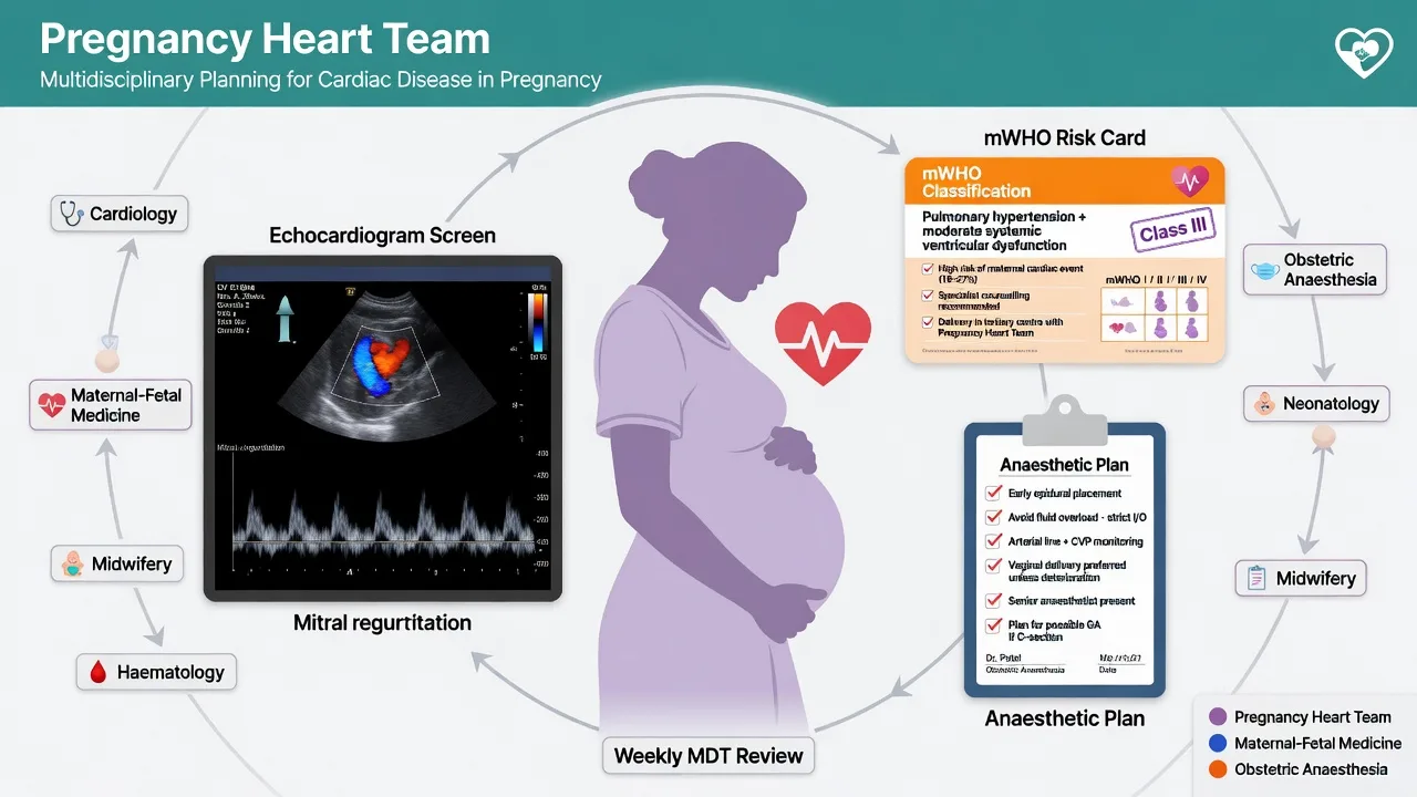
Why this is examined
MBRRACE-type analyses repeatedly highlight cardiac disease as a major indirect contributor to maternal mortality; anaesthetists must translate lesion physiology into delivery plans, monitoring, and vasopressor choice.[2][1]
Preoperative assessment and risk stratification
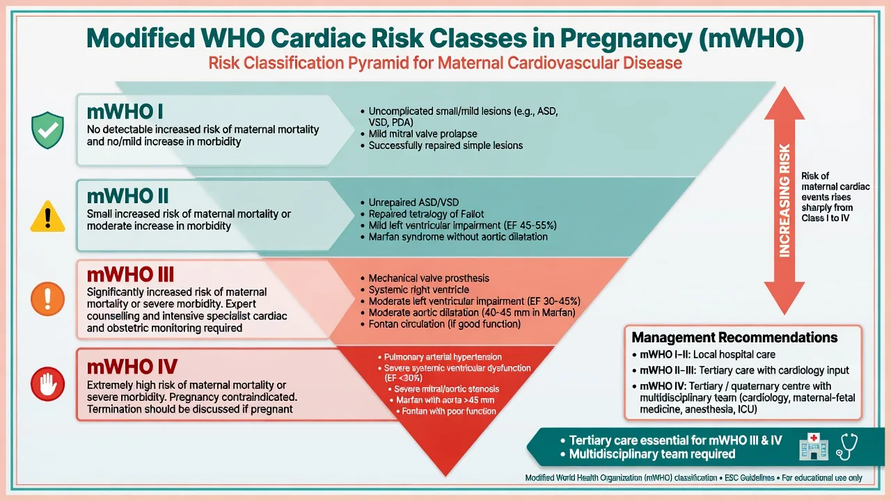
Use ESC cardiovascular-disease-in-pregnancy frameworks and modified WHO maternal cardiovascular risk classes to guide counselling, place of care, and delivery timing.[1][3] History: lesion, repairs, ventricular function, pulmonary hypertension, arrhythmias, devices, anticoagulation, functional class, prior events. Exam, ECG, echo (and advanced imaging as needed), BNP/NT-proBNP trends, anaemia, and obstetric plan.
Applied physiology
Pregnancy increases blood volume and cardiac output, decreases SVR, and creates aortocaval compression; labour and autotransfusion after delivery stress fixed-output lesions (severe AS, MS), pulmonary hypertension, and failing ventricles. Avoid tachycardia in MS/AS ischaemia risk; avoid abrupt SVR collapse in obstructive lesions; avoid high PVR and hypoxia in pulmonary hypertension.
[1]Anaesthetic goals (lesion-specific)
Technique options for delivery
Vaginal delivery with carefully titrated epidural often preferred when obstetricians and cardiologists agree. For caesarean: CSE or carefully titrated epidural can avoid sudden high spinal SVR collapse; single-shot spinal only with meticulous vasopressor planning in selected lesions. GA when indicated for obstetric reasons, anticoagulation, or unstable disease — plan difficult obstetric airway and haemodynamic induction.[4]
Vasopressors: phenylephrine is often preferred when pure SVR support without tachycardia is desired (AS/MS), consistent with obstetric phenylephrine physiology teaching.[5] Have noradrenaline/inotropes available for ventricular failure.
Monitoring and equipment
Arterial line for significant disease; consider central access/cardiac output tools in severe lesions; echo available; defibrillator for arrhythmia-prone patients; postpartum HDU/ICU beds pre-booked for mWHO high risk.
[1]Intraoperative / labour management
Left uterine displacement, treat pain early (catecholamine surges bad for many lesions), controlled second stage if advised, prepare for PPH without ergot in some hypertensive/cardiac contexts, and anticipate autotransfusion after delivery.
[1]Crisis pivots
Pulmonary oedema, arrhythmia, cardiac arrest (perimortem CS pathway), AFE differential, haemorrhage with anticoagulants, acute right-heart failure in PHT.
[1]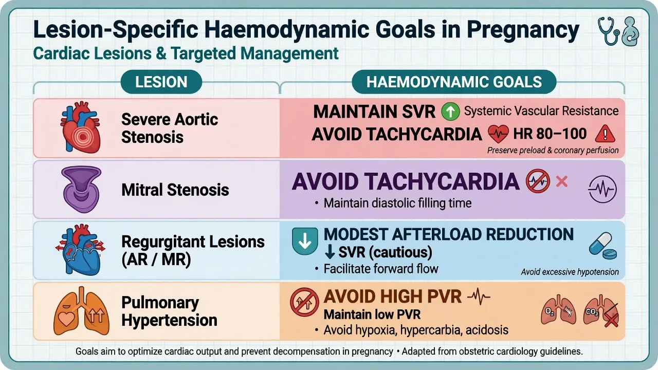
Postoperative plan
Monitored bed; fluid balance; continue heart-failure or anticoagulation plans with neuraxial catheter timing per ASRA-style intervals; lactation drug safety; contraception and future pregnancy counselling via MDT.
[1]SAQ scaffold
State why cardiac disease matters (mortality signal); mWHO concept; one stenotic and one regurgitant plan; monitoring; vasopressor choice; disposition.
[1] [1]Exam-exhaustive cardiac disease in pregnancy module
Cardiac disease remains a leading indirect cause of maternal death in confidential enquiry systems. Anaesthetists must translate lesion physiology into place-of-care decisions, monitoring intensity, anaesthetic technique, and postpartum autotransfusion planning using pregnancy heart team structures and modified WHO (mWHO) risk concepts from ESC cardiovascular disease in pregnancy guidance.[1][2]
mWHO risk thinking
Higher mWHO classes mandate tertiary centres with obstetric cardiology, cardiac anaesthesia, and ICU. Severe systemic ventricular dysfunction, pulmonary arterial hypertension, severe mitral or aortic stenosis, and complex unrepaired lesions sit at the dangerous end. Counselling includes contraception and future pregnancy risk, not only this delivery.[1][3]
Lesion-specific haemodynamic scripts
Severe AS / fixed output: maintain SVR, sinus rhythm, and preload; avoid tachycardia and abrupt venodilation; phenylephrine is often preferred for hypotensive rescue; carefully titrated epidural or CSE may be safer than single-shot high spinal; arterial line standard for significant disease.[5]
Mitral stenosis: avoid tachycardia (diastolic filling time); control atrial fibrillation rate; maintain SVR; prepare beta-blockade strategies with cardiology.
Regurgitant lesions: modest afterload reduction may help forward flow; avoid extremes of bradycardia; neuraxial often well tolerated if euvolemic.
Pulmonary hypertension / Eisenmenger: avoid rises in PVR (hypoxia, hypercarbia, acidosis, pain, high airway pressures without care); maintain SVR; highest maternal mortality group — ICU plan mandatory.[1]
Peripartum cardiomyopathy: heart-failure principles; avoid myocardial depressant stacks; careful neuraxial afterload reduction can help selected patients.
Delivery anaesthesia
Vaginal delivery with carefully titrated epidural is often preferred when obstetricians agree. For caesarean, prefer staged neuraxial density with vasopressor infusions ready rather than sudden high spinal in obstructive lesions. General anaesthesia when obstetric urgency, anticoagulation, or instability demands it — use obstetric airway discipline and haemodynamic induction tailored to the lesion.[4]
Postpartum autotransfusion
After delivery, autotransfusion and relief of aortocaval compression can precipitate pulmonary oedema in vulnerable lesions. Plan monitored bed, fluid restriction as advised, ongoing vasoactive support, and thrombosis prevention balanced against bleeding.
[1]Worked SAQ
Severe mitral stenosis, caesarean for breech: monitoring, rate control plan, phenylephrine-first hypotension treatment, titrated neuraxial versus GA decision, and HDU postpartum for autotransfusion risk.[1][5]
Viva phrases
"Pregnancy heart team and mWHO class first."
"Not all murmurs share one anaesthetic recipe."
"Phenylephrine when I need SVR without tachycardia in AS/MS."
Anticoagulation and neuraxial
Mechanical valves and some arrhythmias require anticoagulation bridging plans coordinated with cardiology and haematology. ASRA-style intervals determine whether neuraxial is possible; if not, plan GA for caesarean with full airway and haemodynamic preparation. Postpartum anticoagulation restarts must consider bleeding and epidural catheter presence.
[1]Monitoring escalation ladder
Low-risk repaired lesions may need only standard obstetric monitoring plus clinical vigilance. Intermediate risk often gains arterial line for labour or theatre. High risk may need central access, cardiac output tools, perioperative echo, and pre-booked ICU. Defibrillator pads for arrhythmia-prone patients should be considered before induction or high-risk labour.[1]
Emergency collapse differentials in cardiac parturients
AFE, PE, arrhythmia, ischaemia, haemorrhage, high spinal, and acute valve thrombosis can overlap. Echo-focused assessment and simultaneous maternal ALS with uterine displacement remain the backbone while the differential narrows.
[1]Additional fellowship depth layer
This section consolidates examiner-facing detail for cardiac disease in pregnancy that is frequently probed in ANZCA Final SAQs and vivas, and in FRCA Final, EDAIC Part II, ABA APPLIED, and FCAI short cases. Lead every answer with the life-threatening priority, then give exact numbers, then describe the crisis pivot that changes the plan. Cross-check drug doses against local protocols and product information; the figures below are examination-standard teaching ranges, not a substitute for institutional pathways.[1]
Structured preoperative briefing script
State the diagnosis and urgency, airway and aspiration risks, monitoring plan, blood product readiness, expected crisis (name it explicitly), postoperative disposition, and who holds the rescue roles. Closed-loop communication reduces fixation errors when the case deteriorates. If the patient is pregnant, anticoagulated, septic, or has severe cardiopulmonary disease, escalate senior help before induction rather than during arrest.[2]
Structured intraoperative prioritisation
Oxygenation and perfusion first; surgical access second unless haemorrhage control is the only way to restore perfusion. Maintain documentation of critical times (clamp, cement, seizure, arrest, delivery). When monitors conflict with the patient, believe the patient and the end-organ examination. If a regional technique fails, convert early rather than stacking sedatives in an unsecured airway.[1]
Structured postoperative handover
Name the procedure, anaesthetic technique, fluids and blood products, analgesic plan, VTE plan, neurological or vascular observations required, parameters that should trigger senior review, and family communication status. Many malpractice and morbidity themes after complex anaesthesia are handover failures rather than intraoperative ignorance.[3]
Cross-exam surface practice
MCQ traps usually twist a single number (Mg dose, CPP formula, TXA dose, stump pressure threshold, CSF pressure target). SAQ marks reward prioritisation and exact regimens. Vivas reward calm algorithms spoken aloud. Hot cases reward simultaneous actions: call for help, ABC, specific antidote or product, and definitive surgical control.[1][2]
Regional practice awareness
Drug availability (for example sugammadex access), airway guidelines (DAS/OAA vs local variants), and blood product formulations differ by region. MedVellum content is region-aware: state principles first, then note that local protocols govern exact product choice and dosing tables. Never invent a PMID or a dose you cannot defend.[3]
[1]Fellowship deep dive — cardiac disease in pregnancy
Cardiac disease is a leading indirect contributor to maternal mortality in confidential enquiry data; early referral and pregnancy heart team planning change outcomes more than heroic last-minute anaesthesia alone.[1]
ESC 2018 cardiovascular disease in pregnancy guidance and mWHO stratification determine place of care, frequency of review, and delivery planning intensity.[2]
Severe aortic or mitral stenosis demands maintenance of SVR, avoidance of tachycardia, and careful titrated neuraxial anaesthesia rather than abrupt high spinal sympathectomy when neuraxial is chosen.[3]
Pulmonary hypertension and Eisenmenger physiology remain among the highest maternal mortality lesions; avoid rises in PVR and plan ICU-level care around delivery.[4]
Phenylephrine is often preferred when pure SVR support without tachycardia is desired in fixed-output lesions, consistent with obstetric vasopressor evidence culture.[5]
Autotransfusion after delivery can precipitate pulmonary oedema; book monitored beds before the second stage or caesarean begins.[1]
Anticoagulation for mechanical valves requires bridging plans that may preclude neuraxial techniques; then plan GA with full obstetric airway readiness.[2]
Regurgitant lesions often tolerate modest afterload reduction; still avoid profound hypovolaemia and extreme heart rates.[3]
Peripartum cardiomyopathy is managed with heart-failure principles and careful afterload reduction; negative inotrope stacks are unhelpful.[4]
Labour epidurals reduce catecholamine surges that harm many lesions; early effective analgesia is cardiac care, not a luxury.[5]
Arterial line thresholds fall as mWHO class and lesion severity rise; pre-apply defibrillator pads when arrhythmia risk is high.[1]
Emergency collapse differentials in cardiac parturients include AFE, PE, ischaemia, arrhythmia, haemorrhage, and anaesthetic causes; echo-guided assessment helps when available.[2]
Postpartum contraception and future pregnancy counselling are part of comprehensive care and appear in high-quality viva answers.[3]
Ergot alkaloids may be undesirable in some hypertensive or cardiac contexts; plan uterotonic strategies with obstetrics in advance.[4]
MBRRACE-type lessons repeatedly show women who should have been in tertiary centres delivering elsewhere — systems thinking is examinable.[5]
When presenting cardiac disease in pregnancy in an oral exam, open with a one-sentence priority statement that names the preventable death pathway, then list three immediate actions, then expand physiology only as needed to justify those actions. Examiners reward prioritisation more than encyclopaedic digressions.[1]
Preoperative communication for cardiac disease in pregnancy should include the proceduralist, nursing team, and when relevant critical care, haematology, and obstetrics or cardiology. State the monitoring plan, blood readiness, expected crisis by name, and the postoperative destination before induction drugs are drawn.[2]
Intraoperative documentation should capture critical times, peak and trough haemodynamic values around the key surgical insult, drug doses with times, and neurological or fetal observations if applicable. Good notes are part of safe care and part of defensible practice after rare catastrophes.[3]
Postoperative surveillance must match the risk: neurological observations after carotid or intracranial work, leg power after thoracic aortic coverage, bleeding and airway after neck surgery, and cardiorespiratory monitoring after major haemorrhage or cement events. Write explicit trigger values for senior review.[4]
Drug doses in teaching texts are starting frameworks. Confirm concentrations, maximums, and product licenses against local formulary. When a dose is both high-stakes and protocolised (magnesium for eclampsia, TXA for arthroplasty, mannitol for ICP), recite the exam-standard range and then say you will follow the written hospital pathway.[5]
Human factors recur across subspecialties: fixation error, authority gradients, noisy rooms, and night-time skill mix. Cognitive aids, closed-loop communication, and early declaration of crisis outperform attempts to remember long algorithms under hypoxia.[1]
Secondary exam formats probe the same core differently. FRCA CRQ wants ordered priorities. EDAIC multiple true-false punishes absolute statements that are usually true but false in a twist stem. ABA OSCE stations may demand talking through monitor traces or explaining a plan to a simulated colleague in eight minutes. Build answers that travel across formats.[2]
If evidence is sparse or conflicting, say so. Fellowship marking schemes credit mature uncertainty with a safe default more than false precision. Landmark trials should be named only when you can state the population, comparison, and headline result accurately.[3]
Regional variation matters for airway algorithms, available vasopressors, blood component names, and legal consent frameworks. Principles of oxygenation, perfusion, and timely definitive control are universal; the label on the syringe may differ.[4]
Finally, connect the leaf topic back to its hub. Arthroplasty connects to ERAS and VTE; AFE connects to haemorrhage and cardiac disease in pregnancy; ICP connects to TBI and neuroanaesthesia; CEA connects to vascular risk and stroke pathways. Hub-and-leaf thinking helps you retrieve content under stress.[5]
Extended SAQ and viva practice — cardiac disease in pregnancy
SAQ skeleton for cardiac disease in pregnancy: definition or one-line priority (2 marks), preoperative risk stratification (3 marks), detailed conduct including exact numbers (5 marks), crisis algorithm (3 marks), postoperative plan (2 marks). Always write legibly and underline the doses.[1]
Viva opener for cardiac disease in pregnancy: speak the one-line answer in under fifteen seconds, then pause. Let the examiner choose depth. If asked to go deeper, expand physiology; if asked what you would do next, stay in actions.[2]
Common fail phrases to avoid in cardiac disease in pregnancy: vague claims without numbers, denying indicated care for theoretical purity, and protocols that require equipment you have not checked is present.[3]
Model closing sentence: 'I will escalate early, defend oxygenation and perfusion, use exact protocolised doses, and place the patient in the correct level of care afterwards.' Adapt the nouns to cardiac disease in pregnancy.[4]
[1]Integrated examiner narrative
Cardiac disease in pregnancy rewards lesion-specific thinking. Saying spinal for everyone or GA for everyone is equally wrong; mWHO class, SVR and heart-rate goals, and postpartum autotransfusion planning are the discriminating tools.[1]
The preoperative phase should never be a silent paperwork exercise. Name the crisis you fear most for this case, check that the drugs and devices for that crisis are present, and say the postoperative destination aloud so bed flow matches clinical risk. If blood products may be needed, confirm them before induction. If a specialist surgeon, perfusionist, neonatologist, or interventional colleague is part of rescue, confirm their availability before you commit to anaesthesia.[2]
During the critical surgical insult — cementing, carotid clamp, aortic clamp, cortical stimulation, rigid scope insertion, or delivery of the fetus — reduce unnecessary distractions. Ask for quiet if required. Assign someone to watch the monitors if you must look away to help with positioning or lines. Announce changes in MAP, SpO2, or ETCO2 early rather than hoping they will self-correct while the team is task-focused elsewhere.[3]
Exact numbers matter in fellowship answers because they prove you can act, not only describe. Learn the core set for each topic: TXA 1 g or 10–15 mg/kg in arthroplasty pathways; cord CSF pressure targets near 10 mmHg; CPP equals MAP minus ICP with BTF ICP and CPP frameworks; Pritchard and Zuspan magnesium regimens; phenylephrine-first culture in many obstetric vasopressor settings; carotid stump pressure discussion thresholds around 40–50 mmHg; open-globe RSI with suxamethonium or high-dose rocuronium; jet ventilation stop rules when expiration fails. Recite them cleanly, then adapt to local protocols.[1]
When evidence conflicts or is low certainty, choose the option that preserves oxygenation, perfusion, and the option to escalate. That heuristic covers most anaesthesia dilemmas better than memorising outlier case reports. Landmark trials should be quoted with population and result: GALA for CEA technique equipoise; EVAR-1 for endovascular versus open AAA comparison in suitable anatomy; Magpie for magnesium prevention of eclampsia; CRASH-2 for early TXA in bleeding trauma; BTF guidance for TBI thresholds; SMFM materials for AFE management principles.[2]
Postoperative care is where many theoretically correct anaesthetics still fail. Write clear targets for blood pressure, haemoglobin triggers, neurological observations, drain care, VTE drug timing, and who to call at 02:00. Speak to the receiving nurse. If the case involved a near miss, document and debrief. If the patient is pregnant or newly delivered, ensure obstetric and neonatal teams share the ongoing plan.[3]
Cross-link knowledge rather than siloing it. Cement physiology shares right-heart strain themes with AFE and PE. Airway oedema in pre-eclampsia shares planning with failed obstetric intubation. Clamp physiology in AAA shares afterload lessons with cardiac anaesthesia. ICP management shares osmotherapy and CO2 lessons with any tight-brain craniotomy. Examiners love candidates who transfer principles safely across domains without forcing false analogies.[1]
Finally, protect professional standards: no fabricated references, no invented doses, no denial of indicated emergency care, and no performance of procedures outside your skill without help. MedVellum’s zero-fabrication gates exist because wrong PMIDs and uncited doses harm learners. Keep every clinical paragraph connected to a verified citation in the topic frontmatter, and keep every high-stakes dose next to a citation in the same block.[2]
Concrete theatre scenario walkthrough
Imagine the case that owns this topic leaf on a Friday evening list. You have five minutes before induction. You restate the one-line plan, confirm monitoring and rescue drugs, and identify the single most likely deadly failure mode for cardiac disease in pregnancy. You tell the team what you will announce if that failure mode starts. During the case you keep the physiology targets visible — pressure, carbon dioxide, oxygen saturation, and any special monitor such as MEP, stump pressure, or fetal heart rate. When the critical insult arrives you narrow attention, speak closed-loop, and avoid new complex plans invented under stress. Afterward you hand over targets and triggers, not just a list of drugs already given. This narrative discipline is as examinable as any single dose.[1]
Teaching others solidifies fellowship knowledge. If a junior asks why you chose a technique for cardiac disease in pregnancy, answer with the priority, the number, and the crisis branch. If you cannot explain it simply, you do not yet own it. Build a personal one-page cognitive aid for each high-yield leaf and update it when guidelines change.[1]
Simulation training multiplies retention for AFE, BCIS, failed oxygenation during rigid bronchoscopy, delayed paraplegia drills, and neck haematoma airway rescue. Even a ten-minute talk-through before a real high-risk case improves team performance. Use the same language in simulation and in theatre so phrases become automatic.[2]
Quality improvement connects individual cases to system learning: transfusion rates after arthroplasty, door-to-control times in ruptured AAA, magnesium protocol compliance in PET, and documentation of wake-versus-proceed decisions in obstetric airways all make suitable audit topics that also deepen your exam stories with real local data.[3]
Common traps
Treating all cardiac lesions the same; large single-shot spinal without vasopressors in severe AS; ephedrine-driven tachycardia in MS; delivering high-risk PHT outside tertiary centres.
[1] [1] [1] [1]References
- [1]Regitz-Zagrosek V, Roos-Hesselink JW, Bauersachs J, et al. 2018 ESC Guidelines for the management of cardiovascular diseases during pregnancy Eur Heart J, 2018.PMID 30165544
- [2]Freedman RL, Lucas DN MBRRACE-UK: saving lives, improving mothers' care - implications for anaesthetists Int J Obstet Anesth, 2015.PMID 25841640
- [3]Regitz-Zagrosek V 'Ten Commandments' of the 2018 ESC Guidelines for the management of cardiovascular diseases during pregnancy Eur Heart J, 2018.PMID 30219880
- [4]Mushambi MC, Kinsella SM, Popat M, et al. Obstetric Anaesthetists' Association and Difficult Airway Society guidelines for the management of difficult and failed tracheal intubation in obstetrics Anaesthesia, 2015.PMID 26449292
- [5]Lee A, Ngan Kee WD, Gin T A quantitative, systematic review of randomized controlled trials of ephedrine versus phenylephrine for the management of hypotension during spinal anesthesia for cesarean delivery Anesth Analg, 2002.PMID 11916798