Anaes · Thoracic anaesthesia
Anaesthesia for lung transplantation
Also known as Bilateral sequential lung transplant anaesthesia · Primary graft dysfunction PGD · LTx ECMO anaesthesia
Exam-pass lung transplant anaesthesia: end-stage lung disease physiology, induction risks, PA clamping and reperfusion, ECMO/CPB triggers, primary graft dysfunction, and ICU ventilation strategies.
On this page & tools
Your progress
Saved locally on this device.
Target exams
Red flags
Why this is examined / the one-line answer
Lung transplantation (LTx) is the extreme thoracic case: end-stage respiratory failure, pulmonary hypertension and right ventricular (RV) risk, OLV and PA-clamp physiology, and primary graft dysfunction (PGD). International multi-society consensus emphasises the anaesthetist across the whole pathway from induction to ICU — this is not “a long lobectomy”.[1]
One-liner: I prepare for haemodynamic collapse at induction and PA clamp, have ECMO/CPB ready when risk is high, reperfuse and ventilate the graft protectively, keep the patient relatively dry, and watch for PGD. [1]
Preoperative assessment
Disease clusters (say them)
| Cluster | Anaesthetic implications |
|---|---|
| COPD / emphysema | Hyperinflation, bullae, dynamic hyperinflation risk, often better RV than IPF/PHT |
| ILD / IPF | Stiff small lungs, pulmonary hypertension common, difficult ventilation |
| CF / bronchiectasis | Secretions, infection, diabetes, liver disease, sinus disease, prior thoracic surgery |
| Pulmonary hypertension | Highest induction/clamp RV risk |
| Retransplant / ECMO bridge | Hostile chest, bleeding, complex support weaning |
| Combined heart–lung | Full cardiac transplant overlap |
Single vs bilateral sequential vs heart–lung changes clamp sequence and native lung issues (single LTx: native lung still participates in gas exchange and complications). [1]
Work-up that changes the plan
RV function and PA pressures (echo/cath), coronary disease, renal/liver function, infection/colonisation (CF), antibodies/crossmatch logistics, airway anatomy, last meal if urgent offer, frailty, previous ECMO, transplant team protocols for immunosuppression induction timing.[2]
Applied physiology by phase
1. Induction
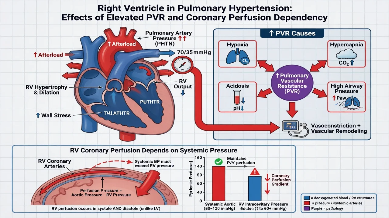
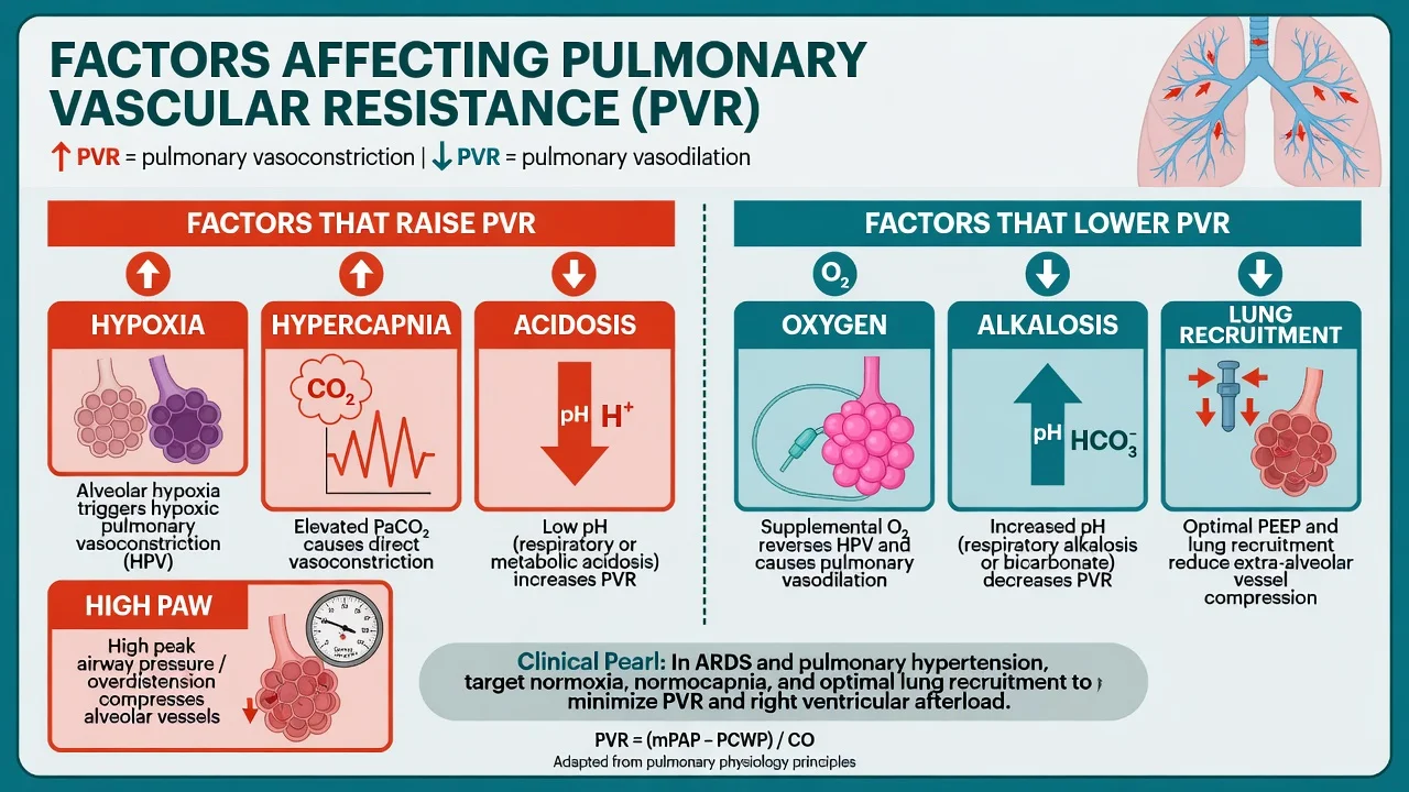
Hypoxia, hypercarbia, loss of sympathetic tone, and positive-pressure ventilation raise PVR, stress a fragile RV, and can cause arrest — especially in severe PHT/ILD. Strategies: optimise oxygenation/ventilation pre-induction when possible, careful titrated induction, pulmonary vasodilator readiness (oxygen, iNO), vasopressor/inotrope plan for RV (maintain coronary perfusion pressure + support contractility), and ECMO/CPB primed readiness in high-risk recipients.[1][2]
2. Dissection / OLV / PA clamp
Clamping the pulmonary artery of the operative side acutely increases RV afterload through the remaining pulmonary vascular bed. A clamp trial may predict need for mechanical circulatory support. Bilateral sequential transplant: implant first lung, then address the second side — the new graft becomes the gas-exchange and afterload partner. [1]
3. Reperfusion
Ischaemia–reperfusion injury produces a spectrum from mild oedema to severe PGD: hypoxaemia, reduced compliance, pulmonary oedema, sometimes pulmonary hypertension. Gentle reperfusion, controlled pulmonary pressures, protective ventilation, and avoidance of fluid overload are the anaesthetic contribution.[1]
4. Chest closure
Compliance falls; bleeding and clamshell pain matter; gas exchange may deteriorate — be ready to reopen or escalate support. [1]
Anaesthetic goals
- Survive induction without RV death.
- Facilitate surgical anastomoses with controlled haemodynamics.
- Support RV during clamp or unload with ECMO/CPB when needed.
- Protect the new graft (ventilation + fluids).
- Hand over a coherent ICU plan for PGD, infection, and immunosuppression. [1]
Technique matrix
Monitoring and equipment
- Arterial line(s); large venous access.
- PA catheter and/or TOE — RV size/function, PA pressures, volume status, exclude tamponade later.
- Lung isolation devices (DLT/blocker) as surgical approach requires; flexible bronchoscope for anastomoses.
- iNO available; vasopressors and inotropes drawn.
- Defibrillator; CPB/ECMO team and circuit readiness per risk stratification.
- Blood products; cell salvage per unit policy.
- Temperature; urine output; ACT if heparinised support.
- Immunosuppression drugs timed with surgical team protocol. [1]
Intraoperative management
Usually GA with full invasive monitoring. Antibiotics and induction immunosuppression per protocol. Fluid strategy: restrictive/goal-directed bias to protect graft, while not underfilling a preload-dependent RV — this is a communication sport with TOE/PAC data, not a single number religion.[1]
After reperfusion: protective tidal volumes, adequate PEEP, bronchial toilet, careful FiO2 (later aim to minimise unnecessary hyperoxia when stable), assess anastomoses bronchoscopically. Transfuse for oxygen delivery and surgical bleeding with awareness that excess volume harms graft oedema; cardiac restrictive transfusion evidence is context for “not all anaemia needs liberal RBC” rather than a direct LTx RCT mandate.[3]
Crisis pivots
Arrest on induction
ACLS plus emergency mechanical support — this is why the circuit plan was spoken aloud before drugs. [1]
RV failure on PA clamp
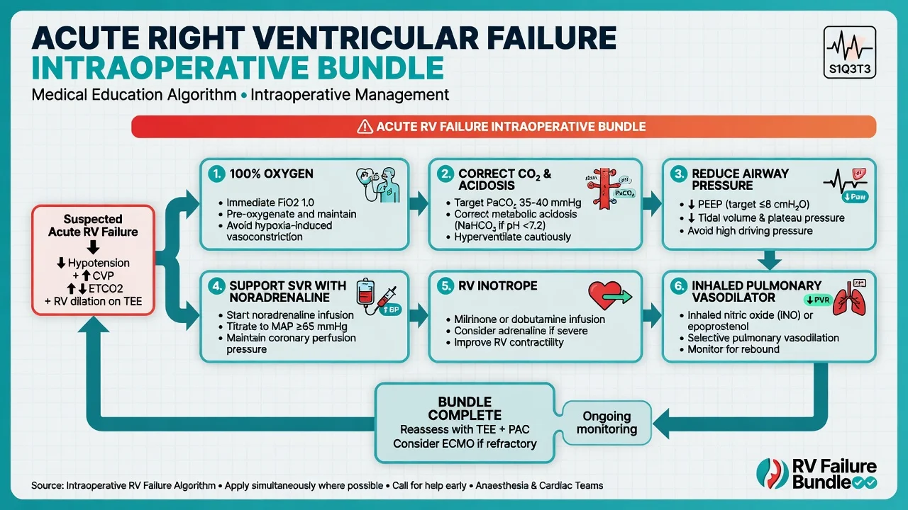
100% oxygen, correct hypercarbia/acidosis, iNO, support systemic pressure for RV coronary perfusion, inotrope for RV, unload with ECMO/CPB if refractory.[2]
Severe graft dysfunction after reperfusion
Protective ventilation, diuresis if overloaded, exclude technical anastomotic problems (surgical/bronch/TOE), escalate to ECMO (often VV for pure gas-exchange failure; VA if concomitant RV/systemic failure). [1]
Bleeding / clamshell
Surgical control + products; watch for haemothorax and impaired venous return. [1]
Dynamic hyperinflation (COPD phenotype)
Suspect if high airway pressures, hypotension that improves with disconnect — allow expiratory time, reduce rate, communicate. [1]
Postoperative / ICU
Protective ventilation, early bronchoscopic checks, PGD grading surveillance, immunosuppression and infection prophylaxis, analgesia for clamshell/thoracotomy, renal protection, glycaemic control (especially CF/steroids), physiotherapy. Consensus documents stress integrated anaesthetic–ICU pathways rather than “throw over the wall”.[1]
Special populations
- CF: secretions, resistant organisms, diabetes, liver disease — plan toilet and infection drugs.
- IPF with PHT: highest vigilance at induction/clamp.
- ECMO bridge to transplant: know configuration (VV vs VA) and weaning goals.
- Single lung transplant: native lung disease still affects ventilation (e.g. native emphysema hyperinflation).
- Retransplant: bleeding, difficult dissection, sensitisation. [1]
SAQ answer scaffold
Discuss the anaesthetic management of bilateral sequential lung transplantation. [1]
- Recipient disease & RV (3): phenotype, PHT, investigations.[2]
- Induction (3): RV protection, support drugs, ECMO readiness.[1]
- Clamp phase (3): afterload, clamp trial, mechanical support triggers.
- Reperfusion/PGD (3): gentle reperfusion, protective vent, dry-ish fluids.
- ICU (2): PGD watch, immuno, infection, analgesia.
Viva stem bank and model phrases
Stem 1: “Why might they arrest on induction?”
Model: “Positive pressure, hypoxia, hypercarbia and loss of sympathetic tone raise PVR and can precipitate acute RV failure in severe pulmonary hypertension.” [1]
Stem 2: “What is PGD?”
Model: “Primary graft dysfunction is ischaemia–reperfusion injury of the allograft — oedema, hypoxaemia, poor compliance — managed with protective ventilation, fluid discipline, and ECMO if needed.”[1]
Stem 3: “Fluids — dry or full?”
Model: “Relatively dry for the graft, but I still fill the RV appropriately using TOE/PAC — empty RV failure is not a virtue.” [1]
Stem 4: “When do you want ECMO?”
Model: “High-risk induction/clamp RV crisis, refractory gas-exchange failure after reperfusion, or planned support in extreme PHT — decided with the surgical team before crisis if possible.” [1]
Stem 5: “iNO role?”
Model: “Selective pulmonary vasodilation to lower PVR and support RV performance and V/Q during critical phases.” [1]
Stem 6: “Single lung transplant specific problem?”
Model: “The native lung still competes — hyperinflation of native emphysema can compress the graft; ventilation strategy must account for both lungs.” [1]
Stem 7: “Immunosuppression timing?”
Model: “I follow the transplant protocol with the team — induction agents are timed to reperfusion/implantation, not improvised.” [1]
Common traps
- Deep induction in severe PHT without support plan
- No mechanical support plan spoken aloud
- Flooding the new lungs
- Missing RV failure on clamp
- Treating LTx like routine lobectomy
- Ignoring native lung issues in single LTx
- Handover without PGD surveillance plan [1]
LTx phases — IRPE
Examiner mental map
- Recipient phenotype and RV/PHT.
- Induction risk and rescue.
- PA clamp physiology and support.
- Reperfusion / PGD.
- Protective graft ventilation + dry-ish fluids.
- ICU integrated pathway. [1]
Phase-based answers win; organ lists without phases fail. [1]
References
- [1]Marczin N, de Waal EEC, Hopkins PMA, et al. International consensus recommendations for anesthetic and intensive care management of lung transplantation. An EACTAIC, SCA, ISHLT, ESOT, ESTS, and AST approved document J Heart Lung Transplant, 2021.PMID 34732281
- [2]Kim HJ, Shin JM, et al. A Review of Anesthesia for Lung Transplantation J Chest Surg, 2022.PMID 35924536
- [3]Mazer CD, Whitlock RP, Fergusson DA, et al. Restrictive or Liberal Red-Cell Transfusion for Cardiac Surgery N Engl J Med, 2017.PMID 29130845