Anaes · Head & neck / trauma airway
Maxillofacial trauma airway management
Also known as maxillofacial trauma airway
Exam-exhaustive maxillofacial trauma airway: Le Fort and mandible threats, C-spine, dual set-up RSI or awake FOI, avoid blind nasal routes with midface/BOFS risk, DAS limits, FONA readiness, IMF extubation safety, and CRASH-2 TXA context for bleeding trauma.
On this page & tools
Your progress
Saved locally on this device.
8 MCQs with explanations
Target exams
Red flags
Why this is examined / the one-line answer
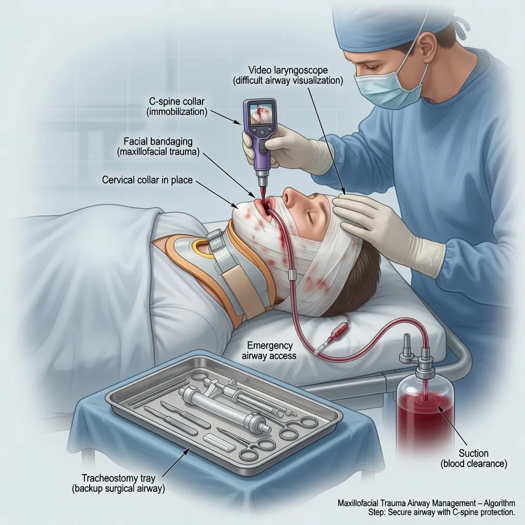
Maxillofacial trauma compresses the fellowship airway syllabus into one case: distorted anatomy, blood and full stomach, C-spine precautions, progressive swelling on a clock, and shared decision-making with surgeons about primary tracheostomy versus oral intubation versus awake flexible techniques. Examiners punish repeated oral attempts while the face swells and reward early declaration of difficulty with a dual set-up for front-of-neck access. [1]
The one-line answer: I treat maxillofacial trauma as a shared difficult airway — suction blood, protect the C-spine, avoid blind nasal intubation when midface or base-of-skull injury is possible, choose awake flexible intubation or video-laryngoscopy RSI with a double set-up for immediate FONA, limit attempts per DAS logic, and do not extubate into progressive oedema without a plan.[1]
Preoperative assessment and risk stratification
Mechanism and time course
Blunt (road traffic, assault, falls) versus penetrating. Time since injury matters: a manageable airway at one hour may obstruct at four hours from haematoma and oedema. Progressive voice change, drooling, stridor, tripod posture, and rising oxygen requirement are hard indications for early definitive airway before prolonged CT delays if the patient is deteriorating. [1]
Fracture pattern and soft tissue
Assess for: [1]
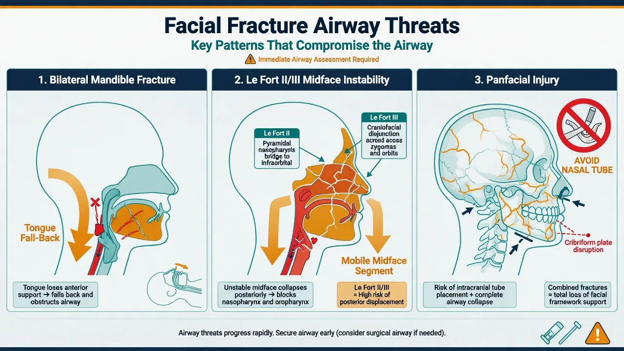
- Bilateral mandibular fractures (“flail mandible”) with posterior tongue fall-back
- Le Fort midface patterns and maxillary mobility
- Panfacial injury combining threats
- Soft-tissue swelling, floor-of-mouth haematoma, expanding neck haematoma
- Dental avulsions and loose teeth (aspiration risk)
- Trismus (mechanical versus pain; may worsen with muscle spasm)
- Base-of-skull signs (Battle’s sign, raccoon eyes, CSF rhinorrhoea/otorrhoea) — nasal route hazard
- Associated TBI, chest injury, cervical spine injury, and haemorrhage [1]
Full stomach and C-spine
Assume full stomach. Assume C-spine injury until cleared — manual in-line stabilisation (MILS) during airway interventions; remove the anterior collar for laryngoscopy while maintaining MILS, then replace. Immobilisation must never delay oxygenation. [1]
Haemorrhage and TXA
Significant bleeding trauma patients benefit from early tranexamic acid in the CRASH-2 paradigm (1 g IV over 10 minutes then infusion protocol as used in trauma systems) when within the evidence time window and without clear contraindications — coordinate with major haemorrhage protocols.[2]
Applied anatomy and fracture airway threats
Le Fort classification (exam skeleton)
- Le Fort I: low maxillary fracture (floating palate) — bleeding, some mobility; airway often manageable but blood is the enemy.
- Le Fort II: pyramidal midface — nasal and infraorbital involvement; epistaxis; increasing instability.
- Le Fort III: craniofacial disjunction — complete midface separation from skull base; major bleeding; base-of-skull association; blind nasal intubation contraindicated. [1]
Mandible
Bilateral body fractures allow the anterior mandible and tongue base to drop posteriorly in the supine patient — classic indication for early definitive airway or careful prone/lateral positioning only as a temporary bridging manoeuvre in extreme field care, not as theatre strategy. [1]
Soft tissue and delayed obstruction
Expanding haematoma in the floor of mouth or neck can convert a “stable for CT” patient into CICO. Serial reassessment beats a single ED glance. [1]
Anaesthetic goals
- Oxygenation and ventilation first — always.
- Definitive protected airway with minimal aspiration.
- Limit trauma from repeated attempts (DAS: declare failure early).[1]
- Maintain C-spine protection without sacrificing the airway.
- Coordinate haemorrhage control and TXA with the trauma team.[2]
- Shared plan for intermaxillary fixation (IMF), throat packs, and extubation or tracheostomy.
- Protect eyes and brain if concurrent injuries exist.
Technique options and decision matrix
Team brief before drugs
Who holds MILS, who suctions (two large-bore Yankauers), who performs laryngoscopy, who is ready for FONA/surgical tracheostomy, what the plan is if Plan A fails, whether primary surgical airway is preferred, blood products available, and head-up if bleeding allows without haemodynamic collapse. [1]
Options
- Awake flexible bronchoscopic intubation — cooperative patient, predicted difficult bag-mask, time available, topicalisation expertise; hard with copious blood (soiling of optic and patient intolerance).
- RSI with video laryngoscopy — common for emergency; dual set-up with scalpel-bougie-tube kit open; bougie loaded; smaller tube sizes ready; gentle first attempt is the best attempt.
- Primary surgical airway / awake tracheostomy under local — when anatomy predicts near-certain failure of oral routes (massive disruption, some penetrating injuries).
- Submental intubation — specialised intraoperative request for panfacial repair when nasal and oral tubes both obstruct the field — not an emergency ED first move. [1]
DAS 2015 adult principles: maximise first-attempt success, limit attempts, maintain oxygenation, second-generation SGA as rescue when appropriate (may sit poorly with disrupted anatomy), declare CICO early, front-of-neck access without delay.[1]
Nasal tubes
Avoid blind nasotracheal intubation with midface fractures or suspected base-of-skull fracture — risk of intracranial tube placement is the classic catastrophic teaching point. If surgeons request a nasal tube later for fixation preference, only after imaging and senior agreement, preferably under vision. [1]
Cricoid and blood
Cricoid force may help or hinder; blood needs suction more than ritual. Full-stomach strategy remains, but oxygenation beats dogma. [1]
Monitoring and equipment
Two large-bore suctions, video laryngoscope, multiple blade geometries, bougies, stylets, second-generation SGAs, flexible scope if awake plan, surgical tracheostomy tray open in the room, front-of-neck kit, blood products and rapid infuser if major haemorrhage, TXA drawn when indicated, difficult airway trolley, and waveform capnography before reliance on tube position.[1][2] Standard AAGBI/ANZCA monitoring; arterial line if major haemorrhage or polytrauma.
Intraoperative management
After secure airway: throat pack with rigorous count, eye protection, head-up if neurosurgically and haemodynamically acceptable for bleeding, antibiotic and tetanus per trauma protocols, blood loss tracking, and communication about IMF wiring. Keep ignition sources and oxidiser controlled if diathermy near the airway (fire triad thinking). Tube exchange only with a plan (airway exchange catheter, surgical backup). If regional adjuncts are used for other injuries, LAST preparedness remains relevant to the overall trauma anaesthetic.[3]
Crisis pivots — what changes the plan
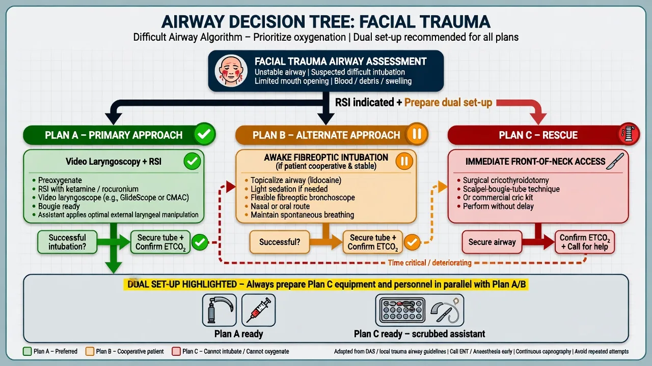
Cannot intubate, can oxygenate: stop repeated attempts, SGA if anatomy allows, wake if appropriate and safe (rare in major trauma), or proceed to definitive surgical airway electively rather than creating CICO with trauma. [1]
CICO: immediate FONA — scalpel-bougie-tube when cricothyroid membrane palpable; surgical tracheostomy by maxillofacial/ENT if anatomy destroyed at cricothyroid level.[1]
Massive facial haemorrhage: packing, TXA, blood products, interventional radiology or surgical ligation pathways, secure airway early before swelling peaks.[2]
Soiled awake FOI failure: abandon and move to planned surgical airway rather than endless topicalisation in a drowning field of blood. [1]
Accidental extubation with wired jaws: cut the wires (cutters at bedside), open mouth, re-secure airway — this is why wire cutters are non-negotiable after IMF. [1]
Postoperative / PACU plan
Do not extubate into progressive oedema. Criteria themes: leak when used as a signal, visualised adequate glottis/supraglottic anatomy if examined, controlled bleeding, patient ability to protect airway, full reversal, ICU bed if wired jaws or tenuous airway. Wire cutters at bedside after IMF. Head-up, antiemesis, careful analgesia. Re-intubation plan documented including surgical backup. Consider delayed extubation or elective tracheostomy after panfacial reconstruction. [1]
Special populations
Children: smaller reserve, different fracture patterns, higher relative tongue size, careful tube sizing. Anticoagulated elderly fall: bleeding plus difficult airway plus cervical disease. Penetrating zone II/III neck: vascular surgery input; primary surgical airway more often. Combined TBI: avoid hypoxia and hypercarbia; IOP and ICP thinking if concurrent ocular injury. Pregnancy: left tilt, two patients, airway oedema of pregnancy stacked on trauma. [1]
Primary survey integration (ABC with a facial injury)
Maxillofacial trauma is not assessed in isolation. Catastrophic haemorrhage (external facial bleeding or associated truncal bleeding), airway obstruction, and cervical spine injury compete for priority. Apply direct pressure and packing to external bleeds, give TXA early in significant haemorrhage per trauma evidence, maintain manual in-line stabilisation during airway manoeuvres, and do not delay the airway for a perfect secondary survey if obstruction is evolving. Hypoxia and hypotension double-hit the injured brain when TBI coexists — airway security enables both oxygenation and controlled ventilation targets for neuroprotection. [1]
Video laryngoscopy, bougies, and tube choice
First-pass success matters more than device brand loyalty. Hyperangulated video blades help when mouth opening is limited, but blood can obliterate cameras — have a standard geometry blade and direct laryngoscopy option, and two suctions running. Load a bougie for the first attempt when you anticipate a grade 3 view. Start with a slightly smaller cuffed tube (for example 6.5–7.0 mm ID in many adults with oedema and blood) rather than forcing a large tube through a swollen glottis. Confirm with waveform capnography, not fogging or auscultation alone. Secure meticulously; facial dressings and blood make tape fail — consider ties with care for venous obstruction and surgical access. [1]
Awake tracheostomy and front-of-neck decision thresholds
Primary awake tracheostomy under local anaesthesia (with surgical team ready, sitting-up if possible, oxygen, careful sedation only if it does not obstruct) is appropriate when oral and nasal routes are predicted to fail: massive tissue loss, some gunshot wounds, or fixed obstruction with stridor and no mouth opening. Do not use awake tracheostomy as a delay tactic while the patient exhausts — if peri-arrest, emergency CICO techniques proceed without the luxury of a calm awake dissection. Scalpel-bougie-tube cricothyroidotomy remains the DAS adult default when the membrane is palpable and time is measured in seconds. [1]
Intermaxillary fixation (IMF) and shared airway surgery
After repair, jaws may be wired or elastics placed. Before extubation: confirm surgical haemostasis, assess swelling trend (often worse at 12–24 hours), consider nasendoscopy or direct look if unsure, ensure wire cutters or elastic scissors are physically on the bed, and prefer ICU delayed extubation after panfacial cases. Submental intubation (tube brought out through the floor of mouth) is an intraoperative solution when surgeons need both nasal and oral fields free — plan tube exchange with airway exchange catheter and surgical backup, never as an ED emergency airway. [1]
Associated injuries that change the anaesthetic
- Base of skull fracture: no blind nasal tubes; CSF leak infection awareness; careful positive pressure if pneumocephalus concerns with neurosurgery.
- Cervical spine fracture: MILS, video laryngoscopy, consider awake techniques if stable enough; hard collar strategy.
- Chest injury / pneumothorax: positive pressure ventilation risks tension — examine and decompress early.
- TBI: target oxygenation and normocapnia to mild hypocapnia only if herniating per neurosurgical advice; avoid nasal tubes if midface/BOFS.
- Globe injury: coordinate with ophthalmology; cough and pressure rules from the OCR/IOP topic apply. [1]
Blood, TXA, and damage control
CRASH-2 supports early TXA in bleeding trauma within the evidence window (classically 1 g IV over about 10 minutes then 1 g infusion over 8 hours in the trial protocol — follow current local major trauma algorithms). Activate massive transfusion for exsanguinating facial or associated injuries, correct hypocalcaemia, and reverse anticoagulants when indicated. Facial bleeding can be underestimated because much is swallowed — serial haemoglobin and haemodynamics matter. [1]
Human factors
Time pressure, blood on the floor, and multiple teams create fixation on CT and delay of airway. Use a 30-second team brief: Plan A VL RSI with MILS, Plan B SGA if it will sit, Plan D FONA with kit open, no nasal route, two suctions, TXA given. Authority gradients must not silence the person watching the saturation. [1]
Le Fort examination tips for the anaesthetist
Gently test maxillary mobility only if it will not worsen injury and the surgeon agrees — often better to read the CT. Look for midface disharmony, malocclusion, infraorbital paraesthesia, and epistaxis. Photograph and document loose teeth before airway manipulation when time allows. Remove dentures carefully; they may help mask seal if left in for bag-mask in edentulous patients — a classic airway pearl. [1]
Preoxygenation and apnoeic oxygenation in the bloody airway
Standard tight mask seal may be impossible. Use high-flow nasal oxygen if the nose is patent and midface injury allows, sitting-up preoxygenation if bleeding permits, and two-person mask techniques with oropharyngeal airways. Apnoeic oxygenation extends the safe apnoea window but does not replace a definitive plan. In the soiled airway, suction first — you cannot preoxygenate through a lake of blood. [1]
Extubation criteria checklist after facial trauma surgery
- Full reversal of neuromuscular blockade (quantitative TOF when available)
- Adequate spontaneous ventilation and oxygenation on modest FiO2
- Haemostasis in the mouth and nose
- Swelling stable or improving, not rapidly progressive
- Patient obeys commands and can protect airway
- Leak test if used as one data point (not sole criterion)
- Wire cutters present if IMF
- Surgical team aware and available for emergency rewire cut / re-intubation
- ICU bed if high risk
- Difficult airway trolley at bedside [1]
NAP4 lessons applied to facial trauma
Head and neck cases are over-represented in serious airway complications. Capnography, planning, and early FONA feature repeatedly. Apply those lessons: waveform capnography always, dual set-up, and no endless attempts. [1]
Communication with radiology and transfer
If the patient must go to CT before the airway is secured, a senior anaesthetist escorts with full airway kit, suction, and a low threshold to intubate in the resus bay first. Never send a stridulous, bleeding midface injury alone with a porter. [1]
Paediatric facial trauma differences
Children have relatively larger tongues, higher oxygen consumption, and faster desaturation. Mandibular growth centres and mixed dentition change surgical priorities. Weight-based drug dosing, smaller tubes, and earlier definitive airway for progressive swelling remain themes. Parental presence for induction is rarely appropriate in bloody trauma resus — prioritise the airway. [1]
Penetrating injury zones (neck) briefly
Zone II neck injuries historically underwent mandatory exploration; modern practice is more selective with imaging in stable patients. Expanding haematoma, stridor, bubbling wounds, and hard signs of vascular injury push toward early definitive airway, often in theatre with surgical airway readiness before extensive imaging. [1]
SAQ answer scaffold
Stem: Assault victim with bilateral mandible fractures, blood in the oropharynx, hard collar in situ, SpO2 91 percent on oxygen. [1]
- Threats (3 marks): tongue fall-back, blood aspiration, C-spine, progressive swelling, full stomach.
- Immediate actions (3 marks): oxygen, suction, MILS, call surgical airway help, prepare dual set-up.
- Technique choice (4 marks): VL RSI vs awake vs primary surgical — justify; no blind nasal.
- DAS failure pathway (3 marks): limit attempts, SGA, FONA.[1]
- Aftermath (2 marks): ICU, wire cutters if IMF, airway alert.
Viva stem bank and model phrases
Stem 1: “Can I put a nasal tube in this Le Fort III?”
Model: “Not blind — midface and base-of-skull risk. Nasal routes only later if imaging and seniors agree and preferably under vision.” [1]
Stem 2: “Your plan for induction?”
Model: “Team brief, dual set-up FONA, MILS, two suctions, video laryngoscopy RSI or awake FOI if cooperative and time, first attempt maximised, early declaration of failure.” [1]
Stem 3: “SpO2 70 percent, no view, SGA will not sit.”
Model: “CICO — immediate front-of-neck access, 100 percent oxygen, concurrent surgical help.”[1]
Stem 4: “When do you give TXA?”
Model: “In significant haemorrhage trauma within the evidence window, CRASH-2 supports early TXA as part of major haemorrhage care.”[2]
Stem 5: “Jaws are wired and the tube comes out in recovery.”
Model: “Cut the wires immediately with bedside cutters, open the mouth, oxygenate and re-intubate — prevention is ICU-level airway planning before extubation.” [1]
Stem 6: “Why not three more laryngoscopies? I almost had it.”
Model: “Repeated attempts cause oedema and bleeding and convert difficulty into CICO. DAS logic limits futile attempts without a strategy change.”[1]
Common traps
- Repeated oral attempts while the face swells
- Blind nasal tube through Le Fort III / base-of-skull injury
- Removing the collar without MILS
- Extubating overnight after major facial reconstruction without oedema assessment
- No wire cutters after IMF
- Sending a deteriorating patient to CT without an airway
- Treating maxillofacial trauma as “just a short nasal intubation for the surgeons” [1]
Facial trauma airway
ANZCA Final candidates should integrate DAS 2015, local major trauma and massive transfusion protocols, and explicit IMF/wire-cutter safety into the same answer — not airway algorithms alone.
References
- [1]Frerk C et al. Difficult Airway Society 2015 guidelines for management of unanticipated difficult intubation in adults Br J Anaesth, 2015.PMID 26556848
- [2]CRASH-2 trial collaborators Effects of tranexamic acid on death, vascular occlusive events, and blood transfusion in trauma patients with significant haemorrhage (CRASH-2): a randomised, placebo-controlled trial Lancet, 2010.PMID 20554319
- [3]Neal JM et al. The Third American Society of Regional Anesthesia and Pain Medicine Practice Advisory on Local Anesthetic Systemic Toxicity: Executive Summary 2017 Reg Anesth Pain Med, 2018.PMID 29356773