Anaes · Neuroanaesthesia
Neuroanaesthesia
Also known as Neuroanaesthesia · Intracranial pressure anaesthesia · Awake craniotomy · Venous air embolism sitting · SS_NS specialised study unit
Exam-pass neuroanaesthesia hub (SS_NS): Monroe-Kellie, CPP=MAP−ICP, agent effects on CBF/CMR, BTF TBI targets, VAE in sitting position, aneurysm rupture plan, awake craniotomy, and leaf links.
On this page & tools
Your progress
Saved locally on this device.
Target exams
Red flags
Why this is examined
SS_NS (neurosurgery and neuroradiology) contributes 29 FEx outcomes with heavy applied physiology. Examiners test whether you can link intracranial compliance to drug choice, ventilatory targets, positioning risks, and crisis algorithms. TBI is a high-yield emergency stem; sitting-position VAE is a classic oral; awake craniotomy tests communication and airway contingency.[1][2][3]
One-liner: I defend CPP, keep the brain relaxed with propofol-lean technique when compliance is poor, avoid secondary insults in TBI, run a VAE drill for sitting cases, and never start an awake craniotomy without an emergency airway conversion plan. [1]
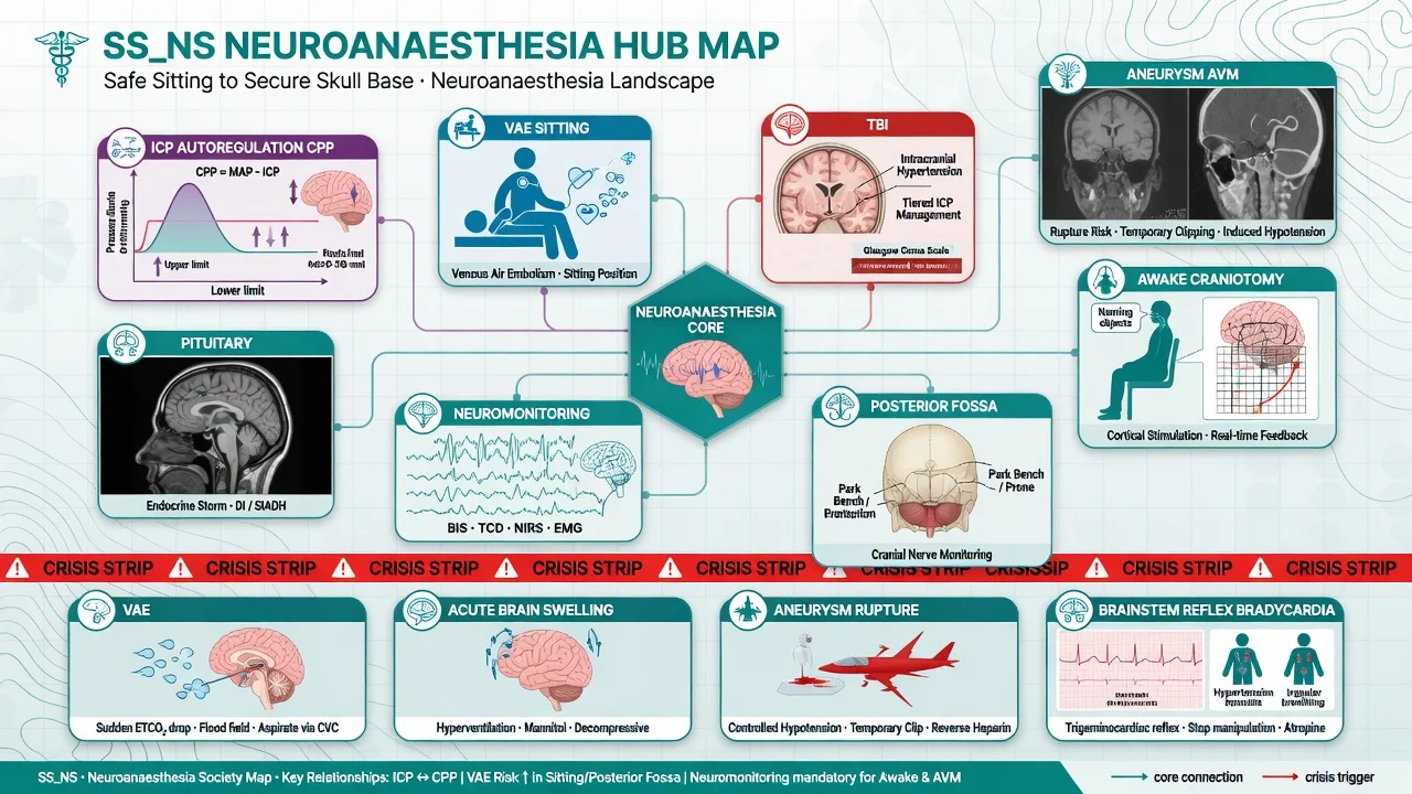
SSU framework and hub map
| Leaf / domain | Examiner focus |
|---|---|
| ICP, autoregulation, CPP | Monroe-Kellie, Lassen curve, CO2 reactivity |
venous-air-embolism-sitting-position | Detection hierarchy, management, PFO |
| TBI management | BTF targets, osmotherapy, avoid secondary insults |
| Aneurysm / AVM | Temporary clipping, blood pressure strategy, rupture |
| Awake craniotomy | Asleep-awake-asleep, mapping, seizures |
| Posterior fossa | Brainstem reflexes, cranial nerve monitoring |
| Neuromonitoring | SSEP/MEP, EEG, oximetry implications for anaesthetic |
| Pituitary / transsphenoidal | DI, cortisol, airway shared with surgeon |
Core physiology (must recite)
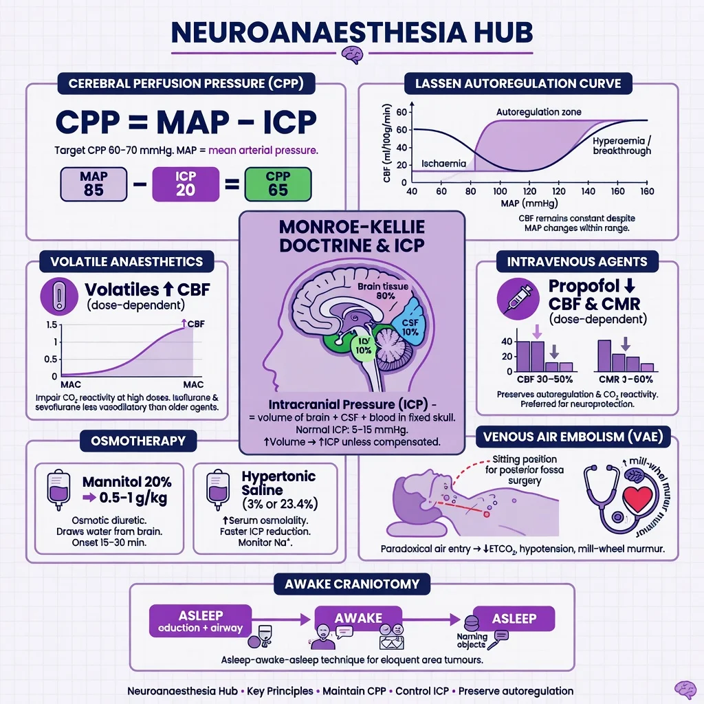
Monroe-Kellie: intracranial volume is fixed (brain + blood + CSF ± mass). Increase in one compartment requires decrease in another or ICP rises. [1]
CPP = MAP − ICP (or MAP − CVP if CVP > ICP). Target CPP commonly 60–70 mmHg in TBI protocols (individualise). Avoid hypotension and hypoxia — the two secondary insults that dominate outcome messaging. BTF-aligned care still centres on preventing those insults and treating intracranial hypertension systematically.[1][3]
Autoregulation (Lassen): CBF roughly constant over CPP ~50–150 mmHg in health; right-shifted in chronic hypertension; impaired after TBI/SAH. CO2 reactivity: hypocarbia vasoconstricts (reduces CBF/ICP temporarily); hypercarbia dilates. [1]
Anaesthetic agents and the brain
| Agent class | CBF | CMR | ICP tendency | Exam note |
|---|---|---|---|---|
| Propofol / barbiturates | ↓ | ↓ | ↓ | Workhorse for tight brains |
| Opioids | minimal if ventilated | ↓ modest | neutral if no hypoventilation | Watch rigidity/chest wall |
| Volatiles | ↑ dose-dependent | ↓ | may ↑ | Keep MAC modest; prefer <1 MAC often |
| Ketamine | historically feared | — | modern data more nuanced | Myth debunking in acute brain injury literature; still context-specific.[4] |
| Dexmedetomidine | variable | ↓ | often favourable | Adjunct; autoregulation studies exist.[5] |
| N2O | ↑ CBF | — | may ↑ | Avoid in raised ICP / air spaces / VAE risk |
Raised ICP toolkit
Head-up 15–30°, neutral neck (venous drainage), adequate sedation/analgesia, normoxia, normocapnia (PaCO2 ~35–40 mmHg) unless herniating, treat seizures/fever/hyponatraemia, surgical decompression when indicated. Osmotherapy: mannitol 0.25–1 g/kg or hypertonic saline (e.g. 3%) — watch sodium, osmolarity, volume status. Temporary hyperventilation only as bridge to definitive care for herniation signs.[1][2]
Traumatic brain injury anaesthetic priorities
Early airway if GCS ≤8; avoid hypoxia and hypotension during RSI (haemodynamically thoughtful induction); target SpO2 ≥94%, normocapnia after intubation, MAP high enough for CPP goal; glucose control; temperature control; coordinate with neurosurgery for haematoma evacuation.[1][3] Neurocardiac injury (catecholamine surge, ECG changes, wall-motion abnormalities) is a recognised axis in acute intracranial stress — treat the brain and the heart as one conversation.
Sitting position and VAE
Risk when surgical field above heart and veins cannot collapse (diploic/emissary/sinus veins). Detection sensitivity hierarchy: TOE (most sensitive) > precordial Doppler > PAP rise > EtCO2 fall > SpO2 fall. Precordial Doppler is common routine monitor; TOE for high risk. [1]
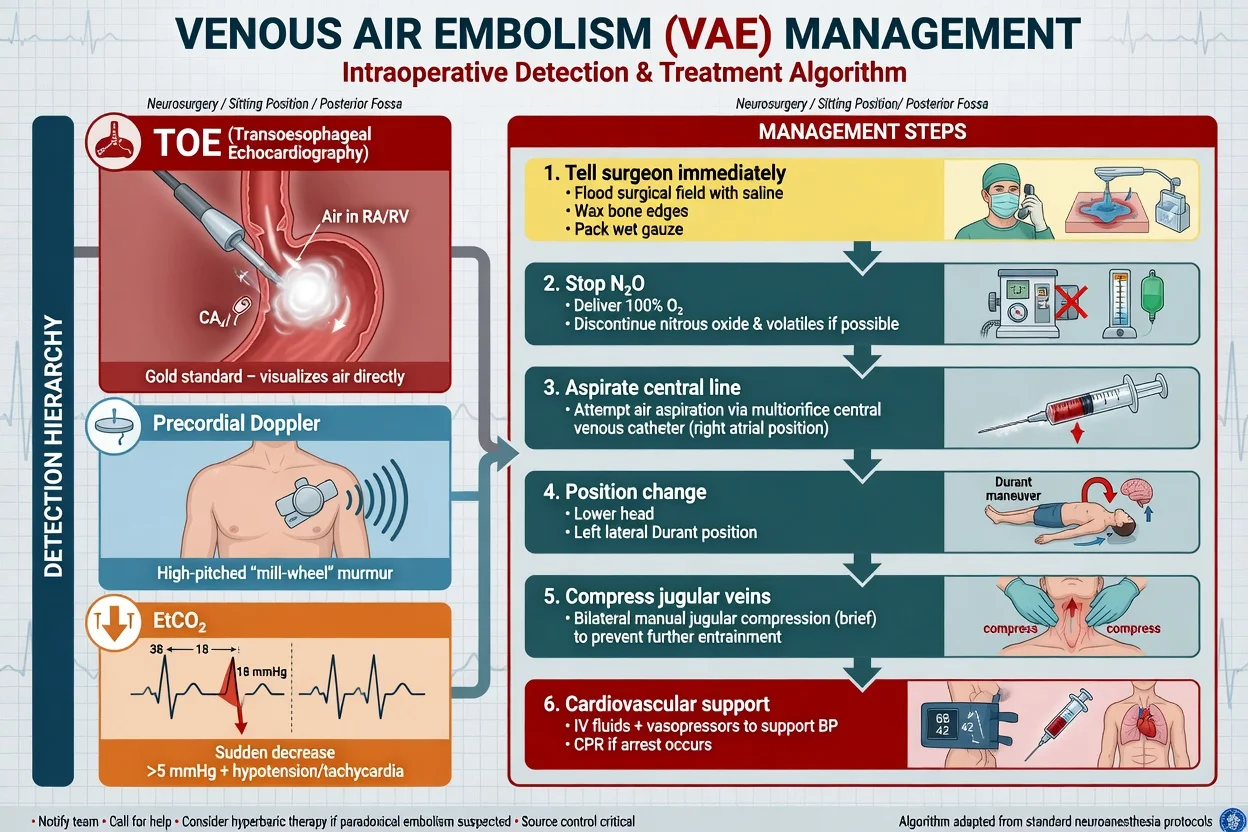
Management: tell surgeon (flood field, wax bone, compress veins), 100% O2, stop N2O, aspirate CVP, Durant manoeuvre (left lateral head-down), jugular compression carefully, ACLS if arrest, consider hyperbaric in selected arterialised embolism — leaf topic owns full depth: venous-air-embolism-sitting-position. [1]
Aneurysm and AVM surgery
Goals: smooth haemodynamics, brain relaxation, preparedness for rupture (adenosine/temporary clip communication, blood available). Induced hypotension is selective and team-agreed, not reflexive. Temporary clipping may prompt burst-suppression strategies in some centres. [1]
Awake craniotomy
Asleep-awake-asleep or conscious sedation throughout depending on centre. Airway plan for the asleep phases and for emergency conversion; map language/motor function; treat intraoperative seizures (cold saline, propofol/benzodiazepine carefully); patient coaching is part of the anaesthetic. [1]
Neuromonitoring implications
SSEP/MEP: total IV anaesthesia often preferred when MEPs used; limit volatiles and neuromuscular blockade as protocol demands; communicate signal changes as possible ischaemia. EEG/processed EEG for depth and burst suppression. Cerebral oximetry adjunctive. [1]
Crisis bank
- VAE — detection + surgical field control + 100% O2 + CVP aspirate + position.
- Acute brain swelling — CO2, depth, osmotherapy, head position, surgical decompression.
- Intraoperative aneurysm rupture — communication, blood pressure strategy, temporary clip, resuscitation.
- Trigeminocardiac / brainstem reflex bradycardia — stop stimulus, atropine, communicate.
- Tension pneumocephalus after sitting cases — late deterioration, scan, release.
- Seizure during awake mapping — cold saline, short-acting agents, protect airway.
- Herniation on induction/transfer — osmotherapy, temporary hyperventilation, urgent OR/ICU. [1]
Landmark guidance
Regional practice deltas
ANZ. SS_NS FEx tagging; sitting craniotomy less common than formerly but VAE still examined. Mannitol and hypertonic saline both used — quote local osmotherapy protocol and sodium targets.
SAQ answer scaffold
Stem: "Sitting posterior fossa craniotomy. Sudden EtCO2 fall and hypotension." [1]
- Declare VAE; 100% O2; tell surgeon to flood/wax/compress.
- Stop N2O; aspirate CVP; head-down left lateral if feasible.
- Support BP; ACLS if needed; abandon sitting if unstable.
- Post-event: echo for right heart air/PFO implications, neuro checks, ICU plan.
- Prevention reflection: Doppler/TOE, meticulous surgical haemostasis, avoid N2O. [1]
Viva stem bank
- "Define CPP and your TBI targets."
- "How do volatiles and propofol differ on CBF and ICP?"
- "VAE detection methods ranked by sensitivity."
- "Mannitol versus hypertonic saline — pros/cons."
- "Awake craniotomy — anaesthetic goals for the awake phase."
- "Patient herniating in ED — anaesthetic priorities to theatre." [1]
Common traps
- Routine hyperventilation for every TBI.
- Using N2O in sitting craniotomy.
- Ignoring venous drainage (head rotated, ties tight).
- Treating EtCO2 fall as only "pulmonary embolism thrombus" without VAE in the differential in sitting cases.
- Dropping MAP for "nice surgical field" in the poorly compliant brain without a plan. [1]
BRAIN — tight brain checklist
Propofol-based
- ↓CBF ↓CMR
- Tight brain
- MEP-friendly TIVA
- Infusion care
Volatile-based
- Easy titration
- CBF↑ at higher MAC
- Limit dose
- Not ideal alone for MEP
Sitting risk path
- VAE ready
- Doppler/TOE
- No N2O
- CVP aspirate plan
Positioning catalogue (exam list)
Supine (most craniotomies), lateral (some temporal), prone (posterior fossa/spine — airway, eyes, abdomen free, VAE less than sitting but still possible), sitting/semi-sitting (VAE risk maximised), park-bench. Each position has nerve-injury and access implications. Document pressure points, eyes, and the shared airway with pins (Mayfield) — pin stimulation can cause hypertensive surge; deepen or bolus before pins. [1]
Interventional neuroradiology crossover
Coiling of aneurysms, stroke thrombectomy, and AVM embolisation move the anaesthetist to the angiography suite: remote airway, contrast reactions, anticoagulation for wires, need for immobility, and sudden rupture/perforation crises. GA vs conscious sedation for thrombectomy is centre-dependent; the exam wants a clear airway plan and BP targets matched to ischaemic penumbra versus haemorrhage risk. [1]
Spine surgery interface
Major spine cases share neuromonitoring rules, prone physiology, blood loss, and multimodal analgesia with the orthopaedic spine pathway. Wake-up test is largely historical where MEP is available; still know the concept. Visual loss after prolonged prone spine surgery is a feared complication (ION) — avoid prolonged severe hypotension and direct eye pressure. [1]
Red flags
[1] [1] [1] [1] [1]References
- [1]Hawryluk GWJ, Rubiano AM, Totten AM, et al. Guidelines for the Management of Severe Traumatic Brain Injury: 2020 Update of the Decompressive Craniectomy Recommendations Neurosurgery, 2020.PMID 32761068
- [2]Bou Sleiman V, et al. Anesthetic management of neurosurgical emergencies Curr Opin Anaesthesiol, 2025.PMID 40071979
- [3]Blacker SN, et al. Intraoperative Anesthetic Care During Emergent/Urgent Craniotomy or Craniectomy for Intracranial Hypertension or Herniation: A Systematic Review J Neurosurg Anesthesiol, 2026.PMID 39793097
- [4]Haywood S, et al. Ketamine Use in Acute Brain Injury: Debunking the Myth Emerg Med Clin North Am, 2026.PMID 42342307
- [5]Banik S, et al. Effect of dexmedetomidine on dynamic cerebral autoregulation and carbon dioxide reactivity during sevoflurane anesthesia in healthy patients Korean J Anesthesiol, 2020.PMID 32209963