Anaes · Neuroanaesthesia
Neuromonitoring: EEG, SSEP, MEP and anaesthetic constraints
Also known as SSEP MEP anaesthesia · Intraoperative neurophysiology · Evoked potential TIVA
Anaesthetic effects on EEG and evoked potentials, TIVA vs volatile strategies for SSEP/MEP, neuromuscular blockade rules, warning criteria, and crisis response when signals degrade.
On this page & tools
Your progress
Saved locally on this device.
10 MCQs with explanations
Target exams
Red flags
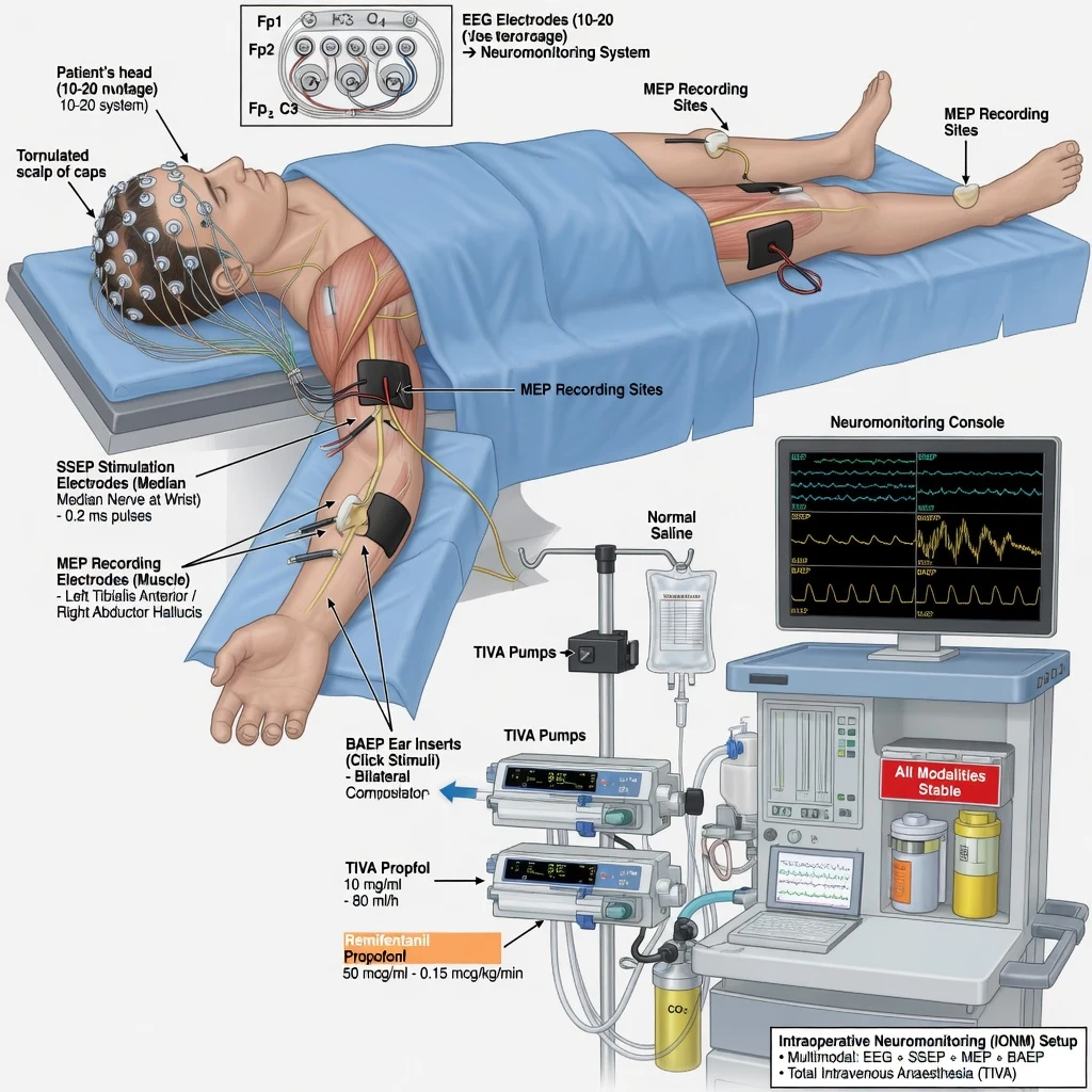
Why this is examined / the one-line answer
Fellowship exams test whether you understand how anaesthetic drugs and physiology distort EEG and evoked potentials, not merely the names of waveforms. Spine deformity, aortic, posterior fossa, and skull base lists all pivot on SSEP/MEP strategy. Signal loss without a structured response fails the hot case. [1]
One-line opener: TIVA for critical MEPs, no ongoing paralysis, stable MAP and CO2, and if signals fall I tell the surgeon and treat ischaemia until proven otherwise. [1]
Preoperative assessment and team brief
Surgical indication for monitoring. Scoliosis and complex spine, spinal tumour, thoracic aortic work, CPA/posterior fossa, epilepsy surgery with ECoG, carotid/cerebrovascular cases with EEG or SSEP adjuncts. [1]
Patient factors. Pre-existing neurological deficits (baseline EP abnormality), neuropathy, severe scoliosis, pacemaker/ICD (MEP electrical interference and safety), seizure history, neuromuscular disease (NMB dosing and MEP interpretation), anaemia, cardiac disease limiting MAP targets. [1]
Team brief must agree: [1]
- Modalities planned (SSEP, MEP, BAEP, EMG, EEG/ECoG)
- Anaesthetic technique (TIVA vs limited volatile)
- NMB plan (intubating dose only vs none after induction)
- MAP targets and blood pressure support plan
- Warning criteria and staged response including wake-up test contingency for spine
- Bite block for MEPs [1]
Awake testing paradigms (awake craniotomy sedation comparisons) reinforce that neurological monitoring quality depends on drug choice and communication, even when the case is not fully awake.[1]
Modalities
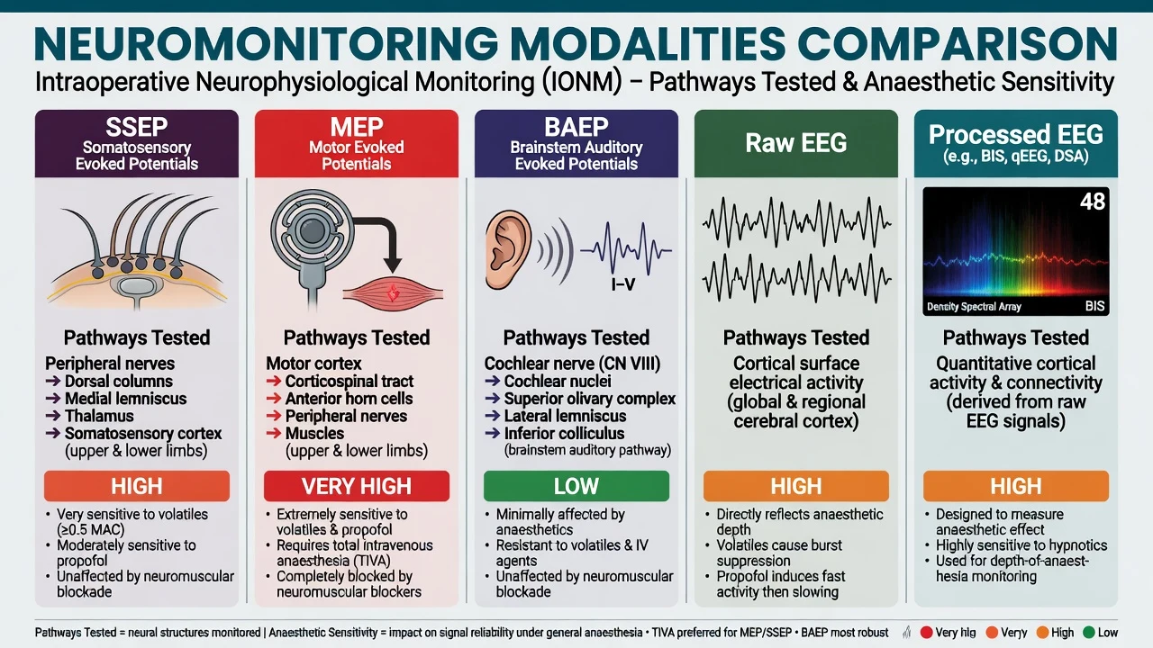
Pathway reminders (viva anatomy)
- SSEP: peripheral nerve stimulation → dorsal columns → brainstem → thalamus → sensory cortex (cortical responses most anaesthetic-sensitive)
- MEP: transcranial electrical stimulation of motor cortex → corticospinal tract → anterior horn → peripheral nerve → muscle (myogenic recording)
- BAEP: auditory nerve → cochlear nuclei → brainstem pathways — robust, useful in CPA surgery [1]
Drug effects (exam core)
Volatile anaesthetics
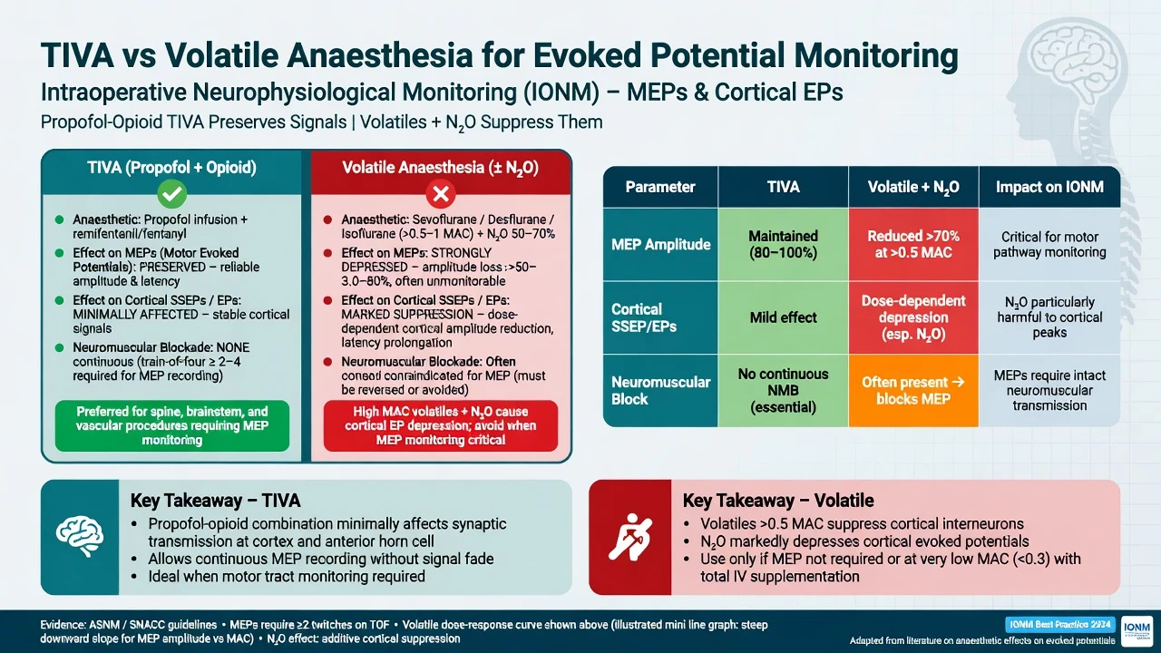
Dose-dependent amplitude decrease and latency increase for cortical SSEP and especially MEP. Many teams keep volatile MAC low (e.g. less than 0.5 MAC) or eliminate volatiles entirely when MEPs are critical. All modern volatiles share this direction of effect. [1]
Nitrous oxide
Further depresses cortical evoked potentials; often avoided during EP-critical windows. [1]
Propofol–opioid TIVA
Standard for spine and many intracranial MEP cases. Propofol still suppresses EEG/EPs at high doses (burst suppression), but is more EP-friendly than equipotent high-dose volatile strategies when titrated carefully. Remifentanil or other opioids have relatively modest EP effects and aid immobility without NMB. [1]
Neuromuscular blockade
- Intubating dose commonly acceptable
- Ongoing NMB is incompatible with myogenic MEPs and with many EMG monitoring goals
- Confirm recovery with quantitative TOF before relying on MEPs
- If absolute immobility needed without NMB: deepen opioid/TIVA, coordinate surgical pauses [1]
Other agents
- Ketamine / etomidate: may enhance some EP amplitudes (context-specific; not a free pass for unstable haemodynamics)
- Dexmedetomidine: used in awake and adjunct settings; effects on EPs are generally more modest than volatiles but know unit experience[1]
- Barbiturates / high-dose propofol: burst suppression for metabolic suppression — abolishes much cortical monitoring utility by design
- Lidocaine infusions: usually minor EP impact at typical doses
Physiology confounds (fix as often as drugs)
- MAP / CPP / cord perfusion: hypotension is a leading reversible cause of signal change
- Haemoglobin / oxygen delivery
- PaCO2: extremes alter CBF and may affect signals
- Temperature: hypothermia increases latencies
- Positioning: stretch, pressure on nerves, shoulder braces, cervical manipulation
- Anaemia, hypoxia, severe anaemia after bleeding
- Technical: electrode displacement, high impedance, electrocautery noise, bite on tube during MEP [1]
Fix physiology before accepting “anaesthetic effect” as the only story. [1]
Anaesthetic goals
- Deliver a stable, EP-friendly anaesthetic without sudden depth swings during critical monitoring windows.
- Maintain agreed MAP (often at or above baseline mean; higher targets if cord at risk).
- Avoid continuous NMB when MEPs/EMG required.
- Communicate early and clearly when signals change.
- Protect the patient from MEP-related injury (bite block, tongue trauma, rare seizures).
- Have a wake-up test plan for selected spine cases if signals remain lost. [1]
Technique: practical TIVA recipe (illustrative, unit-titrated)
- Induction: propofol and opioid; muscle relaxant for intubation if needed; then no further relaxant
- Maintenance: propofol infusion or TCI; remifentanil infusion; air/oxygen; no N2O; volatile off or minimal if team agrees for SSEP-only cases
- Arterial line for spine/aortic/major intracranial lists
- Bite block before MEPs
- Stable temperature and ventilation
- Document baseline signals after positioning and before high-risk surgical steps [1]
When signals degrade — crisis checklist
Signal loss checklist
Expanded response
- Announce signal change to surgeon and neurophysiologist — pause high-risk manoeuvres if safe.
- Check technical factors: leads, impedance, stimulator output, recent diathermy, bite block position.
- Check anaesthetic: recent volatile increase, bolus propofol causing burst suppression, residual NMB (TOF).
- Raise MAP toward or above agreed target (vasopressors as needed).
- Correct anaemia, hypoxia, extreme hypocapnia/hypercapnia, hypothermia.
- Surgeon actions: release distraction, reduce retraction, reverse step that preceded loss, improve cord perfusion surgically.
- If unresolved in spine surgery: consider Stagnara wake-up test (plan analgesia, awareness discussion, secure tube, stepwise lightening with clear motor commands).
- Document timeline meticulously. [1]
Related exam links
- Carotid surgery: GALA compared general vs local anaesthesia for CEA and found no definitive difference in the primary outcome; the broader lesson is that neurological monitoring strategy must be explicit whether by awake testing or under GA adjuncts.[2]
- Paediatric neurodevelopment debates (GAS): intraoperative neuromonitoring for spine is a different question from developmental outcome trials, but examiners may pivot from “anaesthetic toxicity” talk to practical monitoring — keep domains separate and accurate.[3]
Postoperative plan
- Neurological examination as soon as appropriate
- Maintain MAP targets if cord ischaemia was a concern
- Analgesia that does not obliterate examination when serial neurology required (balanced multimodal)
- Handover of any intraoperative signal events and final signal status
- Urgent imaging/surgical review if new deficit [1]
Special populations
- Paediatric spine: TIVA skills, temperature, blood loss, smaller MEP thresholds variability
- Neuromuscular disease: unpredictable NMB; baseline weakness complicates MEP interpretation
- Obesity / prone spine: positioning neuropathies mimicking monitoring changes
- ICD/pacemaker: electrocautery and MEP stimulation precautions with cardiology input [1]
SAQ answer scaffold
- Table of modalities with pathways and anaesthetic sensitivity.
- Why MEPs force TIVA and no continuous NMB.
- Volatile/N2O effects on cortical EPs.
- Physiological confounds list.
- Signal loss algorithm including MAP raise and wake-up test.
- Bite block safety point.
- One-line each on GALA monitoring philosophy and not confusing GAS developmental outcomes with EP technique.[2][3]
Viva stem bank and model phrases
- “How does sevoflurane affect SSEPs and MEPs?”
- “The MEP amplitude has fallen by half after rod distraction.”
- “Can I run a rocuronium infusion for a quiet field?”
- “What MAP will you target and why?” [1]
Model phrases: [1]
- “I am treating this as cord ischaemia until proven otherwise — please pause distraction while I raise MAP.”
- “TOF shows residual blockade — MEPs cannot be interpreted until recovery.”
- “We agreed TIVA without nitrous for this MEP-critical case.” [1]
Common traps
- Running high sevoflurane “because it is easy” during MEPs
- Rocuronium infusion “for stillness” while expecting myogenic MEPs
- Ignoring hypotension when signals fall
- Treating processed EEG numbers as gold-standard brain or cord perfusion monitors
- No bite block → tongue injury
- Changing multiple anaesthetic variables at once so the cause is uninterpretable
- Arguing with the neurophysiologist instead of running the checklist [1]
Cortical versus subcortical SSEP interpretation
Cortical SSEP waveforms are more sensitive to anaesthetics and to cortical ischaemia; subcortical potentials are relatively more robust. A pattern where cortical signals fall while subcortical remain may suggest anaesthetic depth or cortical perfusion issues, whereas loss of both with surgical cord risk timing suggests pathway injury — interpret with the neurophysiologist, not alone. [1]
MEP stimulation safety
Transcranial electrical MEPs cause masseter contraction (bite block mandatory), can rarely trigger seizures, and may interfere with pacemakers/ICDs. Tongue and lip lacerations are preventable. Avoid MEP when absolute contraindications exist per team (e.g. certain epilepsy contexts — individualise). [1]
Wake-up test (Stagnara) practical script
- Pre-agree with patient in preassessment when possible (spine deformity lists).
- When signals lost and not recovering: surgeon ready, room quiet, secure tube, analgesia on board, reverse residual NMB if any.
- Lighten TIVA carefully; ask patient to move hands then feet.
- If feet move: document, re-deepen, proceed with caution.
- If feet do not move: surgical implication — reduce correction, further imaging/exploration as indicated.
- Risk: awareness, tube dislodgement, violent movement — prevent with careful titration and team roles. [1]
MAP targets and spinal cord perfusion
Cord perfusion pressure conceptually relates to MAP minus CSF pressure (or CVP as surrogate in some models). During deformity correction or aortic occlusion contexts, teams often target MAP at or above baseline mean, sometimes 80–90 mmHg bands when signals change — individualise to comorbidities (risk of bleeding vs cord). Raise MAP early in the signal-loss checklist. [1]
Anaesthetic stability beats anaesthetic heroics
Sudden propofol boluses can flatten cortical signals via burst suppression just as volatiles can. Aim for steady effect-site concentrations during instrumentation. If you must deepen for movement, tell the neurophysiologist so they do not call a false surgical alarm. [1]
EEG burst suppression clinical uses
- Deep anaesthesia or metabolic suppression for neuroprotection narratives
- Confirms high anaesthetic depth — EPs will be poor
- Not a goal during MEP windows [1]
Processed EEG (BIS/entropy) helps trend hypnosis but does not measure cord integrity. [1]
Multimodal monitoring philosophy
SSEPs monitor sensory pathways; MEPs monitor motor pathways — they are complementary. Isolated reliance on SSEPs can miss pure motor injury. Free-run EMG adds nerve root irritation data in pedicle screw placement. Choose modality bundles based on surgical risk anatomy. [1]
Documentation for medicolegal and handover quality
Record baseline signals after positioning, times of warning criteria, MAP at events, anaesthetic changes, surgical actions, final signals, and neurological exam on wake. This is part of consultant practice, not optional paperwork. [1]
Worked scoliosis stem
After rod distraction MEPs fall 60%: announce, pause, raise MAP with noradrenaline to agreed target, check TOF 4/4, confirm TIVA stable without recent volatile, surgeon reduces distraction, signals return over minutes — document and proceed with higher MAP. If no return: wake-up test pathway. [1]
Anaesthetic regimen examples (illustrative adult spine MEP case)
- Induction: fentanyl 2–5 microg/kg or remifentanil infusion start; propofol 1–2 mg/kg titrated; rocuronium 0.5–0.6 mg/kg once for intubation
- Maintenance: propofol TCI or 6–10 mg/kg/h band titrated to pEEG and haemodynamics; remifentanil 0.1–0.5 microg/kg/min; oxygen/air; volatile off; N2O off
- After intubation: no further NMB; confirm TOF recovery before critical MEPs
- MAP: maintain at or above baseline; prepare noradrenaline
- Bite block in before first MEP [1]
These numbers are teaching anchors; titrate to patient and unit protocols. [1]
Why volatiles hit MEPs harder than BAEP
Synaptic cortical and anterior horn-related pathways for MEPs involve more anaesthetic-sensitive synapses than the more robust brainstem auditory pathway. Cortical SSEP generators are similarly sensitive. This differential sensitivity is the physiological reason BAEP can tolerate more anaesthetic variety than MEP. [1]
Signal averaging and practical delay
SSEPs require averaging; there is a lag before a change declares itself. MEPs are more instantaneous but intermittent. Plan surgical pauses that allow meaningful checks after high-risk manoeuvres (screw placement, osteotomy, distraction). [1]
Combined carotid and EEG monitoring philosophy
GALA showed no clear outcome superiority of local versus general anaesthesia for CEA in its primary analysis, but awake testing remains a pure neurological monitor when LA/regional is used.[2] Under GA, EEG/SSEP are adjuncts with imperfect sensitivity/specificity — blood pressure management and shunt decisions remain surgical–anaesthetic team calls.
Paediatric spine monitoring extras
Higher propofol sensitivity in some children, temperature drift in long cases, blood loss affecting oxygen delivery, and communication with neurophysiology about baseline deficits in neuromuscular scoliosis. GAS trial neurodevelopmental data address a different question (infant GA vs awake-regional for hernia surgery) and should not be misused as MEP technique evidence.[3]
Troubleshooting matrix
| Finding | Likely causes | Actions |
|---|---|---|
| Global cortical SSEP ↓ after volatile rise | Anaesthetic | Reduce volatile; stabilise TIVA |
| Unilateral change after positioning | Brachial plexus stretch, technical | Reposition, check leads |
| Sudden bilateral MEP loss at distraction | Cord hypoperfusion/stretch | Pause, raise MAP, reverse distraction |
| MEPs absent from start | Residual NMB, deep burst suppression, technical | TOF, lighten carefully, check setup |
| Noisy signals | Diathermy, impedance | Pause diathermy, fix electrodes |
Ethical and consent notes
Discuss wake-up test possibility, risk of neurological injury despite monitoring (monitoring reduces but does not eliminate risk), and bite injuries from MEP. Document discussion for major deformity surgery. [1]
Final viva synthesis paragraph
Intraoperative neuromonitoring is a team sport: the anaesthetist delivers an EP-friendly stable anaesthetic, the neurophysiologist interprets signals, and the surgeon acts on warnings. Your unique responsibilities are drug choice, NMB avoidance for MEPs, physiological optimisation, bite block safety, and leading the structured response when signals fail. [1]
Team brief script (30 seconds)
“This is an MEP-critical case. We will run propofol–remifentanil TIVA without nitrous or volatile. Intubating dose of rocuronium only, then no further relaxant. Bite block before MEPs. MAP target at or above the patient’s baseline mean, raise early if signals drop. If MEPs are lost we pause, run the checklist, and have a wake-up test plan.” [1]
Pharmacology revision bullets
- Volatiles: cortical EP amplitude ↓ latency ↑ dose-dependently
- N2O: further depression — avoid in critical windows
- Propofol: EP-friendlier but high dose → burst suppression
- Opioids: minimal EP effect at usual ranges
- NMB: abolishes myogenic MEPs
- Ketamine/etomidate: may increase amplitudes selectively [1]
Physiology revision bullets
- Hypotension → ischaemic signal loss
- Hypothermia → latency ↑
- Hypocapnia extremes → CBF changes
- Anaemia/hypoxia → delivery failure
- Positioning stretch → focal changes [1]
Warning criteria teaching numbers
Many centres use SSEP amplitude fall of about 50% or latency rise of about 10% as warning thresholds; MEP loss or major amplitude drop triggers immediate response. Always use local neurophysiology criteria. [1]
Wake-up test risks and mitigation
Awareness distress — counsel preop when possible. Violent movement — secure tube, staged lightening. Failure to cooperate — children/cognitive impairment may preclude. Residual NMB — check TOF first. [1]
Documentation checklist
Baseline after positioning; times of warnings; MAP; anaesthetic changes; surgical actions; final signals; post-op neuro exam. Essential after any intraoperative loss event. [1]
Cross-links
Posterior fossa facial EMG rules; scoliosis MEP lists; carotid monitoring philosophy (GALA); do not misuse GAS developmental outcomes as EP technique evidence.[2][3]
Final consultant sentence
“I treat acute evoked potential loss as ischaemia until proven otherwise, while I simultaneously exclude anaesthetic and technical causes in a spoken checklist.” [1]
Worked anaesthetic error stem
Colleague runs 1.2 MAC sevoflurane during scoliosis MEPs and wonders why signals are poor. Correction: convert to TIVA, eliminate N2O, ensure TOF recovered, allow time for volatile washout, re-establish baselines before critical instrumentation, maintain MAP. Educate without delaying surgical safety. [1]
Electrocautery and signal noise
Monopolar diathermy obliterates EP recordings during use. Coordinate quiet periods for checks. Bipolar causes less interference. Plan “monitoring pauses” into the operative flow. [1]
Pacemaker and ICD considerations
MEP and diathermy can inhibit or trigger devices. Preoperative device plan with cardiology: magnet use, asynchronous modes, postoperative interrogation as indicated. Document. [1]
Cord perfusion pressure mental model
Raising MAP is the anaesthetist’s fastest lever when signals fall at distraction. CSF drainage is a surgical/ICU adjunct in some aortic protocols — know it exists for cross-specialty vivas even if not used in routine scoliosis. [1]
Closing synthesis for marks
Modalities → drug effects → physiology confounds → TIVA/MEP rules → signal loss checklist → wake-up contingency → bite block. If you hit those seven beats, you pass the monitoring viva. [1]
One-line monitoring closer
Stable propofol–opioid TIVA, recovered neuromuscular function for MEPs, protected teeth and tongue with a bite block, and an ischaemia-first response to signal loss are the four non-negotiables of exam-safe neuromonitoring anaesthesia. [1]
[1] [1]References
- [1]Goettel N et al. Dexmedetomidine vs propofol-remifentanil conscious sedation for awake craniotomy: a prospective randomized controlled trial Br J Anaesth, 2016.PMID 27099154
- [2]Lewis SC et al. General anaesthesia versus local anaesthesia for carotid surgery (GALA): a multicentre, randomised controlled trial Lancet, 2008.PMID 19041130
- [3]Davidson AJ et al. Neurodevelopmental outcome at 2 years of age after general anaesthesia and awake-regional anaesthesia in infancy (GAS): an international multicentre, randomised controlled trial Lancet, 2016.PMID 26507180