Anaes · Thoracic anaesthesia
Anaesthesia for oesophagectomy
Also known as Esophagectomy anaesthesia · Ivor Lewis anaesthesia · MIE OLV oesophagectomy
Exam-pass oesophagectomy anaesthesia: two-cavity physiology, lung isolation, fluid and anastomotic concerns, thoracic analgesia, aspiration risk, and ERAS-leaning postop care for ANZCA Final.
On this page & tools
Your progress
Saved locally on this device.
Target exams
Red flags
Why this is examined / the one-line answer
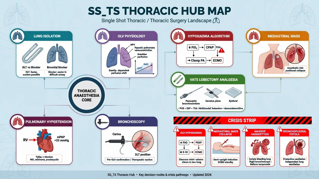
Oesophagectomy packages thoracic one-lung ventilation (OLV), upper GI aspiration risk, major fluid and haemodynamic shifts, and anastomotic vulnerability into one long-case. It is a classic Final SAQ/viva because failure modes are memorable: hypoxia on OLV, fluid overload, weak analgesia with sputum retention, recurrent laryngeal nerve (RLN) injury, atrial fibrillation, and delayed anastomotic leak sepsis.[1][2]
One-liner: I isolate the lung for the thoracic phase, protect the anastomosis with adequate perfusion pressure and balanced fluids, run multimodal thoracic analgesia that still allows cough, and plan HDU-level recovery with leak and aspiration vigilance. [1]
Preoperative assessment and risk stratification
Oncological and physiological fitness
- Diagnosis/staging and planned approach (open vs MIE/RAMIE).
- Neoadjuvant chemoradiotherapy — fitness, marrow, nutrition, possible cardiomyopathy if relevant agents historically; more often general deconditioning.
- Malnutrition / weight loss — albumin, sarcopenia, need for prehab and feeding routes (jejunostomy often planned).
- Reflux, dysphagia, regurgitation — treat as high aspiration risk.
- Smoking, COPD, VO2/CPET or pragmatic stair climb if used locally — pulmonary complications dominate morbidity.
- Cardiac risk — AF risk later; ischaemic disease; ECG baseline.
- Airway — difficult airway plus full-stomach thinking is a bad combination; plan RSI/modified RSI and VL.
- Anaemia and PBM, VTE risk, diabetes, CKD.
- Obesity — combines thoracic OLV difficulty with SOBA-style airway and dosing rules when BMI high.[3]
Optimise what is modifiable: smoking cessation, respiratory prehab, nutrition, anaemia treatment when time allows, CPAP if OSA. [1]
Applied anatomy / physiology
Surgical approaches (state cleanly)
| Approach | Cavities | Lung isolation | Exam pearl |
|---|---|---|---|
| Ivor Lewis | Abdomen + right thorax | Usually yes (right OLV) | Common two-stage |
| McKeown | Abdomen + thorax + neck | Usually yes | Neck anastomosis; RLN risk high |
| Transhiatal | Abdomen + neck blunt mediastinal | Often no formal OLV | Haemodynamic swings on blunt dissection |
| MIE / RAMIE | Thoracoscopy ± laparoscopy | Often still OLV | CO2, steep positions, learning-curve issues |
Approach drives monitoring, isolation device choice, analgesia plan, and complication pattern.[1]
OLV physiology (must be structured)
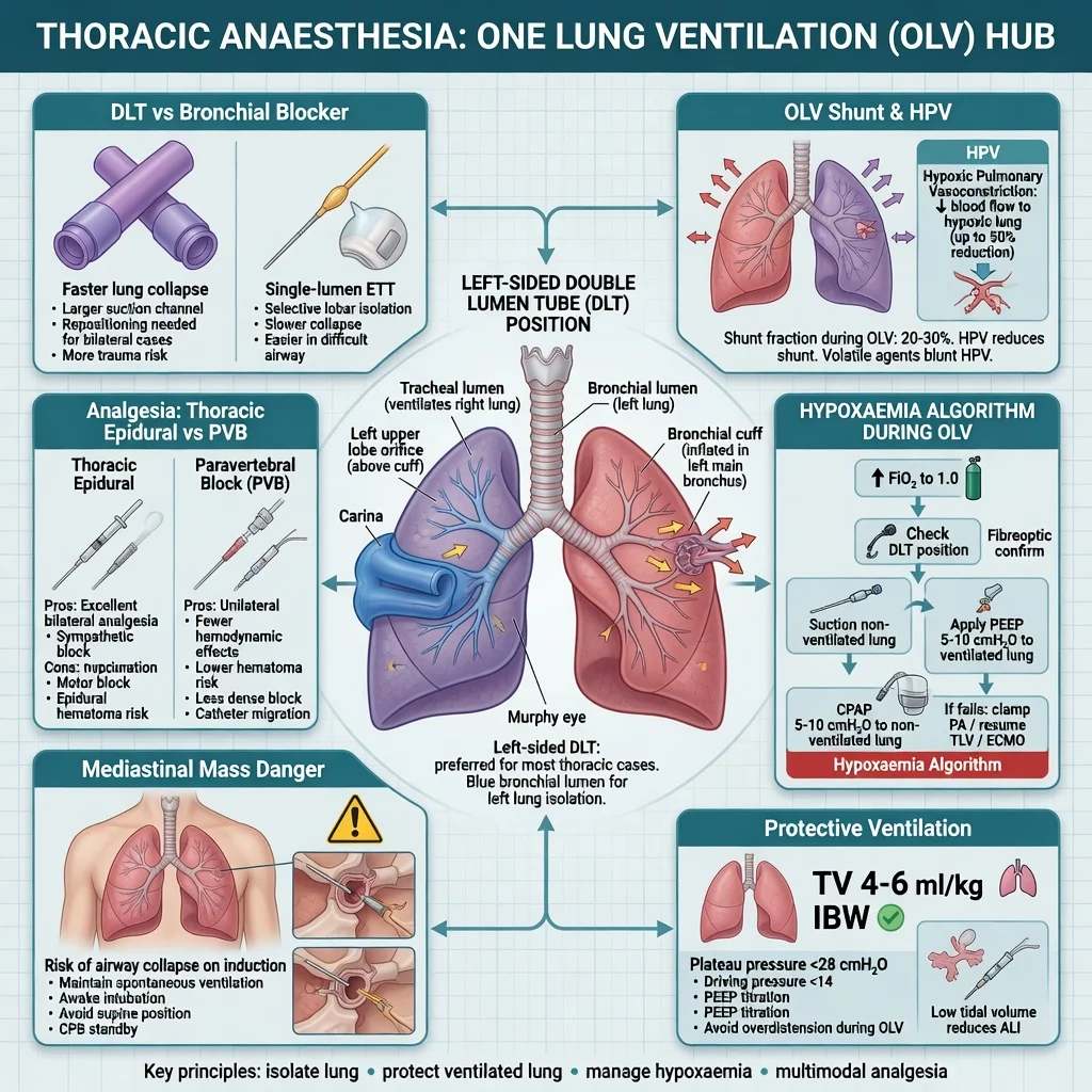
Non-ventilated lung continues to receive blood flow → shunt and hypoxaemia. Hypoxic pulmonary vasoconstriction (HPV) is helpful but blunted by many volatiles at high dose, vasodilators, and infection/atelectasis. Dependent ventilated lung can suffer atelectasis and high pressure. [1]
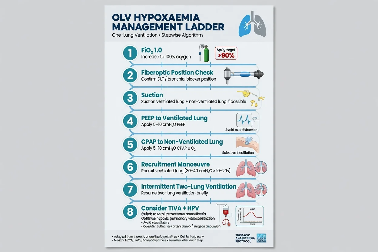
Hypoxaemia algorithm (exam order, not panic): [1]
- Increase FiO2 toward 1.0 temporarily.
- Check tube position with fibrescope — malposition is common after turning lateral.
- Suction blood/secretions; recruit dependent lung carefully.
- Optimise ventilation to dependent lung (protective TV, titrated PEEP).
- Apply CPAP / oxygen insufflation to operative lung if surgical field allows.
- Clamp pulmonary artery temporarily only as surgical/physiological last resort discussion.
- Revert to two-lung ventilation if crisis — life before perfect field. [1]
Conduit perfusion physiology
The gastric conduit is a partially devascularised tube. Hypotension threatens anastomotic perfusion; extreme vasopressor excess and fluid overload (pulmonary oedema, tissue oedema) are also harmful. Teaching aim: adequate MAP with euvolemia, avoid drowning the lungs, communicate with surgeon about pressor choice and targets.[2]
Anaesthetic goals
- Secure airway with aspiration precautions.
- Reliable lung isolation confirmed after every position change.
- Protective ventilation on two-lung and OLV.
- Haemodynamic stability for conduit perfusion.
- Excellent thoracic analgesia enabling physiotherapy.
- Early detection of bleeding, hypoxia, arrhythmia.
- Plan HDU/ICU destination before incision. [1]
Technique options and decision matrix
Monitoring and equipment
- Arterial line (almost always for thoracic phase/major open).
- Large-bore IV access; consider CVC if vasoactive infusions or poor access.
- Lung isolation kit: left DLT common for right thoracotomy, or blocker; flexible bronchoscope mandatory.
- Temperature management; urine output; consider cardiac output monitoring in high-risk.
- Blood available; cell salvage in selected bloody cases.
- Thoracic epidural kit or regional plan (ESP/paravertebral alternatives if epidural unsuitable).
- NG/conduit tube care strictly per surgeon — do not pull “an NG” casually.
- Confirmed HDU/ICU bed. [1]
Intraoperative management
Induction and isolation
Head-up preoxygenation when possible; antacid/prokinetic per local aspiration protocol; RSI or modified RSI with gentle ventilation as modern practice allows when indicated. Place DLT/blocker; confirm with fibrescope. Lateral position: re-confirm isolation immediately.[1]
Ventilation
Two-lung: protective 6–8 mL/kg PBW-style thinking, titrated PEEP. OLV: reduce TV further to ventilated lung, watch driving pressure, avoid stacking. Accept moderate hypercapnia if needed rather than injurious volumes. [1]
Haemodynamics and fluids
Avoid prolonged hypotension during anastomosis. Use vasopressors thoughtfully to defend MAP rather than flooding with crystalloid. Blood for oxygen delivery and ongoing loss; keep warm. Discuss conduit colour/perfusion with surgeon. [1]
Analgesia and ERAS-leaning care
Thoracic epidural remains a classic exam answer for open oesophagectomy analgesia; multimodal systemic analgesia and fascial/paravertebral techniques are alternatives when coagulopathy, refusal, or hypotension risk dominates. PONV prophylaxis matters — vomiting stresses anastomosis. Antibiotics and VTE prophylaxis per protocol.[2]
Extubation
Many centres aim early extubation if stable, warm, fully reversed (quantitative NMM), pain controlled, and gas exchange acceptable. Leave a clear plan for reintubation risk (aspiration, RLN, fatigue). [1]
Crisis pivots
OLV hypoxia
Run the structured algorithm above; communicate with surgeon; do not waste minutes on single interventions repeated without FO tube check. [1]
Massive bleed (azygos, aorta, spleen, short gastrics)
Activate major haemorrhage pathway; surgical control; balanced products; cell salvage if set up. [1]
Aspiration
Suction, head-down if practical, secure airway, lung-protective ventilation, defer elective cases if pre-induction soilage; ICU if severe pneumonitis picture. [1]
New AF (often postoperative but can be intraop)
Correct K+/Mg2+, rate or rhythm control per stability, look for sepsis/hypovolaemia/pain later. [1]
Suspected early conduit ischaemia
Urgent surgical review — anaesthesia role is resuscitation, oxygen delivery, and avoiding further hypoperfusion. [1]
Postoperative plan
Destination: HDU/ICU common after open and many MIE cases. [1]
Pulmonary: analgesia enabling cough and physio; NIV carefully if used (anastomosis/airway team awareness); early mobilisation. [1]
Leak surveillance: fever, tachycardia, pleural contamination, rising CRP/lactate, neck emphysema (cervical anastomosis) — leak until proven otherwise.[2]
RLN injury: hoarse voice, weak cough, aspiration — keep NBM if unsafe swallow, ENT/SLT review. [1]
Nutrition: jejunostomy feeds often; do not rely on oral intake early without clearance. [1]
AF and pneumonia are high-frequency complications — electrolytes, fluids, physio, infection screen. [1]
Special populations
- MIE/RAMIE: still OLV skills; CO2 absorption; less wound pain but same leak biology.
- Salvage oesophagectomy after definitive CRT: hostile tissues, bleeding, poor healing.
- Obese / OSA: ramp, dosing scalars, CPAP plan, HDU bias.[3]
- Elderly frail: pulmonary risk dominates; realistic goals of care.
- Previous gastric surgery: conduit options change (colonic interposition) — longer cases, different perfusion issues.
SAQ answer scaffold
A 62-year-old for Ivor Lewis oesophagectomy after neoadjuvant therapy. Outline your anaesthetic management. [1]
- Assessment (3): staging/fitness, aspiration risk, pulmonary reserve, airway, anaemia, HDU plan.
- Airway & isolation (3): RSI thinking, DLT/blocker, FO confirmation after lateral turn.
- OLV (3): protective ventilation, hypoxia algorithm.
- Perfusion/fluids (3): MAP for conduit, avoid liberal crystalloid, vasopressors thoughtfully.[1][2]
- Analgesia & postop (3): thoracic regional/epidural + multimodal, leak/RLN/AF surveillance, HDU.
Viva stem bank and model phrases
Stem 1: “DLT or bronchial blocker?”
Model: “Either is acceptable if isolation is achieved and confirmed bronchoscopically; left DLT is common for right thoracotomy because of familiarity and suction access.” [1]
Stem 2: “Epidural hypotension — worry about the anastomosis?”
Model: “Yes — I titrate the epidural, use vasopressors to defend MAP, and ensure the conduit is not sacrificed for a pure ‘dry and hypotensive’ dogma.” [1]
Stem 3: “Day 5 fever and pleural sepsis?”
Model: “Anastomotic leak until proven otherwise — resuscitate, broad cultures/antibiotics, imaging and surgical review, airway protection if aspiration.” [1]
Stem 4: “Why re-check the tube after turning?”
Model: “Lateral flexion and surgical manipulation commonly malposition double-lumen tubes; FO check is faster than guessing.” [1]
Stem 5: “Fluid strategy?”
Model: “Balanced, goal-directed thinking — avoid multi-litre crystalloid overload that harms lungs while still maintaining perfusion pressure for the conduit.”[2]
Stem 6: “Transhiatal specific risks?”
Model: “Mediastinal blunt dissection can cause sudden hypotension and arrhythmias even without formal OLV — communicate and be ready with vasopressors and blood.” [1]
Stem 7: “Obese patient for MIE?”
Model: “I combine thoracic OLV skills with obesity airway positioning, dose scalars, and a lower threshold for HDU.”[3]
Common traps
- No fibrescope confirmation of isolation after position change
- Fluid overload “for urine output”
- Ignoring aspiration risk at induction and extubation
- Weak analgesia → sputum retention and pneumonia
- Missing RLN injury post-extubation
- Treating leak sepsis as “simple pneumonia” too long
- Deep extubation without airway protective reflexes in high aspiration risk [1]
Oesophagectomy plan — LEAK
Examiner mental map
- Approach and what cavities/OLV it implies.
- Aspiration and airway plan.
- Isolation + OLV hypoxia algorithm.
- Conduit perfusion / fluids / vasopressors.
- Analgesia that enables cough.
- Postop: RLN, AF, pneumonia, leak. [1]
Hit those six and you sound like a consultant on a thoracic list — not a textbook chapter. [1]
References
- [1]Veelo DP, Geerts BF Anaesthesia during oesophagectomy J Thorac Dis, 2017.PMID 28815066
- [2]Deana C, Vetrugno L, Bignami E, Bassi F Peri-operative approach to esophagectomy: a narrative review from the anesthesiological standpoint J Thorac Dis, 2021.PMID 34795950
- [3]Nightingale CE, Margarson MP, Shearer E, et al. Peri-operative management of the obese surgical patient 2015: Association of Anaesthetists of Great Britain and Ireland Society for Obesity and Bariatric Anaesthesia Anaesthesia, 2015.PMID 25950621