Anaes · Paediatric anaesthesia
Paediatric airway anatomy, equipment sizing, and ETT selection
Also known as Paediatric airway · Paediatric ETT sizing · Cuffed versus uncuffed endotracheal tube · Paediatric laryngoscopy · Difficult paediatric airway
The paediatric airway differs from the adult airway in ways that change every step of airway management: the larynx is high and anterior, the tongue and occiput are large, the epiglottis is long and floppy, and the cricoid is classically the narrowest point. Endotracheal tube selection follows the Cole formulae (uncuffed and cuffed), with depth approximated as three times the internal diameter. Modern cuffed tubes are safe and reduce tube exchange. The defining emergency is laryngospasm progressing to bradycardia and arrest. This topic covers anatomy, equipment sizing, cuffed versus uncuffed evidence, positioning, the difficult and syndromic airway, and laryngospasm management for the ANZCA Final Examination and its cross-exam equivalents.
On this page & tools
Your progress
Saved locally on this device.
Target exams
Red flags
Why this is examined
Airway management is the single most common source of severe adverse events in paediatric anaesthesia, and the paediatric airway behaves differently enough from the adult airway that adult techniques transferred uncritically will fail. The ANZCA Final Examination (and its cross-exam equivalents in the FRCA, EDAIC, ABA APPLIED, FCAI, and FCA(SA)) tests four things repeatedly: the anatomical reasoning that explains why the technique differs, the rote formulae for tube size and depth, the modern evidence on cuffed versus uncuffed tubes, and the crisis pathway for laryngospasm. Each of these is a discriminating question that separates a candidate who has memorised a number from one who understands the physiology. The ANZCA curriculum codes this topic under the Paediatric anaesthesia Specialised Study Unit (SS_PA); it is also foundational AT_AM and BT_AM airway material. [1]
Applied anatomy and physiology
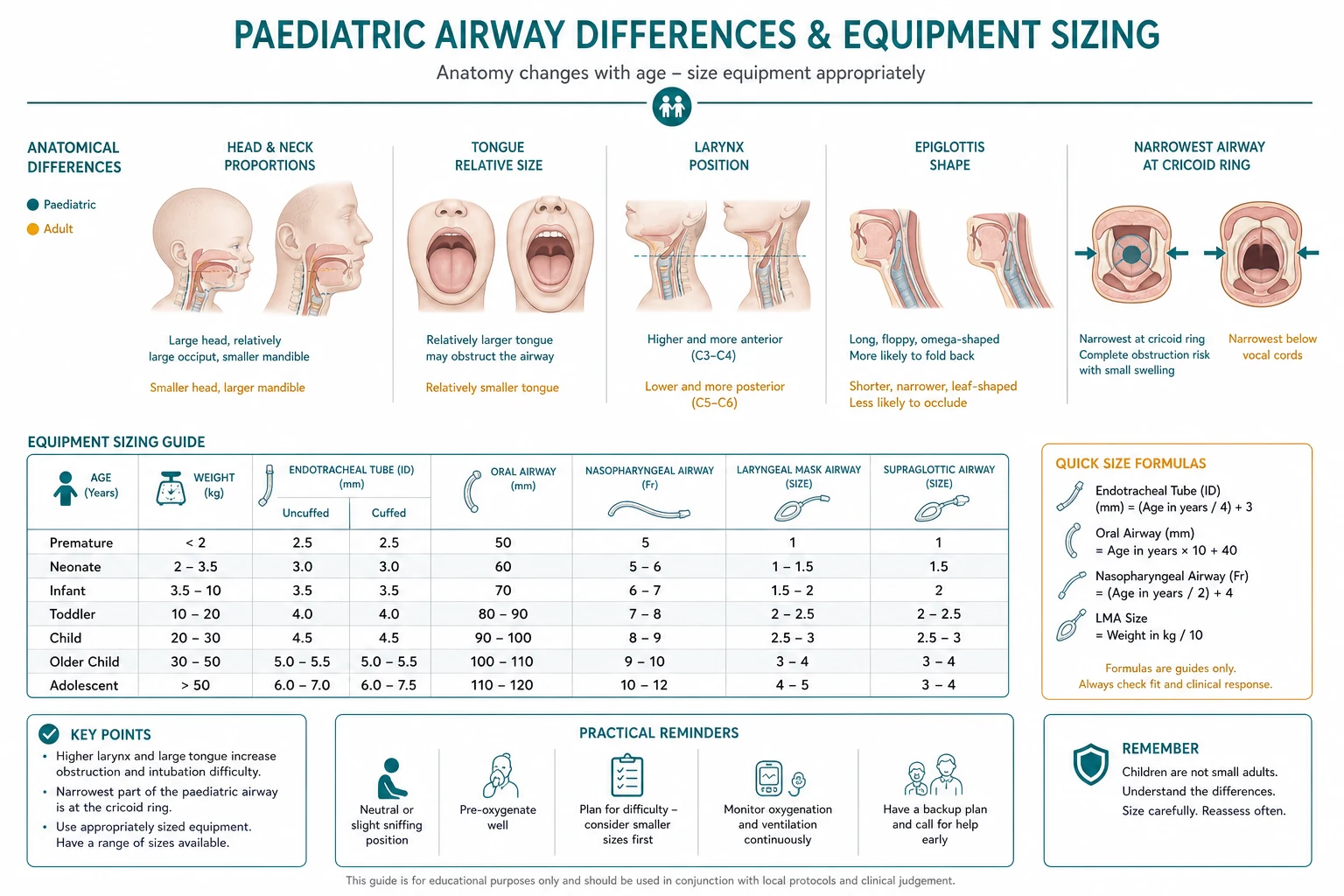
The infant upper airway differs from the adult's in six exam-critical ways. [1]
[1]The physiology is the other half of the answer and explains why airway events in children become emergencies so fast. The functional residual capacity is small, the closing capacity is close to it, and the oxygen consumption is high — around 6 mL/kg/min in the infant, roughly twice the adult value. The result is a safe apnoea time measured in tens of seconds rather than minutes after preoxygenation; the neonate can desaturate below ninety per cent in under a minute of apnoea. The cardiac output is heart-rate dependent, and hypoxaemia produces bradycardia quickly, so an airway event in a child escalates: desaturation, then bradycardia, then cardiac arrest, in a sequence that can complete in under three minutes. [1]
ETT selection, sizing, and depth
Tube selection is built on two formulae and one rule, and the candidate who can reproduce them under viva pressure has answered most of the equipment questions. [1]
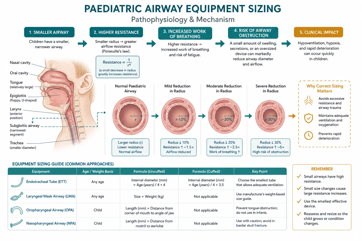
These are the Cole (Motley) formulae. They apply beyond the neonatal period. The depth rule of thumb — three times the internal diameter — gives the insertion depth at the teeth or gums; an alternative is half the age plus twelve. A worked example: a four-year-old takes a cuffed tube of (4/4) plus 3.5 equals 4.5 mm internal diameter, inserted to a depth of 13 to 14 cm at the teeth. Always prepare a tube half a millimetre larger and half a millimetre smaller, because formulae estimate and individuals vary. [1]
For the neonate and infant under one to two years, the formulae do not apply and sizing is by weight and gestational age. A reasonable starting point is a 3.0 mm uncuffed tube for a term neonate, a 3.5 mm for a three- to six-month infant, and progression thereafter. The Broselow tape or a weight estimate of (age plus four) times two kilograms gives the dosing weight when the weight is unknown. [1]
Cuffed versus uncuffed tubes
The historical dogma was that cuffed tubes were unsafe in children because the paediatric cricoid is the narrowest point and a cuff would cause subglottic stenosis. This view came from old data and from cuff designs that are no longer used. The modern evidence is the opposite. [1]
The pivotal meta-analysis pooled two randomised trials and two prospective cohorts totalling 3782 children and found that cuffed tubes did not increase the incidence of post-extubation stridor (risk ratio 0.88, 95 per cent confidence interval 0.67 to 1.16) and dramatically reduced the tracheal tube exchange rate (risk ratio 0.07, 95 per cent confidence interval 0.05 to 0.10)[1]. The clinical message: cuffed tubes are the default in modern paediatric anaesthesia for children over roughly one to two years, used with cuff-pressure monitoring. Uncuffed tubes retain a role in the smallest neonates where a thin-walled polyurethane cuff may still add to outer diameter.
Positioning, preoxygenation, and the modified RSI
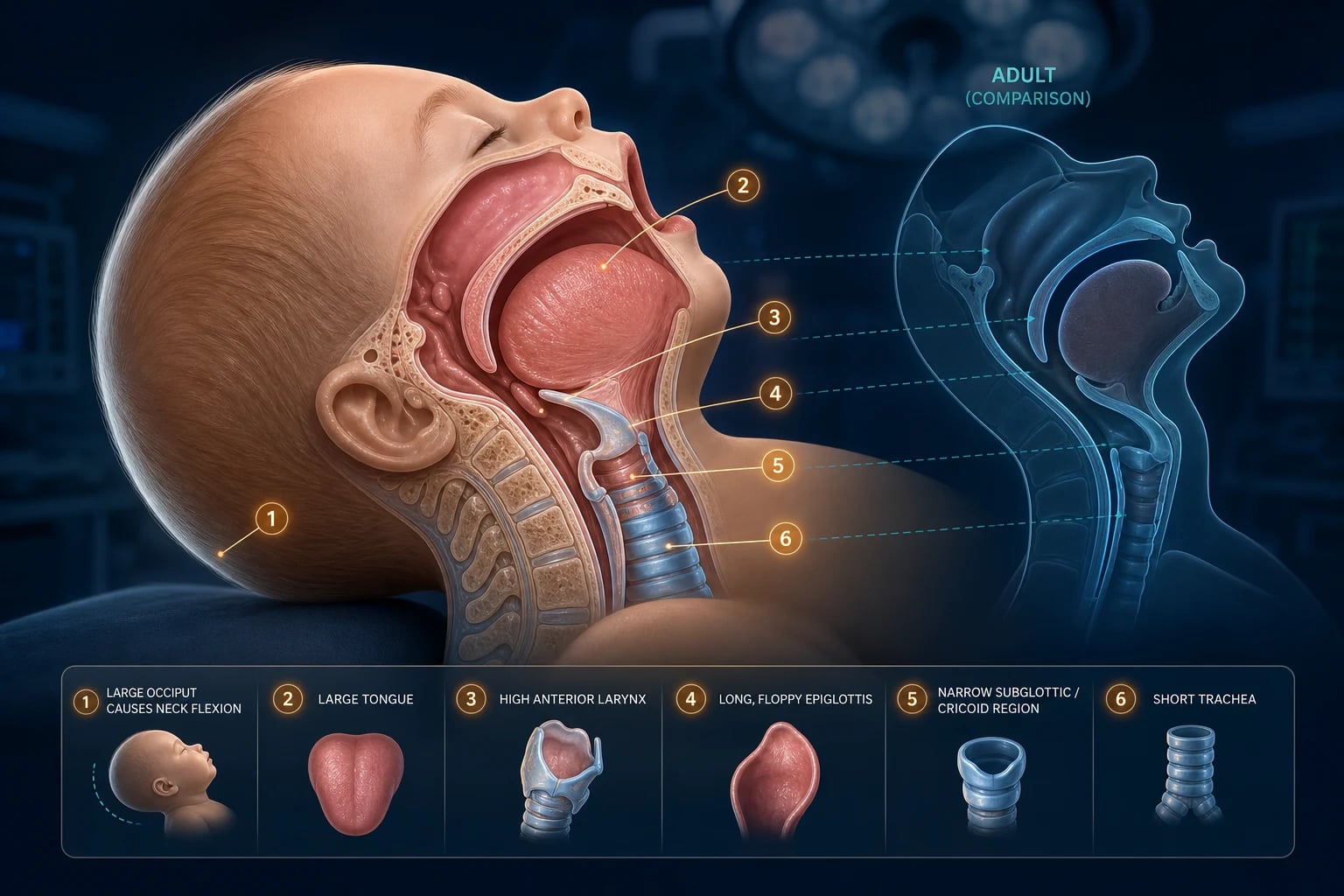
The infant's prominent occiput means the head flexes forward in the supine position; the classic sniffing position is achieved differently than in the adult. Two schools exist. The first places a small shoulder roll to align the laryngeal and pharyngeal axes, with the head resting on the table. The second argues the occiput already provides the sniff and that a shoulder roll over-extends the neck. The defensible position for the exam: position to align the external auditory meatus with the sternal notch in the horizontal plane (the ramped sniffing position), and individualise — the small premature infant often benefits from a shoulder roll, the older child does not. [1]
Preoxygenation is not optional in children. Three minutes of tidal breathing or eight vital-capacity breaths of one hundred per cent oxygen buys the limited safe apnoea time. The modified rapid sequence induction in children reflects two realities: the child desaturates fast, so the apnoeic period must be short, and suxamethonium has a higher rate of adverse effects in children (bradycardia, hyperkalaemia in undiagnosed myopathy, masseter spasm). A common modified RSI uses a high-dose rocuronium (around 1 mg/kg) with a suxamethonium fallback, preoxygenation, and gentle mask ventilation between induction and intubation to maintain oxygenation. Suxamethonium remains the agent when the airway is at risk of being difficult and rapid recovery of spontaneous ventilation is paramount, but it should not be routine in the healthy child for elective surgery. [1]
Apnoeic oxygenation with high-flow nasal cannula (transnasal humidified rapid-insufflation ventilatory exchange, THRIVE) extends the safe apnoea time in children and is increasingly used during laryngoscopy, but it does not eliminate the need for rapid airway securing in the small infant. [1]
Equipment: blades, tubes, and video laryngoscopy
Equipment essentials
The straight (Miller) blade is the infant blade. The high anterior larynx and floppy epiglottis are lifted directly, whereas the curved (Macintosh) blade is inserted into the vallecula and lifts indirectly via the hyoepiglottic ligament — a mechanism less reliable in the infant. Typical sizes are a Miller 0 for the neonate, Miller 1 for the infant and small child, and progression to a Macintosh 2 or 3 in the older child and adolescent. Video laryngoscopy (for example a C-MAC with a pediatric Miller or hyperangulated blade) has become standard backup and first-line for the anticipated difficult airway; it improves the glottic view but does not always make intubation easier, because the tube may still need to be directed around an acute angle. [1]
Other sizing rules the examiner expects: suction catheter size in French is roughly twice the ETT internal diameter (so a 4.0 mm tube takes an 8 Fr catheter); laryngeal mask airway sizes scale with weight (size 1 under 5 kg, 1.5 for 5 to 10 kg, 2 for 10 to 20 kg, 2.5 for 20 to 30 kg, 3 for 30 to 50 kg). Have these on the trolley before induction. [1]
The difficult and syndromic paediatric airway
Anticipated difficulty divides into the syndromic and the acquired. The syndromic airways cluster by mechanism. [1]
The unanticipated difficult paediatric intubation is governed by a guideline, and the candidate must name the algorithm. The Association of Paediatric Anaesthetists of Great Britain and Ireland and the Difficult Airway Society (APAGBI/DAS 2015) paediatric guidelines give three linked algorithms: difficult mask ventilation, unanticipated difficult intubation, and cannot-intubate-cannot-ventilate. The structure mirrors the adult DAS guidance: call for help early, limit attempts, deploy a second-generation supraglottic airway, and in the cannot-intubate-cannot-oxygenate child proceed to age-appropriate front-of-neck access (needle cricothyroidotomy in the younger child, scalpel-bougie in the older). The Vortex cognitive aid is taught alongside as the crisis framework. [1]
Crisis: laryngospasm and upper-airway obstruction
Laryngospasm is the sustained closure of the glottis by the intrinsic laryngeal muscles, and it is the canonical paediatric airway emergency. The recognition is the trigger: stridorous or silent obstruction in a child at light planes of anaesthesia, often at induction (irritant volatile, secretions, blood, a stimulus at the wrong depth) or at emergence. It escalates from partial closure (stridor, tracheal tug) to complete closure (silent, impossible to ventilate), and complete closure leads to bradycardia, negative-pressure pulmonary oedema, and cardiac arrest. [1]
The risk factors are well quantified. A case-control study of 130 children with laryngospasm found that an intercurrent upper respiratory tract infection multiplied the risk (odds ratio 2.03) and the presence of an airway anomaly multiplied it further (odds ratio 3.35); the use of a laryngeal mask airway was independently associated during maintenance and emergence[2]. Passive smoke exposure, asthma, and the child undergoing airway surgery raise the baseline risk. The practical inference the examiner wants: defer elective anaesthesia after a significant upper respiratory tract infection where feasible, and treat the high-risk child with a deep extubation or an intravenous technique.
The management is a ladder, climbed in order and fast. [1]
The ANZCA/APAGBI ladder for laryngospasm
- Remove the stimulus; clear secretions or blood; ask the surgeon to stop.
- Apply one hundred per cent oxygen via a tight-fitting mask with continuous positive airway pressure, and a firm bilateral jaw thrust (Larson's manoeuvre — firm pressure at the laryngospasm notch, behind the ear lobe, between the ascending ramus of the mandible and the mastoid process, while thrusting the jaw forward).
- Deepen the anaesthesia: propofol 0.5 to 1 mg/kg intravenously.
- If refractory, or if bradycardia or desaturation supervenes: suxamethonium, 0.1 to 0.2 mg/kg intravenously (or 4 mg/kg intramuscularly if access is lost); atropine 20 microgram/kg if bradycardic.
- Treat negative-pressure pulmonary oedema if it develops; observe post-event.
Pharmacological prevention is also evidence-based. A systematic review and meta-analysis of nine trials in 787 children found that both intravenous and topical lidocaine reduce the incidence of laryngospasm (risk ratio 0.39, 95 per cent confidence interval 0.24 to 0.66)[3]. The discriminating detail: the candidate who says "give lidocaine" earns half marks; the candidate who gives the pooled risk ratio and distinguishes intravenous from topical earns the pass.
Postoperative considerations
The child who has had a laryngospasm or a difficult airway event needs postoperative observation for negative-pressure pulmonary oedema, which develops in a small fraction of cases and presents with pink frothy sputum and desaturation after an apparently resolved event. Post-extubation stridor (subglottic oedema) is treated with cool humidified oxygen, nebulised racemic adrenaline (0.5 mL of 2.25 per cent in 3 mL saline), and dexamethasone 0.5 mg/kg. Emergence delirium in the preschool child after sevoflurane is distinguished from pain using the Paediatric Anaesthesia Emergence Delirium (PAED) scale, a five-item instrument (eye contact, purposeful action, awareness, restlessness, inconsolability) with strong internal consistency (Cronbach alpha 0.89)[4]; the discriminating detail is that the first three items are reverse-scored, and a score of 10 or more supports the diagnosis.
Special populations
The neonate is the highest-risk airway. The NAP7 audit identified infants under one year and those with congenital heart disease as the groups at highest risk of perioperative cardiac arrest, with hypoxaemia the commonest mechanism — a direct consequence of the short safe apnoea time. The neonatal emergency (congenital diaphragmatic hernia, tracheo-oesophageal fistula, abdominal wall defect) demands the most experienced anaesthetist present, a pre-induction plan, and equipment checked. The airway in congenital heart disease is complicated by cyanosis, by the haemodynamic consequences of positive-pressure ventilation in single-ventricle physiology, and by the need to avoid prolonged apnoea. [1]
The anaesthesia-induced developmental neurotoxicity question is unavoidable in paediatric anaesthesia and is part of the consent. The GAS trial, a multicentre randomised controlled equivalence trial in 722 infants undergoing inguinal herniorrhaphy, found equivalence between awake-regional and brief sevoflurane general anaesthesia at two years (Bayley cognitive composite score)[5] and at five years (full-scale IQ; mean difference 0.23, 95 per cent confidence interval minus 2.59 to 3.06, strong evidence of equivalence)[6]. The clinical position: a single brief exposure is reassuring; the risk-benefit of delaying elective surgery is individualised, and unnecessary exposure is avoided.
SAQ answer scaffold
A fifteen-mark SAQ on paediatric airway equipment and sizing is answered in this structure: [1]
- Anatomical differences (5 marks): high anterior larynx (C3 to C4), large tongue, prominent occiput, long floppy U-shaped epiglottis, cricoid the narrowest point, elliptical subglottis.
- ETT sizing (3 marks): uncuffed (age/4) plus 4; cuffed (age/4) plus 3.5; depth at teeth ID times 3; valid over one to two years; have plus or minus half a millimetre ready.
- Cuffed versus uncuffed (3 marks): modern cuffed preferred; cite the meta-analysis (no increase in stridor, risk ratio 0.88; reduced exchange, risk ratio 0.07); cuff pressure below 20 to 25 cmH2O.
- Laryngospasm (4 marks): recognition; risk factors (URTI odds ratio 2, airway anomaly odds ratio 3.4); the ladder (remove stimulus, Larson's CPAP and jaw thrust, propofol, suxamethonium); lidocaine prevention (risk ratio 0.39). [1]
Viva stem bank
- "Describe the anatomical differences between the infant and adult airway and how each changes your technique." (Open with the six points, each tied to a technique.)
- "What size endotracheal tube would you use for a four-year-old, and why might you choose a cuffed tube?" (Cole formulae, worked number, the meta-analysis.)
- "A previously well two-year-old develops stridor and then silent obstruction during induction with sevoflurane. Manage." (Laryngospasm ladder, climbed fast, with the doses.)
- "Discuss the evidence that general anaesthesia harms the developing brain." (GAS at two and five years, equivalence, the consent.) [1]
Common traps
- Stating the paediatric airway is narrowest at the vocal cords — it is the cricoid (with the elliptical refinement).
- Using the Cole formulae for a neonate — they apply only over one to two years.
- Sizing the depth incorrectly (the common error is age plus ten, which overshoots; the rule is ID times three or age over two plus twelve).
- Treating laryngospasm with repeated suxamethonium before climbing the non-pharmacological ladder — Larson's manoeuvre and CPAP resolve many episodes.
- Forgetting the cuff pressure when using a cuffed tube — check it stays below 20 to 25 cmH2O.
- Equating a difficult view with a difficult intubation in video laryngoscopy — the view improves but the tube delivery may not. [1]
Preoperative equipment briefing (exam spine expansion)
Before any paediatric induction, state the airway plan aloud: predicted easy or difficult; first device (mask, SGA, or tube); backup device; who will help; where the difficult airway trolley is. Measure or estimate weight; calculate ETT size and depth on paper or app; draw emergency drugs by weight (atropine 20 microg/kg, adrenaline 10 microg/kg for arrest, suxamethonium 1.5 to 2 mg/kg IV and 4 mg/kg IM, propofol 0.5 to 1 mg/kg for laryngospasm deepening). Confirm suction works. For shared airway ENT lists, agree tube type (RAE, microlaryngoscopy tube, laser-safe protocols) and who controls the airway during suspension laryngoscopy. [1]
Neonatal and infant sizing beyond Cole
The Cole formulae apply over approximately one to two years of age. For neonates and small infants, start with fixed sizes and clinical leak testing: [1]
- Extreme preterm: often 2.5 mm ID uncuffed (or microcuff designs where available)
- Term neonate: 3.0 to 3.5 mm ID
- 1 to 6 months: 3.5 to 4.0 mm ID
- 6 to 12 months: 4.0 mm ID [1]
Oral insertion depth in neonates is often estimated as 6 plus weight in kilograms (cm at the lips) as a cross-check alongside ID times 3. Always auscultate bilaterally and use capnography; short tracheas make endobronchial intubation easy. Nasal tubes add roughly 10 to 20 percent extra length depending on local practice — confirm clinically. [1]
Supraglottic airways, suction, and adjunct sizing
Mask size should cover nose and mouth without pressure on the eyes. Two-person mask ventilation (one holds the mask with jaw thrust, one squeezes the bag) is the default when single-handed ventilation fails. [1]
Monitoring, cuff pressure, and ventilation targets
After intubation: continuous waveform capnography is mandatory confirmation. Maintain cuff pressure below 20 to 25 cmH2O with a manometer when a cuffed tube is used — finger-feel is unreliable. Ventilate with age-appropriate rates (neonates often 25 to 40 breaths/min; older children lower) and tidal volumes around 6 to 8 mL/kg, avoiding high peak pressures that barotrauma a small airway. Peak pressure rise after intubation may signal endobronchial placement, light anaesthesia with resistance, or tube obstruction from secretions or kinking. [1]
Crisis pivots unique to equipment choices
Wrong tube size: large leak with inability to ventilate → upsize half a millimetre or inflate cuff carefully; high pressure with no leak and difficulty passing suction → tube too large or endobronchial — pull back or downsize. Cuff herniation or overinflation: obstructs the lumen or damages mucosa — deflate, check manometer. SGA failure: poor seal, laryngospasm around the cuff, gastric insufflation — remove, deepen or paralyse, mask oxygenate, intubate. Cannot intubate after relaxant: APAGBI/DAS pathway — help, oxygenate, SGA, limit attempts, age-appropriate CICO. Bradycardia during laryngoscopy: stop, oxygenate, atropine — hypoxia until proven otherwise. [1]
Special populations (equipment focus)
Burns and facial trauma: expect difficult mask seal; early advanced airway with smaller tubes if oedema progressing. Cardiac infants: balance FiO2 and ventilation with shunt physiology; equipment same but haemodynamic plan different. Airway infection (croup/epiglottitis): start smaller than formula; spontaneous ventilation induction; ENT present. Laser airway surgery: laser-safe tube protocols or intermittent apnoeic techniques with total intravenous anaesthesia in specialist practice. [1]
Cross-links to GAS and consent
When parents ask whether the endotracheal tube or the anaesthetic will harm the developing brain, separate the issues: airway trauma is minimised by correct size, cuff pressure control, and gentle technique; neurodevelopmental risk after a single brief sevoflurane anaesthetic in infancy was not different from awake-regional anaesthesia at two and five years in the GAS trial — communicate honestly without over-generalising to prolonged repeated exposures.[5][6]
Extended viva phrases
- “Uncuffed size is age over four plus four; cuffed is age over four plus 3.5; depth is three times the internal diameter.”
- “I prefer a cuffed tube with manometry because the meta-analysis shows no increase in stridor and far fewer tube exchanges.”[1]
- “If the child develops silent obstruction at induction I apply 100 percent oxygen with CPAP and Larson, then propofol, then suxamethonium — four milligrams per kilogram intramuscular if I have lost the drip.”
- “Bradycardia is hypoxia until proven otherwise.”
References
- [1]Shi F, Xiao Y, Xiong W, Zhou Q, Huang X Cuffed versus uncuffed endotracheal tubes in children: a meta-analysis J Anesth, 2016.PMID 26296534
- [2]Flick RP, Wilder RT, Pieper SF, et al. Risk factors for laryngospasm in children during general anesthesia Paediatr Anaesth, 2008.PMID 18315633
- [3]Mihara T, Uchimoto K, Morita S, Goto T The efficacy of lidocaine to prevent laryngospasm in children: a systematic review and meta-analysis Anaesthesia, 2014.PMID 24992191
- [4]Sikich N, Lerman J Development and psychometric evaluation of the pediatric anesthesia emergence delirium scale Anesthesiology, 2004.PMID 15114210
- [5]Davidson AJ, Disma N, de Graaff JC, et al. Neurodevelopmental outcome at 2 years of age after general anaesthesia and awake-regional anaesthesia in infancy (GAS): an international multicentre, randomised controlled trial Lancet, 2016.PMID 26507180
- [6]McCann ME, de Graaff JC, Dorris L, et al. Neurodevelopmental outcome at 5 years of age after general anaesthesia or awake-regional anaesthesia in infancy (GAS): an international, multicentre, randomised, controlled equivalence trial Lancet, 2019.PMID 30782342