Anaes · Neuroanaesthesia
Posterior fossa surgery: cranial nerves, brainstem reflexes, and positioning
Also known as CPA surgery anaesthesia · Posterior fossa craniotomy · Trigeminocardiac reflex
Anaesthesia for posterior fossa and CPA surgery including positioning trade-offs, cranial nerve and brainstem monitoring constraints, trigeminocardiac reflex, VAE risk, and emergence planning.
On this page & tools
Your progress
Saved locally on this device.
10 MCQs with explanations
Target exams
Red flags
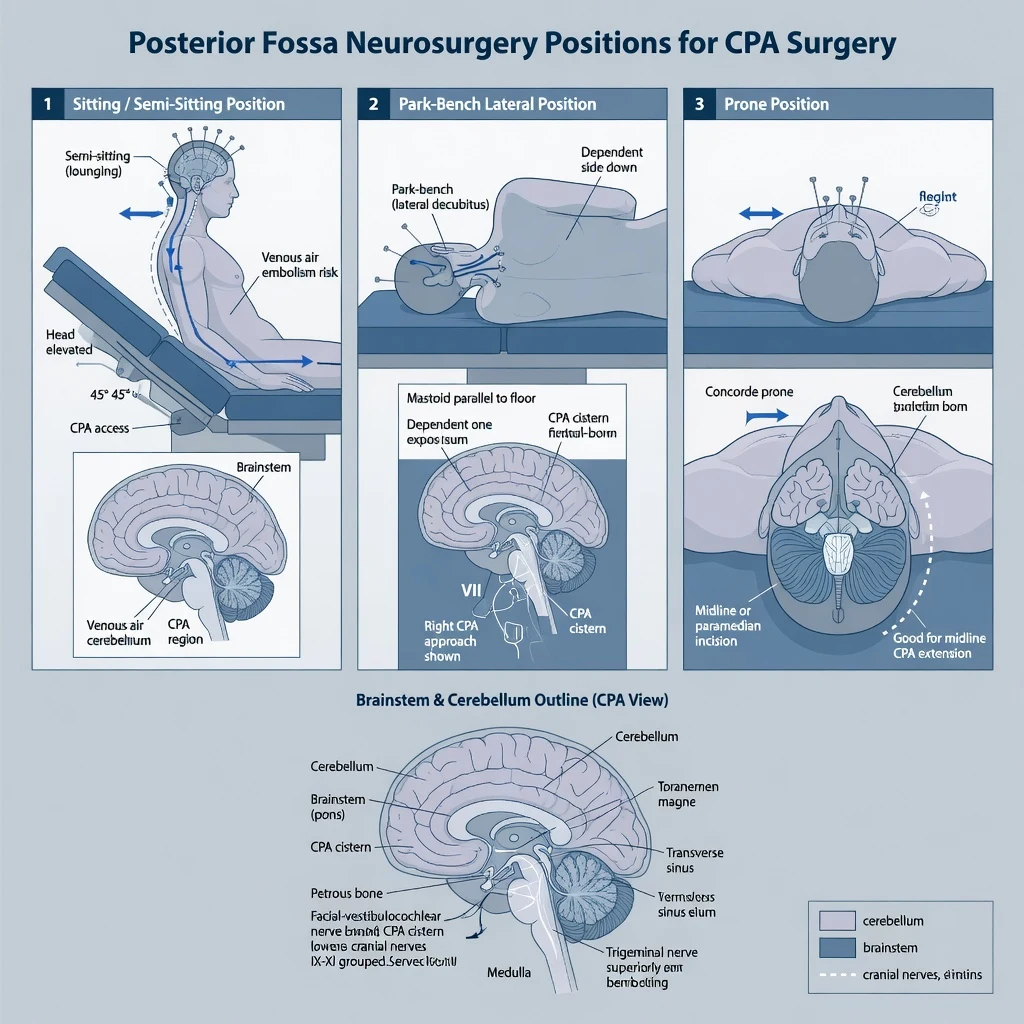
Why this is examined / the one-line answer
Posterior fossa and cerebellopontine angle (CPA) surgery combines positioning physiology, venous air embolism (VAE) risk, brainstem cardiovascular reflexes, and cranial nerve (CN V–XII) injury that changes extubation safety. It is high-yield SS_NS Final material because a single stem can test VAE, trigeminocardiac reflex, facial nerve monitoring constraints, and delayed airway failure from bulbar dysfunction. [1]
One-line opener: Stop surgical stimulus first for sudden bradycardia, keep VAE on the differential when the head is up, and do not extubate a patient who cannot protect the airway because lower cranial nerves are injured. [1]
Preoperative assessment and risk stratification
Pathology. Tumour type and location (vestibular schwannoma, meningioma, cerebellar metastasis, Chiari, vascular lesions), brainstem compression, hydrocephalus, raised ICP signs, lower CN deficits already present (hoarse voice, weak cough, aspiration, reduced gag), hearing and facial function baseline. [1]
Comorbidities. PFO or right-to-left shunt (paradoxical embolus risk if sitting), cardiovascular disease, OSA, reflux, cervical spine disease limiting positioning, anticoagulant use. [1]
Investigations. MRI anatomy, ECG, echo if PFO screening or cardiac risk indicated, baseline swallow assessment if bulbar symptoms, bloods and crossmatch as indicated. [1]
Airway. Anticipate difficult airway if prior radiotherapy, restricted neck, or large tumours with secondary issues; plan extubation strategy before induction (DAS culture for unanticipated difficulty).[2]
Positioning decision matrix
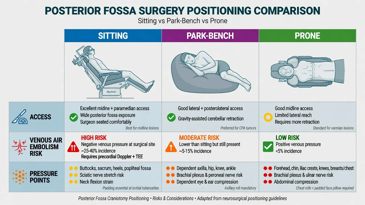
Sitting position specifics
- Improved surgical field and venous drainage; lower blood loss historically claimed
- Open venous sinuses above the heart → air entrainment
- Need VAE detection plan: precordial Doppler and/or TOE, EtCO2, PA catheter selectively
- Multi-orifice CVP with tip near SVC–RA junction if aspiration strategy planned
- Relative contraindication discussions around known large PFO (risk of paradoxical embolus) [1]
Applied physiology and anaesthetic goals
Intracranial and brainstem goals
- Maintain CPP (MAP − ICP or RAP as context); avoid hypotension especially in sitting position
- Controlled ventilation to normocapnia unless surgeon requests brief hypocapnia for relaxation (avoid extreme hypocapnia)
- Avoid venous obstruction: extreme neck flexion/rotation, tight ties, high intrathoracic pressure
- Smooth induction and emergence to limit ICP and bleeding swings
- Enable neuromonitoring: often facial nerve EMG, BAEP, sometimes SSEP/MEP → TIVA preferred when motor pathways monitored; minimise ongoing neuromuscular blockade after intubation [1]
Cardiovascular goals
- Have anticholinergics drawn (atropine or glycopyrrolate) for trigeminocardiac/brainstem bradycardia
- Vasopressors ready for sitting-position hypotension (metaraminol/phenylephrine/noradrenaline as practice)
- Continuous arterial pressure monitoring standard for major posterior fossa work [1]
Technique options
Induction. Standard IV induction with attention to ICP if raised; secure reinforced ETT carefully for prone/park-bench; bite block if MEPs planned; eye protection meticulous in prone. [1]
Maintenance. Propofol–opioid TIVA is the default when MEP/SSEP quality matters; low-dose volatile may be acceptable when only BAEP/facial EMG are used, but know dose-dependent EP depression. Remifentanil common for rapid titration. Avoid N2O in sitting/VAE-risk cases (expands air bubbles). [1]
Neuromuscular blockade. Intubating dose acceptable; do not run continuous NMB if facial nerve EMG or myogenic MEPs are required — confirm recovery with quantitative TOF. [1]
Access and monitoring. Arterial line; large-bore IV; central access for sitting/VAE protocol or vasoactive infusions; urinary catheter; temperature; precordial Doppler for elevated head; TOE if high VAE concern; processed EEG optional; nerve monitoring leads coordinated with neurophysiology. [1]
Propofol–remifentanil and dexmedetomidine sedation literature in awake craniotomy illustrates broader neuroanaesthesia TIVA/sedation fluency but posterior fossa macro-resections are usually under GA with monitoring constraints.[1]
Cranial nerve and brainstem considerations
CN V — trigeminocardiac reflex
Afferent trigeminal pathways; efferent vagal cardioinhibition. Triggers: stimulation of trigeminal sensory territory, including CPA/brainstem work and skull base. Clinical: sudden bradycardia, hypotension, asystole. [1]
Management order: [1]
- Stop surgical stimulus immediately
- Ensure oxygenation/ventilation
- Atropine (e.g. 0.6–1.2 mg IV adult, titrate) or glycopyrrolate if persists
- Deepen anaesthesia if lightly anaesthetised and appropriate
- Keep VAE on the differential if sitting with EtCO2 fall and no clear surgical trigeminal trigger [1]
CN VII / VIII — CPA (vestibular schwannoma)
Facial nerve monitoring limits muscle relaxant after induction. BAEP relatively robust to anaesthetics. Postoperative facial weakness and hearing outcomes are surgical but anaesthetic facilitation of monitoring matters. [1]
CN IX–XII — lower cranial nerves
Gag, swallow, laryngeal sensation/motor, tongue. Injury → aspiration, ineffective cough, airway obstruction at extubation. Plan delayed extubation or tracheostomy discussion if bilateral or severe dysfunction expected. [1]
Brainstem handling
Hypertension or hypotension, arrhythmias, irregular respiratory patterns if spontaneous modes used (rare under GA but relevant conceptually). Communicate early; temporary cessation of retraction may restore stability. [1]
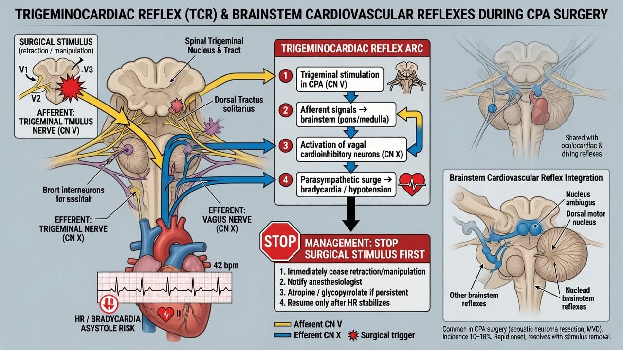
Crisis pivots
Posterior fossa bradycardia
VAE (elevated head)
Detection hierarchy commonly taught: TOE (most sensitive) greater than precordial Doppler greater than rising PA pressure greater than EtCO2 fall greater than SpO2 fall. [1]
Immediate management: [1]
- Notify surgeon — flood field, bone wax, compress veins, lower head if possible
- Stop N2O if used; FiO2 1.0
- Aspirate air from multi-orifice CVP if correctly placed
- Support circulation; Durant manoeuvre (left lateral head-down) when feasible
- TOE assessment; consider paradoxical embolus if PFO and systemic signs
- ACLS if arrest [1]
Other crises
- Tension pneumocephalus postoperatively: delayed emergence, deterioration — imaging and surgical review
- Brainstem injury with unstable haemodynamics: support, stop insult, ICU ventilation plan
- Failed extubation / aspiration from bulbar palsy: re-intubate early; consider delayed airway plan
- Prone airway loss: prevent with secure tube; if lost, follow difficult airway principles with positioning constraints.[2]
Intraoperative haemodynamic and ventilation targets (exam phrasing)
- MAP individualised to baseline and CPP goals; treat sitting hypotension aggressively
- PaCO2 typically normocapnic (~35–40 mmHg / ~4.7–5.3 kPa range as unit practice)
- Avoid hypoxia (raises CBF and VAE injury risk)
- Glucose control; temperature management; smooth blood pressure for haemostasis at closure [1]
GALA (carotid anaesthesia RCT) is not a posterior fossa trial but is often cited in neurovascular viva conversations about GA vs local neurological monitoring philosophy; for posterior fossa, neuromonitoring under GA is the usual paradigm rather than awake testing.[3]
Postoperative / ICU plan
- HDU/ICU for airway vigilance, especially after CPA and lower CN risk cases
- Neurological observations; early swallow assessment before oral intake
- High PONV risk after posterior fossa — multimodal prophylaxis
- CSF leak, wound haematoma, hydrocephalus surveillance
- Blood pressure control to protect the surgical bed without inducing ischaemia
- Delayed emergence differential: residual drug, hypothermia, seizures, haematoma, pneumocephalus, brainstem injury [1]
Special populations
- Paediatric posterior fossa (e.g. medulloblastoma): blood loss, positioning, VAE, opioid-sensitive respiration
- Chiari decompression: often prone; pain and respiratory monitoring
- Reoperations / radiotherapy: difficult closure, CSF leak, friable tissues
- Known PFO for planned sitting: multidisciplinary discussion of position alternatives [1]
SAQ answer scaffold
- Position table with anaesthetic trade-offs.
- Trigeminocardiac reflex: mechanism and stop-stimulus-first management.
- Lower CN extubation risk and delayed extubation criteria.
- VAE detection hierarchy and immediate management.
- Why TIVA and limited NMB for facial EMG/MEP.
- Postoperative complications list including pneumocephalus and PONV. [1]
Viva stem bank and model phrases
- “Sudden asystole during CPA dissection — what do you do?”
- “Why might you refuse continuous rocuronium infusion for this case?”
- “EtCO2 falls sharply in the semi-sitting position.”
- “When will you not extubate after vestibular schwannoma surgery?” [1]
Model phrases: [1]
- “Surgeon, please stop stimulation — treating as trigeminocardiac reflex.”
- “I am not running neuromuscular blockade because facial nerve monitoring is planned.”
- “This is a neck-elevated case — VAE is on my differential for any sudden EtCO2 drop.” [1]
Common traps
- Continuous NMB during facial nerve monitoring
- Treating asystole only as “light anaesthesia” without stopping brainstem/trigeminal stimulation
- Ignoring VAE when EtCO2 falls in semi-sitting
- Extubating despite bulbar dysfunction
- Using N2O in high VAE-risk positions
- Missing pneumocephalus as a cause of delayed wake
- Inadequate eye and pressure-point protection in park-bench/prone [1]
Neurophysiology monitoring constraints in CPA detail
- Facial nerve EMG: continuous free-run EMG detects irritation; triggered EMG maps nerve. No continuous NMB after induction dose wears off.
- BAEP: useful for cochlear nerve/brainstem auditory pathway; relatively anaesthetic-resistant; still document baseline after positioning.
- SSEP/MEP: used when long tracts or brainstem motor pathways at risk; forces TIVA discipline. [1]
Agree electrode placement before sterile drapes; protect wires during position changes. [1]
Sitting position cardiovascular physiology
Venous pooling in the lower body reduces preload; anaesthesia blunts compensatory vasoconstriction. Treat with fluids judiciously, vasopressors, and leg compression devices. Excessive fluid to chase CVP without TOE may not fix the problem and can worsen oedema. May need higher vasopressor support than supine cases even without bleeding. [1]
Pneumocephalus and delayed emergence
Air enters the intracranial space during surgery especially in sitting positions. Tension pneumocephalus can cause deterioration after closure — “reverse brain herniation” narratives, hypertension, fixed deficits, poor wake-up. Management: ABC, imaging, surgical review, sometimes twist-drill or re-exploration. Do not keep giving naloxone alone for a structural problem. [1]
Lower cranial nerve examination before extubation
Before removing the tube after CPA/posterior fossa work: [1]
- Adequate spontaneous ventilation and neuromuscular recovery
- Protective airway reflexes present
- Consider leak test only as a limited data point — not a guarantee
- If surgeon reports CN IX/X concern, or patient had preoperative aspiration, plan delayed extubation or ICU intubation overnight
- Suction ready; re-intubation plan if stridor/aspiration [1]
Haemostasis and blood pressure at closure
Posterior fossa haematoma is a neurosurgical emergency (brainstem compression). Smooth emergence: avoid coughing bucking on the tube when possible (remifentanil or lidocaine strategies), control MAP within surgeon-agreed band, treat hypertension promptly (labetalol, esmolol, clevidipine/GTN per practice) without crashing CPP. [1]
PONV after posterior fossa
Among the highest risk surgeries. Multimodal prophylaxis: ondansetron, dexamethasone (if not contraindicated), consider second agent (droperidol/cyclizine/aprepitant per formulary). Uncontrolled vomiting raises ICP and patient distress. [1]
Venous air embolism expanded detection
- Precordial Doppler: high-pitched change — sensitive screening
- EtCO2: sudden fall — physiological impact marker
- EtN2 if available: rises with air
- TOE: bubbles visualised — most sensitive
- SpO2 fall and hypotension: late [1]
Treat early at the Doppler/EtCO2 stage, not after PEA. [1]
Worked asystole stem
During vestibular schwannoma dissection HR falls from 70 to 0: tell surgeon stop, look at field, atropine 0.6–1.2 mg IV, start chest compressions if needed per ALS, check for VAE if sitting (EtCO2), resume only when stable, consider glycopyrrolate prophylaxis discussion for recurrent episodes, document. [1]
Full case narrative: vestibular schwannoma park-bench
Preassessment notes unilateral hearing loss and mild facial numbness; MRI shows CPA mass without hydrocephalus. Discuss facial weakness and lower CN risk. Induce TIVA, secure reinforced ETT, place arterial line and two large IVs, position park-bench with meticulous padding of dependent arm and hip, neutral neck as possible, eyes taped and padded, bite block if MEPs planned. Neurophysiology connects facial EMG and BAEP. Maintain propofol–remifentanil, MAP near baseline, normocapnia. During dissection, HR halves: stop stimulus, atropine, resumes. Closure smooth; extubate awake after confirming adequate cough and planned ICU step-down observation for cranial nerve function and PONV control. [1]
Sitting position packing list
- Precordial Doppler and/or TOE
- Multi-orifice CVP aimed for SVC–RA junction
- Arterial line
- Vasopressors drawn
- N2O off permanently for case
- VAE drill verbalised in brief
- PFO risk discussed if known
- Graduated compression / careful positioning to help venous return [1]
Brainstem auditory and facial outcomes — anaesthetic contribution
You cannot guarantee facial preservation; you can avoid burying the monitoring in NMB and volatile-heavy regimes when MEPs are used, maintain stable perfusion, and treat crises without denying the surgeon stop-moments for nerve mapping. [1]
Hydrocephalus and raised ICP overlays
Some posterior fossa masses obstruct the fourth ventricle. Induction should avoid hypercapnia and extreme venous obstruction. Surgeons may place EVD. Coordinate MAP with ICP drainage. Postoperative deterioration may be hydrocephalus or haematoma — urgent CT and neurosurgical review. [1]
Emergence hypertension control options (adult order-of-magnitude)
- Propofol/remifentanil residual titration
- Beta-blockers: esmolol boluses/infusion, labetalol titrated
- Hydralazine or clevidipine/GTN where used
- Adequate analgesia (scalp blocks/local by surgeon, paracetamol, careful opioids) Avoid hypercapnia from hypoventilation as a driver of high ICP and bleeding. [1]
Cranial nerve map quick revision
- V: trigeminocardiac, facial sensation
- VII: facial movement, monitoring
- VIII: hearing/balance, BAEP
- IX/X: gag, larynx, aspiration risk
- XI: shoulder
- XII: tongue [1]
Examiners love “which nerves determine safe extubation?” → primarily IX/X protective reflexes and overall bulbar function. [1]
Paradoxical embolism
Air or debris crossing a PFO into the arterial system can cause coronary or cerebral embolism. If sitting case with VAE and new coronary ischaemia or stroke signs, consider paradoxical pathway. Prevention: minimise VAE; screen high-risk patients; avoid sitting when possible if large shunt. [1]
MAP and sitting hypotension worked example
Baseline MAP 95 mmHg in hypertensive patient; after sitting, MAP 55 mmHg. Raise legs if possible, vasopressors (phenylephrine/metaraminol boluses then noradrenaline infusion), careful fluid, check for bleeding and VAE. Do not accept MAP 55 as “fine for ICP” without context — cord and cerebral perfusion still need adequate driving pressure, and surgical bleeding control needs balance. [1]
Trigeminocardiac versus VAE versus light anaesthesia
| Feature | TCR | VAE | Light anaesthesia |
|---|---|---|---|
| Trigger | Nerve/brainstem handling | Open veins, head up | Surgical stimulus, tachycardia often |
| EtCO2 | Unchanged early | Falls | May rise if hypoventilating |
| First step | Stop stimulus | Flood field, stop entrainment | Deepen |
Always stop stimulus first when bradycardic during CPA work — covers TCR immediately. [1]
Extubation decision tree
If lower CN function uncertain → delay extubation.
If facial weakness only → usually can extubate if bulbar OK.
If posterior fossa haematoma signs → do not extubate; scan/theatre.
If sitting case with large pneumocephalus risk and poor wake → investigate before blaming opioids alone. [1]
Monitoring package summary
Arterial line, appropriate venous access, TOE/Doppler if elevated head, nerve monitoring coordination, urinary catheter, temperature, optional pEEG. Standard ASA monitors always. [1]
PONV prophylaxis example
Dexamethasone 4–8 mg IV at induction (if appropriate) + ondansetron 4 mg IV near end ± second agent for high-risk posterior fossa. Avoid excessive opioids that worsen nausea and depress airway protection. [1]
High-yield one-liners
- Stop stimulus first for TCR
- Sitting = VAE plan
- No continuous NMB for facial EMG
- Lower CNs gate extubation
- Pneumocephalus delays wake [1]
Closing statement
“Posterior fossa anaesthesia is positioning, perfusion, cranial nerve respect, and a VAE-ready mindset.” [1]
CSF leak and wound issues
Posterior fossa approaches can leak CSF postoperatively — headache, wound leak, meningitis risk. Anaesthetic relevance: smooth emergence, avoid violent coughing when possible, manage PONV, and escalate early postoperative neurological change rather than attributing everything to residual anaesthesia. [1]
Blood loss and air entry relationship
As venous pressure falls and sinuses open, both bleeding and air entrainment can occur. Surgical haemostasis with bone wax and wet fields reduces VAE risk. Anaesthetist maintains adequate but not excessive venous pressure balance — not so low that air enters freely, not so high that bleeding obscures the field. [1]
Cranial nerve monitoring drug summary
| Goal | Drug implication |
|---|---|
| Facial EMG | No continuous NMB |
| MEP | TIVA, no continuous NMB, bite block |
| BAEP | More forgiving; still document baseline |
| Smooth TCR management | Anticholinergic ready |
ICU observation priorities first 12 hours
Airway protection, neurological observations, BP control, PONV, CSF drain care if present, swallow assessment before oral intake, urgent CT for deterioration. [1]
Position-specific pressure injury prevention
Park-bench and prone cases demand meticulous padding of the dependent shoulder, hip, knees, and ankles; neutral axial neck alignment as surgically allowed; frequent checks of the dependent ear and eye; and secure ETT taping that will not slip with flexion. Facial and orbital compression injuries are preventable anaesthetic complications. Document the positioning checklist before knife-to-skin. [1]
VAE drill card (speak in under 15 seconds)
“Air embolism drill: surgeon floods the field and waxes bone, nitrous is already off, FiO2 one hundred percent, I aspirate the central line, we support the blood pressure, and we consider head-down left-side position if feasible.” [1]
Why examiners pair this topic with neuromonitoring
CPA lists require facial EMG rules and sometimes BAEP/MEP constraints; sitting lists require VAE physiology; extubation requires lower cranial nerve judgment. A single posterior fossa stem can examine three domains — that is why the leaf must be exam-exhaustive rather than a short position table. [1]
[1] [1]References
- [1]Goettel N et al. Dexmedetomidine vs propofol-remifentanil conscious sedation for awake craniotomy: a prospective randomized controlled trial Br J Anaesth, 2016.PMID 27099154
- [2]Frerk C et al. Difficult Airway Society 2015 guidelines for management of unanticipated difficult intubation in adults Br J Anaesth, 2015.PMID 26556848
- [3]Lewis SC et al. General anaesthesia versus local anaesthesia for carotid surgery (GALA): a multicentre, randomised controlled trial Lancet, 2008.PMID 19041130