Anaes · Neuro / orthopaedic anaesthesia
Prone spine surgery and perioperative visual loss
Also known as spine prone visual loss
Exam-exhaustive prone spine anaesthesia and POVL: ASPF registry risk factors for ION, CRAO vs posterior ION, positioning checklist, blood pressure and anaemia targets, neuromonitoring constraints, and vision emergency response for ANZCA Final.
On this page & tools
Your progress
Saved locally on this device.
8 MCQs with explanations
Target exams
Red flags
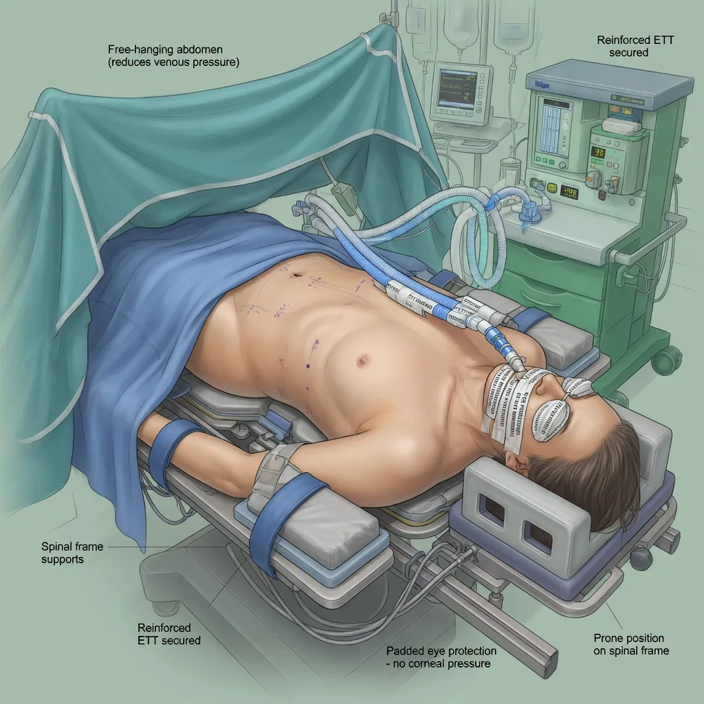
Why this is examined / the one-line answer
Prone spine surgery is a fellowship favourite because it stacks airway vulnerability, major haemorrhage, neuromonitoring constraints, and the rare but devastating complication of perioperative visual loss (POVL). Examiners want a consultant who can run a positioning checklist, name the dominant mechanism after spinal fusion (posterior ischaemic optic neuropathy), list ASPF registry risk factors accurately, and respond to sudden vision loss without confusing it with simple direct eye pressure every time. [1]
The one-line answer: I secure a reinforced tracheal tube, free the abdomen, protect the eyes from pressure, plan blood loss and neuromonitoring, counsel high-risk patients about POVL, and avoid stacking prolonged prone time, profound anaemia, and extreme hypotension — recognising that post-fusion POVL is often bilateral posterior ION and may be irreversible.[1]
Preoperative assessment and risk stratification
Surgical map
Levels (cervical vs thoracic vs lumbar), number of levels fused, expected duration, revision surgery, deformity correction, osteotomies, combined anterior–posterior approaches, and expected blood loss. Wilson frame versus Jackson table versus other frames changes abdominal compression and venous congestion profiles — Wilson frame was associated with higher ION odds in the ASPF analysis.[1]
Patient risk factors for POVL and general morbidity
Male sex, obesity, vascular disease, diabetes, hypertension, smoking, pre-existing optic neuropathy or glaucoma (discussion with ophthalmology when relevant), anaemia, and obstructive sleep apnoea. Baseline visual acuity if practical. Consent for major spine surgery in high-risk patients should explicitly include the possibility of partial or complete visual loss — rare, but life-altering when it occurs. [1]
Airway and cervical pathology
Cervical myelopathy, limited neck extension, rheumatoid disease, and unstable fractures change intubation strategy (video laryngoscopy, awake techniques, manual in-line stabilisation). Prone dislodgement risk must be anticipated before induction. [1]
Blood conservation and VTE
Plan cell salvage, tranexamic acid per institutional spine protocols (trauma TXA evidence informs the broader antifibrinolytic safety narrative), type and screen or crossmatch, and postoperative VTE prevention compatible with neurology examination needs.[2]
Applied physiology of the prone position
Respiratory
Functional residual capacity often improves compared with supine if the abdomen hangs free and the chest is supported on bolsters or a frame that unloads the belly. A hanging pannus forced upward, tight bolsters, or a poorly padded Wilson frame increases intra-abdominal pressure, reduces venous return, increases surgical bleeding from epidural veins, and worsens ventilatory pressures. [1]
Cardiovascular
Venous pooling in dependent parts, reduced preload if abdomen compressed, and facial/orbital venous congestion if the head is dependent or neck veins are obstructed. Mayfield pins versus soft headrests change facial pressure patterns. [1]
Ocular and optic nerve
Two mechanisms dominate teaching: [1]
- Central retinal artery occlusion (CRAO) — typically unilateral, associated with external pressure on the globe (malpositioned headrest, horseshoe headrest errors historically). Fundoscopy may show a classic cherry-red spot pattern in true CRAO (exam description, not something you invent as a fake image).
- Ischaemic optic neuropathy (ION), especially posterior ION after prone spinal fusion — often bilateral, associated with haemodynamic and haemodilutional factors rather than direct globe pressure; vision loss may be complete and irreversible. [1]
Cortical blindness from occipital infarction and other causes complete the differential but are less classic for the ASPF spine story. [1]
ASPF registry risk factors (must-recite)
In a multicenter case-control study of ION after spinal fusion surgery, independent risk factors included male sex, obesity, Wilson frame use, longer anaesthetic duration, greater estimated blood loss, and lower percent colloid in non-blood replacement.[1] Note the colloid signal carefully: it is an association from that analysis, not a licence to flood with starch products against modern restrictive colloid practice — discuss as registry evidence, then apply current fluid practice thoughtfully.
Anaesthetic goals
- Secure airway for the entire prone duration with zero unplanned extubation.
- Protect eyes, face, brachial plexus, genitals, breasts, and pressure points.
- Keep abdomen free to reduce bleeding and improve ventilation.
- Maintain spinal cord perfusion (and avoid optic nerve ischaemic stacking: long duration + hypotension + anaemia).
- Enable reliable neuromonitoring (SSEP/MEP) with an anaesthetic technique compatible with signals.
- Prepare for massive transfusion and coagulopathy.
- Assess vision as soon as the patient can cooperate postoperatively. [1]
Technique options and decision matrix
Airway
Intubate on a trolley with full preoxygenation; use a reinforced (armoured) tracheal tube well secured (tape plus ties as local practice; avoid excessive neck venous obstruction from tight ties). Confirm position before and after prone turn with capnography and auscultation/depth check. Disconnect circuits carefully during the flip; enough people to turn log-roll style. Prone rescue airway is hostile — prevention dominates; DAS principles apply if re-intubation needed after flipping supine.[3]
Positioning
Chest and pelvic supports with free abdomen; arms tucked or abducted within brachial plexus safe zones (avoid greater than 90 degrees abduction extremes and posterior shoulder displacement); head neutral or slightly elevated if surgical access allows; eyes free of pressure with documented checks after positioning and at intervals; genitalia and breasts free; knees and toes padded; reverse Trendelenburg slight if it reduces facial oedema without compromising surgical field. Avoid horseshoe headrest errors that compress globes. [1]
Anaesthetic maintenance and neuromonitoring
Total intravenous anaesthesia (propofol ± opioid infusion) is often preferred when motor evoked potentials are used because volatiles and nitrous oxide depress signals in a dose-dependent fashion; neuromuscular blockade must be managed per monitoring protocol (often avoided or kept minimal/stable for MEPs). Communicate continuously with the neuromonitoring team. [1]
Blood management
TXA as per protocol, cell salvage, restrictive yet safe transfusion thresholds individualised when ongoing losses and cord/optic perfusion concerns coexist, temperature control, and surgical haemostasis.[2]
Monitoring and equipment
Standard monitoring plus arterial line for major multilevel fusion, large-bore IV access, blood warmer, urine output, temperature, processed EEG optional, and neuromonitoring liaison. Document intermittent eye checks. Central access for very large cases or vasoactive infusions. Immediate ability to flip supine for airway catastrophe. [1]
Intraoperative management
Maintain mean arterial pressure targets agreed with the surgeon for cord perfusion (often higher in myelopathy or deformity correction — institutional). Avoid pure deliberate hypotension as a blood-sparing strategy in high POVL-risk patients. Track cumulative losses and haemoglobin. At cementation of any implants, remember orthopaedic embolisation physiology if relevant to the construct. [1]
Crisis pivots — what changes the plan
Neuromonitoring signal loss
Check blood pressure, haemoglobin/haematocrit, anaesthetic depth, neuromuscular blockade status, positioning (shoulders, neck), and surgical field (distraction, screw malposition). Treat reversible causes before irreversible cord injury. [1]
Airway dislodgement prone
Call for help, attempt careful reconnection and tube advancement if partially displaced, prepare to flip supine for definitive management if ventilation fails — do not persist with blind heroic prone intubation while the patient dies.[3]
Massive haemorrhage
Activate major haemorrhage protocol, TXA if not given, cell salvage, balanced transfusion, surgical packing/staging. [1]
Suspected intraoperative eye compression
Immediately relieve pressure, document, ophthalmology postoperatively even if uncertain. [1]
Postoperative plan
Ask about vision as soon as cooperation allows — before deep sedation masks the complaint. Any deficit: urgent ophthalmology, optimise blood pressure and haemoglobin, head-up if safe, investigate (ION vs CRAO vs cortical), and avoid false reassurance. ICU for unstable major deformity corrections. Analgesia that permits neurological assessment. Face and airway oedema may progress for hours after long prone cases — extubation criteria must respect this (leak test where used, visualised airway, fully reversed, ICU backup). [1]
Special populations and comorbidities
Obesity and Wilson frame: counsel highest POVL risk narrative; consider alternative frames when possible.[1] Cervical prone: worst airway stakes; reinforced tube and secure fixation. Combined anterior–posterior: staged anaesthetic plans and cumulative duration awareness. Paediatric scoliosis: massive blood loss potential, neuromonitoring, wake-up test historical context. Known glaucoma: multidisciplinary discussion; avoid prolonged extreme hypotension.
Detailed positioning checklist (consultant level)
- Team and roles: enough people to turn; airway doctor owns the tube; second person owns lines and monitors; surgeon owns timing of flip.
- Pre-flip: suction on, airway secured, eyes taped lightly then protected after prone, bite block if indicated, all connectors long enough, arterial line zeroed plan after turn.
- Flip: disconnect circuit briefly if needed, log roll as one unit for unstable spines, reconnect, confirm EtCO2 immediately, check bilateral chest expansion, re-check tube depth.
- Supports: chest rolls or frame pads that unload the abdomen; pelvis supported without crushing femoral nerves; knees flexed slightly on pillows; shins and toes free; male genitalia free; breasts medialised carefully in women; iliac crests padded.
- Upper limbs: tucked at sides with padding of ulnar nerves, or abducted on boards with less than extreme abduction and neutral shoulders; avoid brachial plexus stretch from caudal shoulder depression plus rotation.
- Head and eyes: neutral cervical alignment if cord at risk; Mayfield pins versus cushion — if cushion, confirm globes free by lifting lids after positioning; recheck every hour and after any reposition; document “eyes checked, no pressure.”
- Venous congestion control: slight reverse Trendelenburg if surgical access allows; avoid tight ETT ties that obstruct jugular veins; free abdomen. [1]
Spinal cord perfusion versus optic nerve — the trade-off conversation
Surgeons may request higher mean arterial pressure for myelopathy or deformity correction (for example targets at or above baseline, sometimes MAP greater than 80 mmHg institutional). That same hypertension management must still avoid wild swings. Optic nerve perfusion is not directly measured; the ASPF lesson is to avoid the stack: long duration + deep anaemia + relative hypotension + venous congestion from frame and head position. Cell salvage and TXA reduce the need for extreme anaemia; staging bilateral or multi-stage deformity corrections may be safer than heroic single-session marathon cases in the highest-risk body habitus. [1]
Fluid strategy nuance after ASPF
The registry associated higher colloid fraction of non-blood replacement with lower ION odds. Modern practice has reduced starch use because of renal and coagulation concerns in other critical care contexts. Exam answer: I acknowledge the ASPF colloid signal, avoid extreme crystalloid overload that worsens facial and orbital oedema, use balanced crystalloid thoughtfully, employ albumin where institutional practice supports it in large loss cases, and prioritise treating anaemia and hypotension over any single fluid brand. Do not claim starch is mandatory. [1]
Wake-up test and neuromonitoring pharmacology
Historical wake-up tests required lightening anaesthesia to command move feet — risk of tube dislodgement, awareness, and violence. Modern SSEP/MEP monitoring reduces wake-up frequency but imposes drug constraints: volatiles and nitrous oxide depress amplitudes; propofol TIVA is friendlier for MEPs; ketamine is sometimes used as an adjunct that may preserve signals; neuromuscular blockade must be absent or stable and minimal for myogenic MEPs. Bolus propofol or sudden deep volatile can mimic surgical injury on the monitor — communicate every syringe with the monitoring team. [1]
Postoperative visual loss pathway (stepwise)
- Confirm the complaint with simple bedside tests (count fingers, light perception, fields confrontation).
- Urgent ophthalmology — do not wait for routine morning rounds.
- Optimise oxygen delivery: correct anaemia, support blood pressure to high-normal for the patient, head elevation if safe for the spine construct.
- Differentiate CRAO (often unilateral, pressure story) from posterior ION (often bilateral after long fusion) from cortical causes (stroke workup).
- Imaging and neuro-ophthalmology as directed; prognosis for complete bilateral posterior ION is often poor — honest counselling and support services.
- Incident reporting and team debrief; review positioning documentation and haemodynamic charts. [1]
Massive transfusion and coagulopathy in deformity surgery
Long fusions can behave like major trauma. Activate institutional major haemorrhage protocols early, use TXA, cell salvage, temperature control, ionised calcium replacement with citrated products, and viscoelastic testing where available. Hypotension from hypovolaemia is not a POVL prevention strategy — replace losses while avoiding pure waterlogging of the face. [1]
Cervical versus lumbar prone differences
Cervical prone cases combine myelopathy cord risk with the most unforgiving airway if the tube displaces. Mayfield pin fixation is common; pin sites can bleed and rarely cause venous air embolism if sinuses are open in head-up positions. Lumbar deformity corrections dominate the POVL ASPF literature because of duration and blood loss. Thoracic cases add one-lung ventilation only if anterior thoracic approaches are used — then thoracic anaesthesia principles stack on prone issues. [1]
Pressure injuries beyond the eyes
Cheeks, lips, breasts, iliac crests, genitalia, knees, and toes all ulcerate during long cases. Tongue macroglossia after prolonged prone with hard bite blocks or swelling can threaten extubation. Document skin checks after positioning. Neuropraxias (ulnar, brachial plexus, lateral femoral cutaneous) are common claims — padding and neutral joint positions are preventive. [1]
Deliberate hypotension — historical versus modern
Older teaching used deliberate hypotension to reduce spine bleeding. Modern high-POVL-risk and myelopathy patients should not receive deep deliberate hypotension as routine blood-sparing. Prefer surgical haemostasis, TXA, cell salvage, and careful table and frame setup that frees the abdomen (which itself reduces bleeding more than many drug tricks). [1]
Consent script for high-risk fusion
There is a rare risk of partial or complete loss of vision after prolonged prone spine surgery, which may be permanent and can affect both eyes. We reduce risk by protecting your eyes from pressure, keeping your blood pressure and blood count in safe ranges, and limiting the time face-down when possible. Please tell recovery staff immediately if your vision is not normal when you wake. [1]
Research literacy for the viva
Cite the Postoperative Visual Loss Study Group Anesthesiology 2012 ASPF analysis for ION risk factors after spinal fusion. Distinguish association from proven interventional targets. ASA has practice advisories on POVL — mention that institutional protocols should track current advisory language without inventing false certainty about exact MAP numbers for every patient. [1]
Intraoperative haemoglobin and transfusion decision-making
There is no single universal haemoglobin number that prevents ION. The ASPF signal for greater blood loss and anaemia risk means treating severe anaemia rather than tolerating extreme haemodilution for the sake of avoiding transfusion. In a long multilevel fusion with ongoing losses, maintain oxygen delivery with timely red cells, control surgical bleeding, and avoid stacking low Hb with low MAP for hours. Discuss transfusion thresholds with the team preoperatively for Jehovah’s Witness patients and document accepted minima and cell salvage plans. [1]
Temperature and venous air embolism footnotes
Hypothermia worsens coagulopathy and bleeding, prolonging the case — active warming is POVL-relevant indirectly. If the head is elevated and large veins are open, venous air embolism can occur even outside classic sitting craniotomy; sudden EtCO2 drop and hypotension need the VAE differential alongside haemorrhage. [1]
SAQ answer scaffold
Stem: Eight-hour multilevel instrumented lumbar fusion in an obese man on a Wilson frame. [1]
- POVL types (3 marks): posterior ION vs CRAO vs other.
- ASPF risks present (3 marks): male, obesity, Wilson, long duration, likely high EBL.[1]
- Prevention checklist (4 marks): eyes, abdomen free, BP, anaemia, fluids, duration, consent.
- Airway (2 marks): reinforced ETT, flip plan.
- Vision loss in recovery (3 marks): urgent ophtho pathway.
Viva stem bank and model phrases
Stem 1: “What causes POVL after spine fusion?”
Model: “Most often posterior ischaemic optic neuropathy, which may be bilateral and irreversible — not always direct eye pressure. CRAO is the pressure story.” [1]
Stem 2: “List ASPF risk factors.”
Model: “Male sex, obesity, Wilson frame, longer anaesthesia, greater blood loss, and lower percent colloid in non-blood replacement.”[1]
Stem 3: “How do you check the eyes?”
Model: “After prone positioning and at intervals I confirm the globes are free of pressure from the headrest, document the check, and recheck after any head repositioning.” [1]
Stem 4: “Tube comes out prone.”
Model: “Call for help, attempt oxygenation, and if I cannot ventilate I flip supine for airway rescue per difficult airway principles.”[3]
Stem 5: “MEPs vanishing.”
Model: “I verify blood pressure, haemoglobin, depth, paralysis status, and positioning, and ask the surgeon to pause while we correct reversible factors.” [1]
Stem 6: “Does TXA help POVL?”
Model: “TXA reduces bleeding in many protocols; less blood loss may reduce one ASPF risk factor, but TXA is not a specific POVL antidote.”[2]
Common traps
- Assuming all POVL is from direct pressure (confuses CRAO with ION)
- No vision check in recovery
- Tight bolsters causing venous congestion and bleeding
- Forgetting neuromonitoring anaesthetic constraints
- Deliberate deep hypotension plus anaemia in a long obese Wilson-frame fusion
- Unsecured non-reinforced tube for a long prone case
- Extubating into progressive facial and airway oedema overnight without a plan [1]
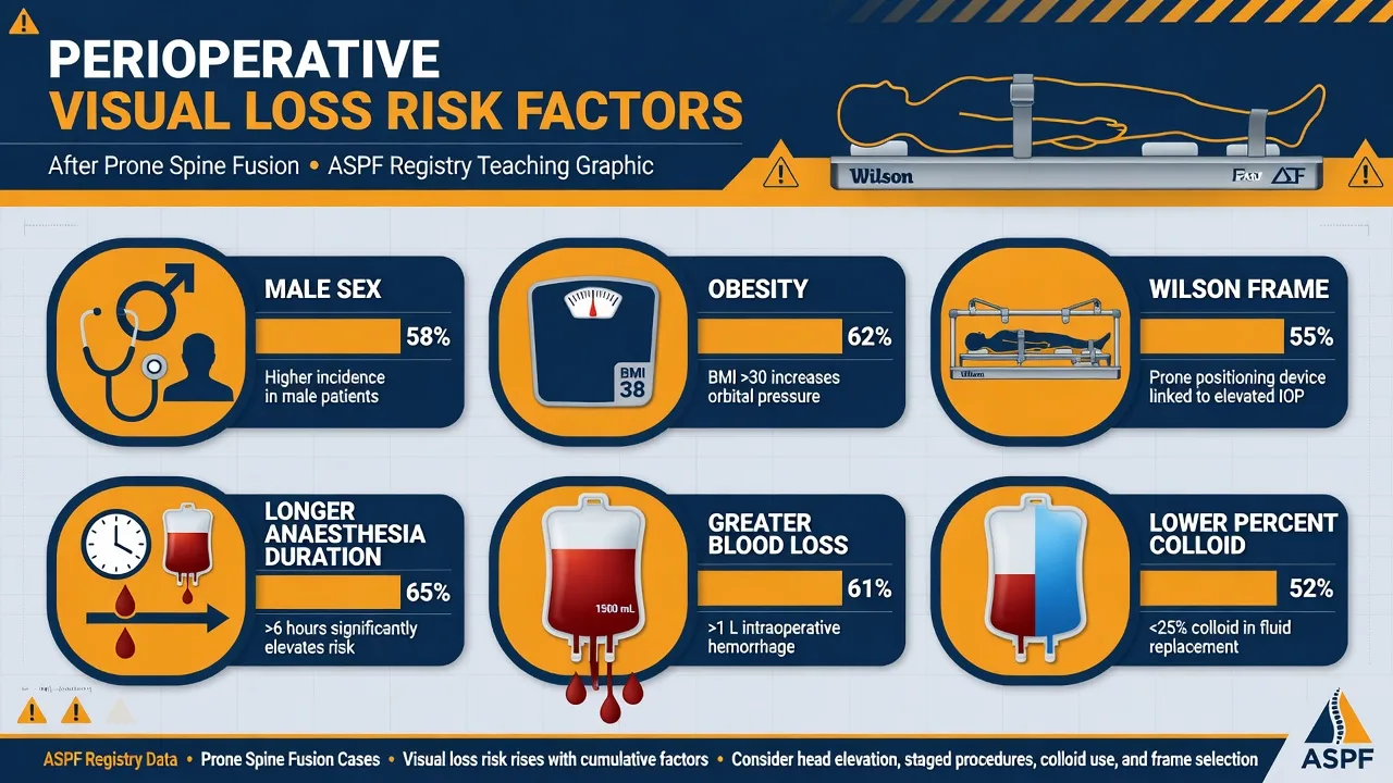

Prone spine checklist
References
- [1]The Postoperative Visual Loss Study Group Risk factors associated with ischemic optic neuropathy after spinal fusion surgery Anesthesiology, 2012.PMID 22185873
- [2]CRASH-2 trial collaborators Effects of tranexamic acid on death, vascular occlusive events, and blood transfusion in trauma patients with significant haemorrhage (CRASH-2): a randomised, placebo-controlled trial Lancet, 2010.PMID 20554319
- [3]Frerk C et al. Difficult Airway Society 2015 guidelines for management of unanticipated difficult intubation in adults Br J Anaesth, 2015.PMID 26556848