Anaes · Thoracic anaesthesia & one-lung ventilation
Thoracic anaesthesia and one-lung ventilation
Also known as One-lung ventilation · Double-lumen tube · Bronchial blocker · OLV hypoxaemia · SS_TS specialised study unit
Exam-pass thoracic anaesthesia hub (SS_TS): left DLT vs bronchial blocker, HPV and OLV hypoxaemia algorithm, protective ventilation, mediastinal mass rescue plan, and leaf links for VATS analgesia and lung isolation.
On this page & tools
Your progress
Saved locally on this device.
Target exams
Red flags
Why this is examined
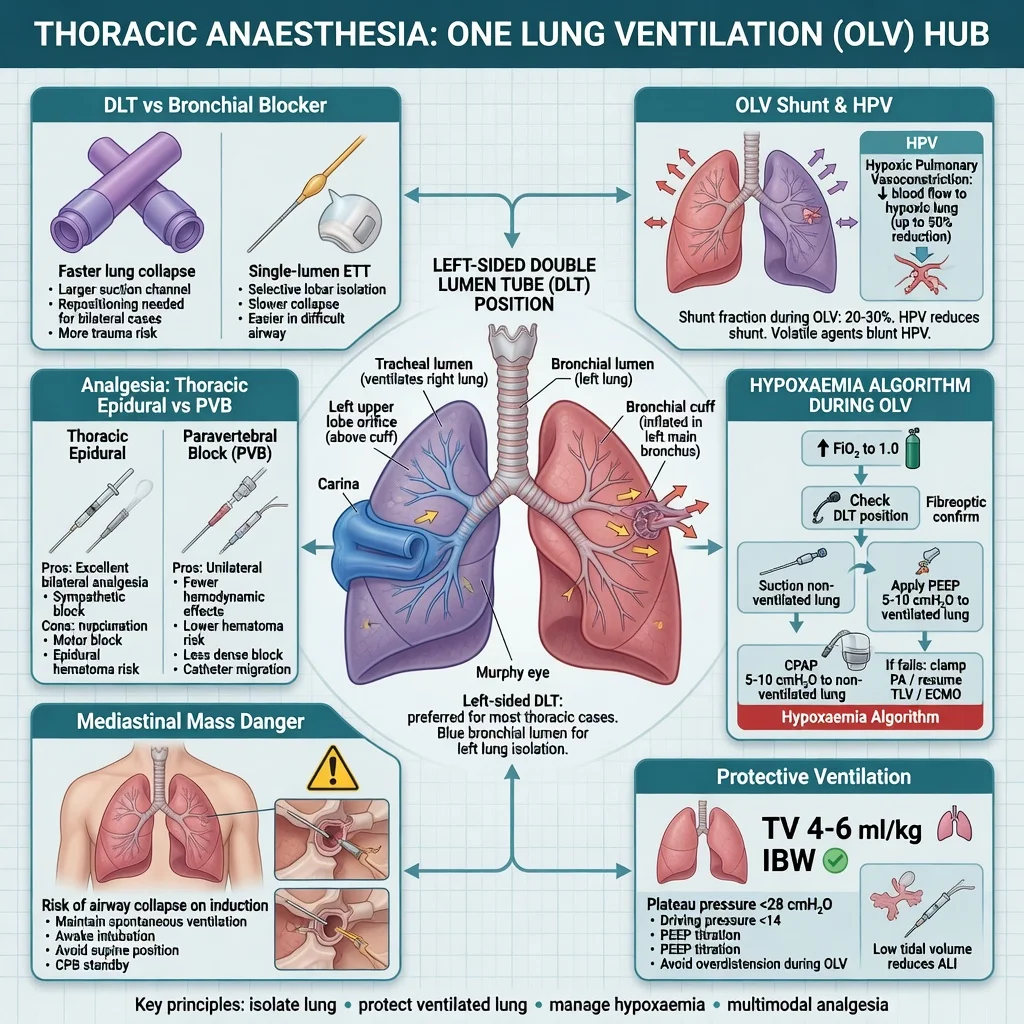
SS_TS contributes 24 FEx outcomes. It is a classic equipment-physiology-crisis triad: how you isolate the lung, why shunt and HPV determine oxygenation, and what you do when SpO2 falls on OLV. EACTAIC consensus keeps modern isolation technique examinable; HPV physiology explains most of the drug choices when the patient goes blue.[1][2] FRCA and ABA love DLT sizing conventions, fiberoptic confirmation steps, and mediastinal mass stems.
SSU framework and hub map
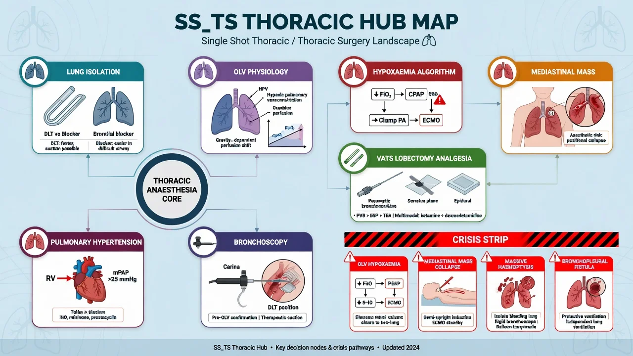
| Leaf / domain | Depth |
|---|---|
| Lung isolation DLT/blocker | Sizing, left vs right, placement, FO confirmation |
olv-physiology-hypoxaemia-algorithm | Shunt, HPV, step ladder for desaturation |
| Mediastinal mass | Spontaneous ventilation, rescue CPB/rigid scope |
| VATS / lobectomy analgesia | Epidural vs PVB vs ESP, ERAS thoracic |
| Bronchoscopy / airway surgery | Shared airway, rigid scope, jet ventilation themes |
| Pulmonary hypertension / COPD thoracic | RV protection, ventilation strategy |
| Oesophagectomy overlap | OLV + two-cavity physiology |
Lung isolation techniques
Double-lumen tube (DLT). Workhorse for most adult lung resection. Left-sided DLT preferred (margin of safety — only one bronchial orifice at risk versus right upper lobe take-off proximity on right-sided DLT). Starting sizes commonly ~35 Fr female, ~37 Fr male (convention; individualise by height/tracheal width). Confirm placement with fiberoptic bronchoscopy every time after placement and after repositioning.[1]
Bronchial blocker. Via single-lumen tube (≥7.5 mm ID typically): Arndt, Cohen, Fuji, EZ-blocker. Preferred when: difficult airway already intubated with SLT, tracheostomy, paediatric isolation, need for postoperative ventilation on SLT, selective lobar blockade. [1]
Left DLT
- Default adult isolation
- FO confirm mandatory
- Good surgical flexibility
- Bulkier airway
Bronchial blocker
- Difficult airway friendly
- SLT remains
- Slower deflation sometimes
- Migration risk
Right DLT
- When left bronchus surgery
- RUL orifice risk
- FO essential
- Less margin of safety
OLV physiology in one page
Lateral decubitus + collapse of operative lung → the operative lung still receives blood flow initially → transpulmonary shunt and hypoxaemia risk. Hypoxic pulmonary vasoconstriction (HPV) diverts blood from hypoxic alveoli to ventilated lung, improving V/Q.[2][4] HPV is inhibited by volatiles (dose-dependent), vasodilators (GTN, nitroprusside, some inotropes), and extreme hypocapnia/alkalosis patterns. TIVA may better preserve HPV when hypoxaemia is stubborn. Dependent ventilated lung can also atelectase — needs PEEP and recruitment carefully.[3]
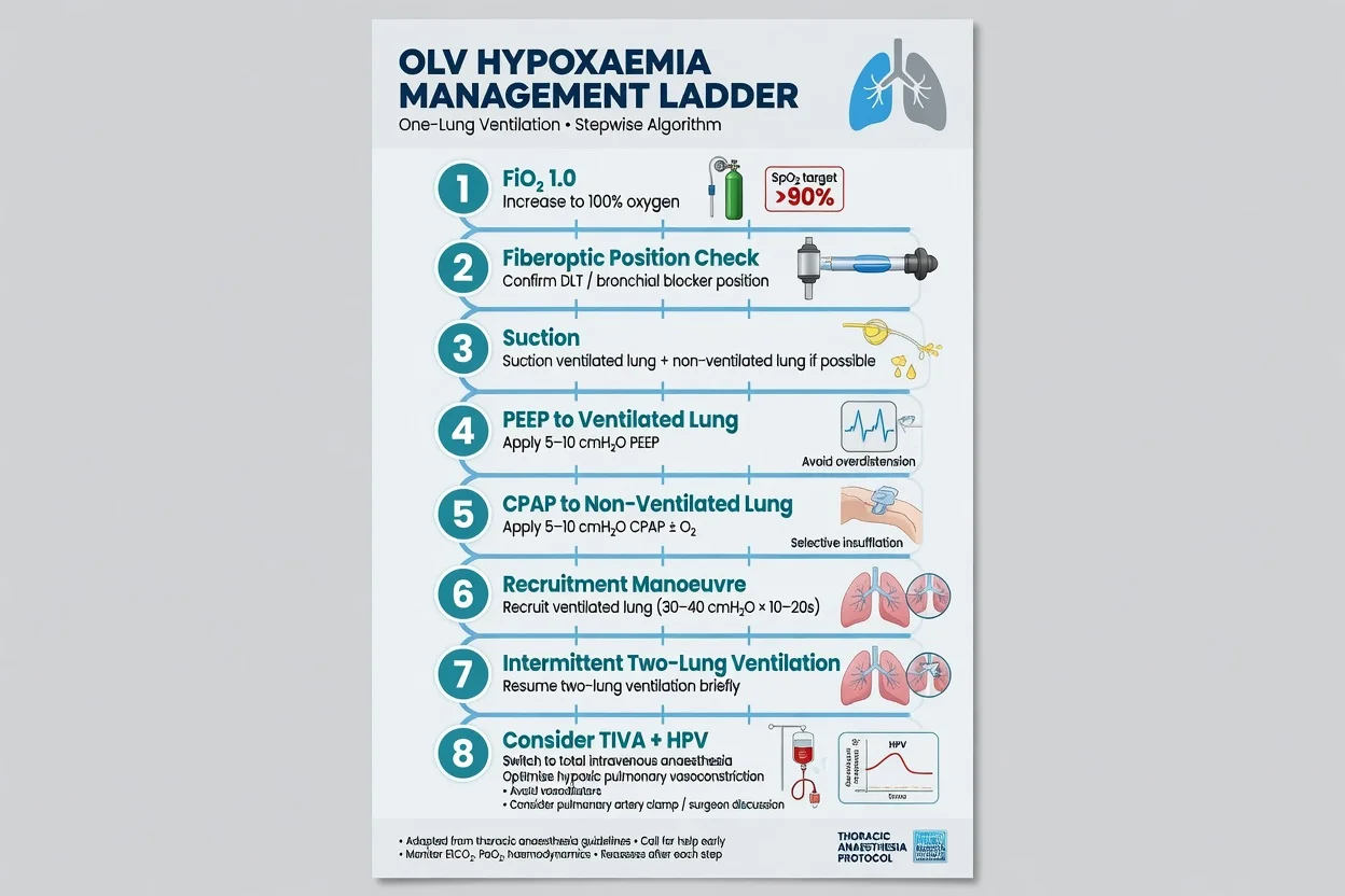
OLV hypoxaemia algorithm (order matters)
Leaf depth: olv-physiology-hypoxaemia-algorithm. Hub ladder: [1]
- Increase FiO2 to 1.0
- Fiberoptic check of DLT/blocker position (malposition is common)
- Suction blood/secretions
- Optimise ventilation of dependent lung — PEEP, adequate rate, check for high pressures
- Apply CPAP (or oxygen insufflation) to the non-ventilated operative lung
- Recruitment manoeuvre to ventilated lung (watch BP)
- Intermittent two-lung ventilation / clamp pulmonary artery surgically if open thoracotomy and life-threatening
- Consider TIVA, treat low cardiac output, rule out pneumothorax on ventilated side [1]
Protective ventilation and monitoring
Tidal volume on OLV ~4–6 mL/kg predicted body weight with appropriate PEEP; keep plateau/driving pressures reasonable (peak often kept <35 cmH2O as a crude ceiling). Arterial line for blood gas. Standard EtCO2 under-reads true PaCO2 sometimes — check ABG.[3]
Anaesthetic technique sketch
Thoracic epidural or paravertebral catheter before induction for open thoracotomy in many units; ESP/PVB single-shot for VATS ERAS pathways. Induction and DLT placement with neuromuscular blockade; FO confirmation; lateral positioning with careful attention to eyes, brachial plexus, padding; start OLV when chest open or as surgical needs dictate; fluid-restrictive relative to historical liberal practice for lung resection (avoid fluid overload → pulmonary oedema risk especially post-pneumonectomy). [1]
Non-intubated VATS exists in selected centres with spontaneous ventilation — not for the unprepared; conversion plan mandatory. [1]
Mediastinal mass (cannot-miss crisis)
Anterior mediastinal mass can compress trachea, bronchi, and right heart/PA. Induction of anaesthesia and loss of spontaneous negative-pressure ventilation may cause irreversible obstruction. High-risk features: orthopnoea, stridor, SVC obstruction, >50% tracheal compression on imaging, pericardial effusion. Plan: multidisciplinary, possible awake FO assessment, maintain spontaneous ventilation, rigid bronchoscope ready, femoral wires/CPB on standby for extreme risk, avoid muscle relaxant until airway proven. [1]
Postoperative analgesia and complications
Thoracic epidural gold standard historically for open thoracotomy; paravertebral comparable analgesia with less hypotension in many comparisons; fascial plane blocks for VATS. Multimodal systemic analgesia. Complications to know: post-pneumonectomy pulmonary oedema, AF, air leak, bronchopleural fistula, respiratory failure, haemorrhage. [1]
Crisis bank
- Hypoxaemia on OLV — algorithm above.
- Mediastinal mass collapse — reposition, reinstate spontaneous effort if possible, rigid scope, crash onto femoral bypass in extreme.
- Massive haemoptysis — isolate bleeding lung, FO/rigid toilet, surgical control.
- Bronchopleural fistula — avoid high pressure to fistula, isolation strategy.
- DLT malposition after turning — FO recheck always.
- Cardiac herniation (rare post-pneumonectomy) — surgical emergency.
- Tension on ventilated side — decompress. [1]
Landmark / consensus anchors
Regional practice deltas
ANZ. Left DLT default language standard; FO confirmation expected. Thoracic epidural still common for open cases; PVB/ESP rising for VATS. Metaraminol/phenylephrine for epidural hypotension.
SAQ answer scaffold
Stem: "During VATS lobectomy on OLV SpO2 falls to 88%." [1]
- FiO2 1.0; tell surgeon; check EtCO2/BP.
- FO position check and suction.
- PEEP dependent lung; CPAP operative lung.
- Recruitment; consider TIVA; intermittent two-lung.
- Exclude pneumothorax, tube migration, haemodynamic cause.
- If refractory open conversion / PA clamp discussion. [1]
Viva stem bank
- "Left versus right DLT — why prefer left?"
- "When would you choose a bronchial blocker?"
- "Explain HPV and what inhibits it."
- "Stepwise management of hypoxaemia on OLV."
- "Anterior mediastinal mass for biopsy — anaesthetic plan."
- "Protective ventilation settings on OLV." [1]
Common traps
- Assuming desaturation is always "HPV failure" without checking tube position.
- Right DLT without FO vigilance for RUL obstruction.
- Paralysing a critical mediastinal mass patient on induction as default.
- 8–10 mL/kg tidal volumes on OLV (historical harmful practice).
- Forgetting to reconfirm isolation after lateral positioning.[3]
Thoracic hub numbers
Protective OLV volumes and isolation confirmation are the thoracic hub numbers examiners expect.[1][3]
[1] [1]Preoperative thoracic assessment
History of dyspnoea, sputum, smoking, prior thoracic surgery, chemo/radiotherapy, and exercise tolerance. Spirometry and DLCO guide resectability discussions (surgeons lead operability; anaesthetists flag who will not ventilate or wean). ABG on air if hypoxic; CT for airway anatomy and mass effect; echo if PH or right-heart disease suspected. Optimise COPD (bronchodilators, physiotherapy, smoking cessation when time allows). Discuss thoracic epidural/PVB risks and benefits. [1]
Positioning and one-lung practicalities
Lateral decubitus with axillary roll, head neutral, eyes taped and checked, dependent arm padded, bean-bag or supports secure. After turning: reconfirm DLT/blocker with FO, re-zero arterial line if needed, check breath sounds and pressures. Start OLV with FiO2 high initially then titrate if saturations allow; some units begin at FiO2 1.0 until stable. Communicate with surgeon before any prolonged desaturation algorithm step that needs surgical pause. [1]
Pneumonectomy specifics
Higher risk of post-pneumonectomy pulmonary oedema and cardiac herniation (especially intrapericardial). Fluid restriction relative to liberal historical practice, protective ventilation of remaining lung, careful emergence, ICU or HDU destination. Sudden shock and mediastinal shift after pneumonectomy may be herniation — surgical emergency. [1]
Red flags
[1] [1] [1] [1] [1]References
- [1]Granell M, et al. Airway Management in Thoracic Anesthesia: EACTAIC Consensus Document J Cardiothorac Vasc Anesth, 2026.PMID 41353026
- [2]Lumb AB, Slinger P Hypoxic pulmonary vasoconstriction: physiology and anesthetic implications Anesthesiology, 2015.PMID 25587641
- [3]Shum S, Huang D, Parotto M Hypoxaemia during one lung ventilation BJA Educ, 2023.PMID 37600211
- [4]Licker M, et al. The hypoxic pulmonary vasoconstriction: From physiology to clinical application in thoracic surgery Saudi J Anaesth, 2021.PMID 34764832