Anaes · Trauma and massive haemorrhage
Trauma and massive haemorrhage
Also known as Damage control resuscitation · Massive transfusion protocol · Permissive hypotension · Haemostatic resuscitation · Trauma-induced coagulopathy · The lethal triad · Damage control surgery · Tranexamic acid in trauma · Viscoelastic testing in trauma
Trauma and massive haemorrhage is the prototypical time-critical anaesthetic emergency and the commonest cause of preventable trauma death is uncontrolled haemorrhage. The modern response is damage control resuscitation — the integration of permissive hypotension (systolic 80 to 90 mmHg until haemostasis), haemostatic resuscitation with blood products in a 1:1:1 ratio rather than crystalloid, early tranexamic acid (1 g IV within 3 hours, CRASH-2), the activated massive transfusion protocol, damage control surgery (control bleeding, pack, temporary closure, ICU, definitive surgery when stable), and the active prevention of the lethal triad of acidosis, hypothermia and coagulopathy. The anaesthetist is central to the trauma team — the airway with cervical-spine precautions, the breathing, the circulation with haemorrhage control, and the coagulation.
On this page & tools
Your progress
Saved locally on this device.
Target exams
Red flags
Overview and definition
Trauma is the leading cause of death worldwide in the first four decades of life, and exsanguinating haemorrhage is the dominant mechanism of preventable death in the first few hours after injury. The trauma patient who reaches the operating theatre or the resuscitation bay alive but bleeding is the single most time-critical emergency the anaesthetist faces, and the conduct of that anaesthetic — the airway, the breathing, the circulation, and above all the resuscitation and the coagulation — is the difference between survival and death. The modern discipline that frames the whole response is damage control resuscitation (DCR), articulated for the military by Holcomb and colleagues in 2007 and codified for civilian practice in successive editions of the European guideline on the management of major bleeding and coagulopathy following trauma.[4][3]
DCR is not one intervention but an integrated, protocolised strategy with five interlocking pillars: permissive hypotension until surgical haemostasis; haemostatic resuscitation with blood products in a balanced ratio rather than clear fluid; damage control surgery, the abbreviated operation to control bleeding and contamination followed by a planned return; the active prevention of the lethal triad of acidosis, hypothermia and coagulopathy; and the massive transfusion protocol (MTP) that delivers blood products to the bedside without delay. The anaesthetist owns the resuscitation end of this strategy and must understand the surgery, the coagulation, and the physiology as fluently as the airway.[4]
The examination of trauma anaesthesia at fellowship level is reliable and predictable. The examiner expects a structured primary survey, an exact knowledge of the doses and the evidence (the CRASH-2 and CRASH-3 trials, the PROPPR trial, the European guideline recommendations), a mechanistic account of the lethal triad and trauma-induced coagulopathy, and a defensible anaesthetic plan for the shocked, bleeding, possibly head-injured patient. The candidate who can link the physiology to the action — the permissive hypotension to the cerebral perfusion pressure, the crystalloid to the coagulopathy, the citrate to the calcium — passes comfortably. [1]
The trimodal distribution and the preventable death
Trauma mortality follows a trimodal distribution. The first peak is within seconds to minutes of injury — the great-vessel laceration, the devastating brain injury, the high-cord transection — and most of these patients die at the scene. The second peak is within minutes to a few hours — the exsanguinating haemorrhage, the evolving intracranial haematoma, the tension pneumothorax — and THIS is the peak where organised, protocolised care saves lives, and where the anaesthetist is central. The third peak is days to weeks later — sepsis and multiple organ failure — the domain of critical care. The entire DCR strategy is aimed at the second peak: the rapid, simultaneous control of haemorrhage and the prevention of the physiological exhaustion that drives the third peak. [1]
The concept of the preventable trauma death drives the audit and the systems: a death from a treatable airway problem, a tension pneumothorax, or an uncontrolled but surgically-addressable bleed is a failure of the system, not an inevitable outcome. The trauma team, the MTP, the pre-alert, the senior decision-maker at the bedside, and the rehearsed handover all exist to eliminate these deaths. The anaesthetist's role is hard-wired into that system — the airway, the breathing, the lines, the resuscitation, the induction, and the co-ordination with surgery and the blood bank. [1]
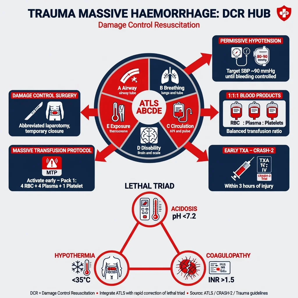
ATLS primary survey: A, B, C, D, E
The Advanced Trauma Life Support (ATLS) framework structures the first minutes into a prioritised survey that treats the greatest threat to life first. The survey is performed in order and is simultaneous once the team is assembled — the airway doctor secures the airway while the circulatory doctor places the lines and activates the MTP. The order reflects the principle that an airway problem kills before a breathing problem, which kills before a circulatory problem. [1]
ABCDE — the primary survey in order
The survey is iterative. The team re-evaluates A, B and C continuously, because a patient who was stable can deteriorate the moment the induction agent is given or the chest drain is placed. The senior member of the team — the team leader — stands back, does not perform tasks, and maintains the shared mental model through closed-loop communication and a regular verbal state-of-the-patient. [1]
The trauma team and the communication
Trauma resuscitation is a team sport. The well-formed trauma team has a team leader (who does not perform procedures but coordinates and decides), an airway doctor (the anaesthetist), a breathing doctor, a circulation doctor (or two), a nurse on each side of the patient, a scribe, a radiographer, and a porter. The blood bank and the laboratory are pre-warned; the MTP pack is en route the moment it is activated. [1]
The commonest mode of failure in trauma is not a knowledge gap but a communication failure: the unspoken assumption, the ambiguous instruction, the dose that was said but not heard. The defences are closed-loop communication (the instruction is spoken, read back, and confirmed), SBAR (Situation, Background, Assessment, Recommendation) for the handover, the shared mental model stated aloud every few minutes, and the cognitive aid for the MTP and the difficult airway. The team that rehearsed together — the in-situ simulation, the debrief after the real event — performs better under pressure than the team that did not. The anaesthetist is often the team leader in the absence of a dedicated trauma surgeon, and must therefore practise the deliberate non-involvement in the hands-on tasks that the leadership role demands. [1]
A — the airway with cervical-spine precautions
The trauma airway is the highest-risk airway in anaesthetic practice. It is compromised on four independent axes at once: the cervical spine may be injured and must be protected; the stomach is full (and the bleeding patient may have swallowed blood); there is blood, debris and secretions in the mouth; and there may be direct facial or laryngeal injury that distorts the anatomy. To this is added the shocked physiology that makes the standard induction agents dangerous, and — in the head-injured patient — the absolute need to avoid hypoxia and hypotension. [1]
The cervical spine is protected by manual in-line stabilisation (MILS) throughout the airway manoeuvre, NOT by hard-tape-and-collar immobilisation. The hard collar is opened or removed for the laryngoscopy because it forces the head into flexion, impedes mouth opening, and obscures the view; the three-person technique — one assistant holds the head in neutral in-line from below, the intubator stands at the head, and a third holds the cricoid — is the standard. The aim is to hold the occiput on the cervical spine, the cervical spine on the thoracic spine, in a straight line, without traction. MILS reduces but does not abolish intubation difficulty, and the laryngoscopic view is typically a grade worse than in the uninjured patient. [1]
The technique is a rapid sequence induction (RSI), modified for the trauma setting. The patient is preoxygenated with 100 per cent oxygen (recognising that the denitrogenation is incomplete in the shocked, desaturating patient, so apnoeic oxygenation with nasal high-flow oxygen — THRIVE — is a valuable adjunct). The induction agent is chosen for cardiovascular stability in shock (see the anaesthetic technique below). The muscle relaxant is suxamethonium 1 to 1.5 mg/kg (the fastest onset; the agent of choice in the crash airway and the potentially difficult airway where a surgical airway may be needed, with the caveats of hyperkalaemia in the burns, crush and denervation patient after 24 hours) OR rocuronium 1.2 mg/kg (the larger intubating dose that approaches suxamethonium in onset speed and is reversible with sugammadex). Cricoid pressure is applied by a third person from the moment of loss of consciousness and released only on the team leader's instruction or if it impairs the view. Videolaryngoscopy (a hyperangulated blade) improves the first-pass success and is the standard in many trauma systems, with a bougie or a stylet loaded and a second-generation supraglottic airway and a scalpel-bougie-tube cricothyroidotomy set ready at the bedside. [1]
If the airway is lost and cannot be rescued by the supraglottic device or the videolaryngoscope, the team escalates without delay to the front-of-neck access — the scalpel-bougie-tube surgical cricothyroidotomy, the canonical emergency surgical airway. The earlier the decision, the better the outcome; the team that drifts into repeated failed attempts at laryngoscopy while the saturation falls into the 70s has converted a difficult airway into a cardiac arrest. The DAS (Difficult Airway Society) vortex and the ANZCA difficult airway algorithm both converge on this point. [1]
In the patient with a combined traumatic brain injury, the airway is secured EARLY — the patient with a Glasgow Coma Scale of 8 or less is intubated, and the intubated head-injured patient is hyperventilated ONLY if there are signs of impending herniation (a unilateral fixed dilated pupil, a Cushing response, an extensor posturing). The targets are normoxia (the oxygen saturation above 94 per cent) and normocapnia (the end-tidal carbon dioxide 35 to 40 mmHg, about 4.5 to 5.3 kPa), and a mean arterial pressure of 80 mmHg or above to defend the cerebral perfusion pressure (the CPP equals the MAP minus the intracranial pressure). [1]
B — breathing
The immediately life-threatening chest injuries are identified and treated in the primary survey, before and without the chest X-ray. The five are: the tension pneumothorax, the massive haemothorax, the open (sucking) pneumothorax, the flail chest, and the cardiac tamponade. [1]
Tension pneumothorax is the air under pressure in the pleural space that collapses the lung, deflects the mediastinum to the contralateral side, obstructs venous return, and causes obstructive shock. The clinical picture is hypoxia, hypotension, a tracheal deviation (a late sign), unilateral hyperresonance and reduced breath sounds, and distended neck veins (which may be absent in the hypovolaemic patient). The treatment is immediate decompression — traditionally a large-bore needle (14 or 16 gauge) in the second intercostal space, mid-clavicular line, but the modern recommendation is the fifth intercostal space, mid-axillary line (the same site as the chest drain) because the anterior site has a significant failure rate from the chest-wall thickness and the subcutaneous fat. The needle decompression is a bridge to the definitive chest drain (an intercostal catheter with an underwater seal), which is placed immediately after. The decompression converts a tension into a simple pneumothorax and buys the minutes to put in the drain. [1]
Massive haemothorax is more than 1500 mL of blood (or more than 200 mL per hour for several hours) in the pleural cavity. It presents with hypoxia, hypotension, dullness to percussion and reduced breath sounds on the affected side. The treatment is a large-bore chest drain (32 to 36 French) at the fifth intercostal space, mid-axillary line — NOT a needle. The drain does two jobs: it re-expands the lung, and it quantifies the bleeding (the volume drained and the rate guide the decision to thoracotomy). The blood drained can be autotransfused (the cell salvage circuit) in the absence of contamination. An immediate return of more than 1500 mL, or an ongoing loss of more than 200 mL per hour, is an indication for a thoracotomy (the resuscitative or emergency department thoracotomy in the arrested patient, the theatre thoracotomy in the patient with a sustained output). [1]
Open pneumothorax (the sucking chest wound) is managed by a three-sided occlusive dressing (a valve) that allows air to escape but not enter, followed by a formal chest drain remote from the wound, and the surgical closure of the defect. [1]
Flail chest — a segment of chest wall that moves independently, paradoxically inwards on inspiration, from fractures in two or more places on two or more consecutive ribs — produces severe pain, hypoventilation, and an underlying pulmonary contusion. The hypoventilation and the contusion cause hypoxia that worsens over hours. The patient who is hypoxic, tiring, or has a significant head injury is intubated and ventilated with a lung-protective strategy; positive pressure ventilation splints the flail segment and improves the gas exchange. The pain control is multimodal — a thoracic epidural or a paravertebral or an erector spinae plane block in the stable patient, patient-controlled opioid or ketamine in the unstable. [1]
Cardiac tamponade — Beck's triad of hypotension, distended neck veins, and muffled heart sounds, with a pulsus paradoxus — is treated by pericardiocentesis as a bridge or, in the trauma setting, by a resuscitative thoracotomy (the clamshell or left anterolateral thoracotomy) to relieve the tamponade, cross-clamp the descending aorta, and directly repair or control the cardiac wound. The clamshell thoracotomy is also the procedure of choice for the patient in arrest from a suspected thoracic aortic or great-vessel injury, and for the patient in arrest from uncontrolled intra-abdominal haemorrhage (the aortic cross-clamp to redirect the residual blood volume to the heart and brain). [1]
C — circulation and the massive haemorrhage
The third letter of the survey is circulation WITH HAEMORRHAGE CONTROL. The two go together: the resuscitation is futile if the bleeding is not stopped, and the bleeding is harder to stop the longer the coagulopathy is allowed to deepen. The team therefore externalises the compressible bleeding (the limb, the scalp, the trunk wound) with direct pressure and a tourniquet, identifies and stabilises the pelvic fracture (the pelvic binder), decompresses the chest, and prepares for the operating theatre or the angiography suite — all while the resuscitation runs. [1]
The recognition of the depth of shock is the first anaesthetic task. The ATLS classes of haemorrhagic shock frame the vital-sign response to the blood loss: [1]
ATLS classes of haemorrhagic shock (the 70 kg adult)
The critical anaesthetic point is that the blood pressure is maintained until Class III — a normal or near-normal blood pressure in the bleeding patient does not exclude a Class II or early Class III loss. The young, fit, catecholamine-driven patient compensates by vasoconstricting and tachycarding, and the systolic pressure may read 110 mmHg with two litres of blood gone. The reliable early markers of the depth of shock are the pulse rate, the pulse pressure (the diastolic rises with the vasoconstriction and narrows the pulse pressure before the systolic falls), the capillary refill, the lactate and the base excess (the metabolic markers of tissue hypoperfusion), and the mental state. A rising lactate and a worsening base excess in a patient who looks "stable" is a patient who is not stable. [1]
The induction of anaesthesia in the shocked patient unmasks the compensated vasoconstriction — the sympathetic tone is the only thing holding the blood pressure up, and the moment it is removed by the induction agent the pressure collapses. The anticipation of this — the "drawn-up vasopressor, the reduced induction dose, the running fluid, the drawn-up metaraminol or noradrenaline" — is the mark of the prepared trauma anaesthetist. [1]
Damage control resuscitation — the five pillars
DCR is the overarching strategy that begins at the scene (or the moment haemorrhage is recognised) and ends in the intensive care unit with the physiologically-restored patient. It rests on five pillars.[4][3]
Pillar 1 — permissive hypotension
Permissive hypotension is the deliberate maintenance of a lower-than-normal blood pressure until surgical haemostasis is achieved, on the principle that a lower driving pressure dislodges fewer tenuous clots and reduces the ongoing blood loss from disrupted vessels. The target is a systolic blood pressure of 80 to 90 mmHg (equivalently a mean arterial pressure of about 65 mmHg) in the patient without a head injury, maintained by a restricted crystalloid and blood-product resuscitation until the bleeding is controlled. Once the haemostasis is achieved, the resuscitation is completed and the blood pressure is restored to normal. [1]
[1]The contraindications are the situations in which the lower pressure is more dangerous than the clot disruption it prevents: the traumatic brain injury (the cerebral perfusion pressure equals the mean arterial pressure minus the intracranial pressure, and the head-injured brain cannot tolerate a low MAP — the target is a MAP of 80 mmHg or above); the spinal cord injury (the cord ischaemia); the elderly patient and the patient with cardiovascular disease (the coronary and the cerebral perfusion); and the prolonged transport or the delayed haemostasis (the permissive hypotension is a temporary strategy, not a substitute for haemostasis). The patient who has bled for hours at a systolic of 80 mmHg has traded the clot for a deepening acidosis. [1]
Pillar 2 — haemostatic resuscitation with blood products
Haemostatic resuscitation replaces the lost blood with blood products rather than crystalloid, and in a ratio that approximates whole blood. The empirical target is a 1:1:1 ratio of packed red blood cells to fresh frozen plasma to platelets — one unit of plasma and one unit (one adult dose, a pool of four to six donor units) of platelets for every unit of red cells — which delivers oxygen-carrying capacity, clotting factors and platelets together and avoids the dilutional coagulopathy of the crystalloid-heavy resuscitation.[4]
The evidence base for the ratio is the PROPPR trial (Holcomb and colleagues, JAMA 2015), a pragmatic multicentre randomised trial of 680 severely injured patients predicted to need a massive transfusion, randomised to 1:1:1 versus 1:1:2 (red cells to plasma to platelets).[2] The trial found no significant difference in the primary outcomes of 24-hour or 30-day mortality, but it found that the 1:1:1 group achieved haemostasis more often (86 per cent versus 78 per cent) and died less often from exsanguination within the first 24 hours (9.2 per cent versus 14.6 per cent), with no increase in complications (no more acute respiratory distress syndrome, no more multiorgan failure, no more thromboembolism). The 1:1:1 ratio is therefore the empirical starting point, fine-tuned by the viscoelastic testing. The principle is: resuscitate with blood products, give them early, and give them in a balanced ratio.
The crystalloid is restricted because it is the iatrogenic cause of the lethal triad. The large-volume saline or Hartmann's dilutes the clotting factors and the platelets (the dilutional coagulopathy), causes a hyperchloraemic metabolic acidosis (the balanced solutions are less harmful than saline, but any crystalloid in excess contributes), and cools the patient (the cold fluid straight from the fridge). The DCR target is the minimisation of crystalloid — small volumes to keep the line open, the resuscitation done with warmed blood products. [1]
Pillar 3 — early tranexamic acid
Tranexamic acid is the antifibrinolytic that inhibits the conversion of plasminogen to plasmin and so stabilises the clot. The evidence in trauma is the CRASH-2 trial (Shakur and the CRASH-2 collaborators, Lancet 2010), a randomised placebo-controlled trial of 20 211 adult trauma patients with or at risk of significant bleeding in 274 hospitals across 40 countries, treated within 8 hours of injury.[1] The regimen was a loading dose of 1 g over 10 minutes followed by 1 g over 8 hours. The trial found a reduction in all-cause mortality (14.5 per cent versus 16.0 per cent; relative risk 0.91, 95 per cent confidence interval 0.85 to 0.97) and in death due to bleeding (4.9 per cent versus 5.7 per cent; relative risk 0.85), with no increase in vascular occlusive events.
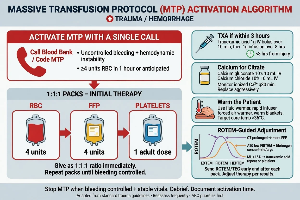
The time-to-treatment analysis was the seminal finding: the benefit was greatest in the first hour (the "golden hour") and persisted up to 3 hours, but tranexamic acid given after 3 hours increased mortality. The clinical message — and the most examined single fact in trauma anaesthesia — is: give tranexamic acid early, ideally within 3 hours of injury, and ideally pre-hospital or at the moment haemorrhage is recognised. The drug is cheap, stable, easy to give, and now embedded in every major trauma guideline and the World Health Organization essential medicines list. [1]
In the traumatic brain injury, the parallel CRASH-3 trial (Lancet 2019, 12 737 patients with TBI treated within 3 hours) found that tranexamic acid was safe and reduced head-injury-related death in the mild-to-moderate head injury group (relative risk 0.78) but not in the severe head injury group, with no increase in vascular occlusive events or seizures.[6] The principle extends: tranexamic acid is safe in the bleeding or head-injured trauma patient and is given early.
Pillar 4 — the massive transfusion protocol
The MTP is the institutional protocol that delivers blood products to the bedside in a pre-defined, rehearsed sequence the moment it is activated, without the delay of individual ordering or cross-matching. The activation is a single call to the blood bank — "activate the MTP" — that triggers the issue of the first pack. The products are delivered in rotating packs every 15 minutes (or per the local protocol): the first pack brings the O-negative packed red cells (universal, immediately available, the emergency units) and the AB plasma (the universal plasma donor); the second and subsequent packs add the platelets (one adult dose, a pool of four to six donor units) and the cryoprecipitate (two pools, for the fibrinogen). The patient is cross-matched in parallel and switched to group-specific and then cross-matched products as soon as the laboratory allows. [1]
The MTP is not just about the red cells. The fresh frozen plasma provides the clotting factors; the platelets provide the platelets; the cryoprecipitate provides the fibrinogen (the first factor to fall to a critical level, the target of which is 1.5 to 2.0 g/L or above in the bleeding patient); and the calcium is replaced because the citrate in the transfused products chelates it and a hypocalcaemia (an ionised calcium below 1.0 mmol/L) worsens the coagulation, the cardiac contractility and the vascular tone. The laboratory and the viscoelastic test (the ROTEM or the TEG) run in parallel and guide the fine-tuning — more fibrinogen if the FIBTEM is low, more plasma if the CT is prolonged, more platelets if the platelet contribution to clot strength is low. The MTP is formally stood down when the haemostasis is achieved and the patient is stable. [1]
[1]Pillar 5 — damage control surgery
Damage control surgery is the abbreviated, staged operation whose only goals are the control of haemorrhage and the control of contamination, followed by a temporary closure and a transfer to the intensive care unit for the restoration of the physiology, with the definitive surgical repair deferred to a planned re-operation (typically 24 to 48 hours later) when the patient is warm, coagulopathy-corrected, and acidosis-free. The philosophy is that the cold, acidotic, coagulopathic patient cannot tolerate the definitive long operation, and that the attempt to complete it kills the patient.[4]
The specific manoeuvres are: the packing of the bleeding liver or the pelvis; the ligation or shunting of the disrupted artery; the resection without anastomosis of the devascularised or perforated bowel (the ends stapled and returned); the temporary closure of the abdomen with a vacuum dressing (to accommodate the inevitable oedema and to avoid the abdominal compartment syndrome); and the rapid transfer to the ICU. The anaesthetist uses this window to warm the patient, to correct the acidosis and the coagulopathy, and to complete the resuscitation; the surgeon returns to the theatre for the definitive repair (the bowel anastomosis, the vascular reconstruction, the formal closure) when the physiology is restored. The principle — life before limb, and physiology before anatomy — is the opposite of the elective surgical mindset and is the single most important conceptual shift for the trainee. [1]
The lethal triad
The lethal triad — acidosis, hypothermia and coagulopathy — is the self-perpetuating death spiral of the uncontrolled haemorrhage and the central target of the DCR strategy. The three derangements feed each other in a vicious circle: the acidosis and the hypothermia each independently impair the coagulation cascade and the platelet function, the resulting coagulopathy deepens the bleeding and the shock, the deeper shock worsens the acidosis, and the circle tightens until the death.[3]
Acidosis. A pH under 7.2 impairs the coagulation factor activity (the enzymes are pH-dependent), reduces the fibrinogen synthesis, depresses the myocardial contractility, and blunts the catecholamine response. It arises from the tissue hypoperfusion (the anaerobic metabolism and the lactate) and is worsened by the hyperchloraemic acidosis of the large-volume saline. The management is the restoration of the perfusion (the haemostasis and the blood-product resuscitation) and the avoidance of the acidifying crystalloid — the bicarbonate is a temporary chemical fix, not a treatment. [1]
Hypothermia. A core temperature under 35 degrees Celsius slows the coagulation enzymes (the reaction rates roughly halve for each degree below 37), impairs the platelet function (the granule release and the aggregation), and shifts the oxyhaemoglobin dissociation curve to the left. The patient cools by exposure, by the cold fluids and blood, by the open body cavity, and by the evaporation from the peritoneum and the pleura. The prevention and the treatment are the same: the active warming — the forced-air warmer, the warmed fluids (every fluid and every unit of blood through the rapid infuser with its built-in heater), the raised ambient temperature of the operating theatre (the "hot trauma theatre"), the warmed humidified gases, and the covering of the patient the moment the survey allows. A core under 32 degrees Celsius is catastrophic and carries a near-100 per cent mortality; the goal is to keep the patient above 36 degrees and to actively rewarm the patient who arrives cold. [1]
Coagulopathy. The third arm — the trauma-induced coagulopathy — is the topic of the next section. The key insight is that the coagulopathy is present on ARRIVAL in the severely injured patient (within minutes of injury), before any dilution or hypothermia, and is therefore an intrinsic part of the injury — not merely a consequence of the resuscitation. [1]
Trauma-induced coagulopathy
Trauma-induced coagulopathy (TIC) is the early, endogenous coagulopathy of the severely injured patient, distinct from (and preceding) the later dilutional coagulopathy of the resuscitation. It is present within minutes of injury in about a quarter of the severely injured patients, it is independent of the dilution (it is there before any fluid is given), and it doubles the mortality. The recognition of TIC as an intrinsic, injury-driven process — rather than a late, iatrogenic, dilutional phenomenon — was the conceptual breakthrough that underpinned the whole DCR strategy.[5]
The mechanism was elucidated by Brohi and colleagues (Annals of Surgery 2007) in a prospective cohort of major trauma patients: the early coagulopathy occurs ONLY in the presence of tissue hypoperfusion (measured by the base deficit), and is mediated by the thrombomodulin–protein C pathway.[5] The shocked, hypoperfused endothelium releases thrombomodulin, which complexes with thrombin and diverts it from its procoagulant role (fibrinogen to fibrin) to its anticoagulant role (the activation of protein C). The activated protein C then consumes the factors Va and VIIIa (systemic anticoagulation) and, by inactivating the plasminogen activator inhibitor-1, unleashes the hyperfibrinolysis (the uncontrolled breakdown of the clot). The result is a systemic anticoagulated, hyperfibrinolytic state — the very opposite of the thrombotic state one might expect from "tissue factor release."
To this endogenous process are added the exacerbating factors of the resuscitation: the dilution (the crystalloid and the red-cell-only transfusion lower the clotting factors and the platelets), the hypothermia (the enzyme and the platelet dysfunction), the acidosis (the factor dysfunction), and the hypocalcaemia (the citrate chelation of the calcium that is factor IV). The lethal triad is therefore the intersection of the endogenous TIC and the iatrogenic resuscitation injury — and the DCR strategy addresses both: the haemostatic resuscitation (the factors and the platelets), the tranexamic acid (the antifibrinolytic, directed at the hyperfibrinolysis), the warming and the acidosis correction, and the calcium. [1]
The clinical consequence is that the bleeding trauma patient is coagulopathic from the start, and the anaesthetist resuscitates with the clotting factors and the platelets alongside the red cells from the first pack. The fibrinogen is the first factor to fall to a critical level (its baseline in the non-pregnant adult is 2 to 4 g/L, and the bleeding target is 1.5 to 2.0 g/L or above) and is replaced early with the cryoprecipitate or the fibrinogen concentrate. [1]
Viscoelastic testing — ROTEM and TEG
The conventional coagulation tests (the prothrombin time, the activated partial thromboplastin time, the INR, the platelet count, the fibrinogen) are plasma-based, slow, and non-functional: they report on a snapshot of the plasma in a tube, not the dynamics of the whole-blood clot formation, and they take 30 to 60 minutes to return — an eternity in the bleeding patient. The viscoelastic tests (the rotational thromboelastometry, ROTEM; and the thromboelastography, TEG) are whole-blood, point-of-care, functional assays that measure the kinetics of the clot formation, the clot strength, and the clot lysis from a single sample in 10 to 30 minutes.[3]
The viscoelastic trace has four phases: the clotting time (the CT or the reaction time, the time to the first clot — a measure of the clotting factor activity); the clot formation time and the alpha angle (the speed of the clot build-up — a measure of the fibrinogen and the platelet contribution); the maximum clot firmness (the MCF or the maximum amplitude, the strength of the final clot — a measure of the platelet number and function and the fibrinogen); and the lysis (the breakdown of the clot over 30 to 60 minutes — the hyperfibrinolysis that tranexamic acid treats). Specific reagents isolate the components: the FIBTEM (ROTEM) or the functional fibrinogen (TEG) suppresses the platelet contribution and isolates the fibrinogen; the EXTEM and the INTEM assess the extrinsic and the intrinsic pathways. [1]
The viscoelastic-guided MTP uses the trace to target the therapy: a prolonged CT gives more plasma (fresh frozen plasma or prothrombin complex concentrate); a low FIBTEM (a low fibrinogen) gives cryoprecipitate or fibrinogen concentrate; a low MCF with a normal FIBTEM gives platelets; and a rapidly lysing trace (the hyperfibrinolysis) gives tranexamic acid. The viscoelastic is therefore an adjunct to the empirical 1:1:1 ratio — the ratio is the starting point in the first minutes (when the trace is not yet available), and the viscoelastic fine-tunes the later packs. The European guideline endorses the goal-directed, viscoelastic-guided approach as the standard once the immediate empirical resuscitation is under way.[3]
Traumatic brain injury — the anaesthetic imperatives
The head-injured trauma patient is the patient in whom the standard DCR strategy must be modified: the permissive hypotension is abandoned (the brain cannot tolerate it), and the priority shifts to the cerebral perfusion pressure (the CPP equals the mean arterial pressure minus the intracranial pressure). The four anaesthetic imperatives, codified by the Brain Trauma Foundation and the European guideline, are:[3]
- Avoid hypotension. A single episode of a systolic below 90 mmHg (or a MAP below 65) in the head-injured patient doubles the mortality. The target is a MAP of 80 mmHg or above, maintained with the blood products (not the crystalloid) and a vasopressor (noradrenaline) as needed. The permissive hypotension is contraindicated here.
- Avoid hypoxia. A single episode of an oxygen saturation below 90 per cent (or a PaO2 below 60 mmHg) worsens the outcome. The patient is intubated EARLY (a GCS of 8 or less is the standard indication, plus the rapidly-declining GCS, the asymmetric pupils, the significant extracranial injury, and the need for transport or a scan), preoxygenated, and ventilated to an oxygen saturation above 94 per cent.
- Maintain normocapnia. The end-tidal carbon dioxide is kept at 35 to 40 mmHg (about 4.5 to 5.3 kPa). Hypocapnia (the over-ventilation) causes cerebral vasoconstriction and ischaemia and is avoided except as a temporary measure in the imminently-herniating patient (the unilateral fixed pupil, the Cushing response); hypercapnia causes vasodilation and a raised intracranial pressure.
- Early imaging and definitive care. The intubated, stabilised patient goes to the CT scan, then to the neurosurgical unit for the evacuation of the surgical lesion (the extradural, the subdural, the depressed fracture) or to the ICU for the medical management of the diffuse injury. The tranexamic acid is given within 3 hours (the CRASH-3 trial), the seizure prophylaxis (levetiracetam) and the deep-vein-thrombosis prophylaxis (deferred until 24 to 72 hours for the intracranial bleeding) are considered. [1]
The induction in the head-injured patient is therefore the modified trauma RSI with a haemodynamically-tolerated agent (a reduced-dose propofol or thiopental, or etomidate, or ketamine — see below), the suxamethonium or the rocuronium, the manual in-line stabilisation, and the anticipation of the post-induction hypotension with a drawn-up vasopressor and a running blood-product infusion. [1]
Specific trauma scenarios
Pelvic fracture. The unstable pelvic ring fracture (the open-book, the lateral compression, the vertical shear) is a major source of blood loss — the pelvic venous plexus and the internal iliac arterial branches bleed into the retroperitoneum, and litres of blood can be accommodated in the expanding pelvis. The first intervention is the pelvic binder (a circumferential sheet or a commercial binder applied at the level of the greater trochanters, NOT at the iliac crests) that closes the pelvic ring, reduces the volume, and tamponades the bleeding. The binder is applied in the primary survey and left in place until the definitive fixation. The angiographic embolisation (the interventional radiology) addresses the persistent arterial bleeding; the external fixation and the pre-peritoneal packing are the surgical adjuncts. The anaesthetist supports the resuscitation, anticipates the massive transfusion, and avoids the crystalloid. [1]
Traumatic aortic injury and the resuscitative thoracotomy. The blunt traumatic aortic injury (the rapid deceleration — the high-speed motor vehicle crash, the fall from height) is a tear at the ligamentum arteriosum that may be contained (the pseudoaneurysm) or free (the exsanguination into the left hemithorax). The contained injury is managed by the controlled, deferred endovascular or open repair; the free rupture is fatal at the scene or presents in arrest. The resuscitative thoracotomy (the left anterolateral or the bilateral clamshell thoracotomy) in the emergency department is the procedure for the patient in arrest from a penetrating thoracic injury (within minutes of the arrest) or, more selectively, for the blunt trauma arrest with signs of life on arrival — it relieves the tamponade, allows the direct control of the cardiac or the pulmonary hilum injury, and permits the cross-clamping of the descending aorta to redistribute the residual blood volume to the heart and the brain and to control the sub-diaphragmatic bleeding. The anaesthetist's role is the airway, the large-bore access, the rapid infuser, the activated MTP, and the ventilation once the circulation is restored. [1]
Trauma in pregnancy. The pregnant trauma patient is two patients, and the physiology of pregnancy modifies the response. The aortocaval compression by the gravid uterus (from about 20 weeks) mandates the left lateral tilt (or the manual left uterine displacement) in every supine resuscitation — the supine hypotensive syndrome is worsened by the blood loss, and the tilt restores the venous return. The pregnant patient is a "full stomach" (the delayed gastric emptying and the relaxed lower oesophageal sphincter of pregnancy) and a difficult airway (the engorged airway, the reduced functional residual capacity, the rapid desaturation) — a careful RSI. The blood volume is increased by about 40 per cent (the patient compensates further and the signs of shock appear later), but the fetal distress appears earlier (the uterine blood flow is the last to be sacrificed in the maternal shock, and the mother can be "stable" with a compromised fetus). The Rh-D-negative mother with a sensitising event (the abdominal trauma, the placental abruption) receives the anti-D immunoglobulin, guided by the Kleihauer-Betts test. The perimortem caesarean section (the resuscitative hysterotomy) is considered at 4 minutes into a maternal cardiac arrest (above 20 weeks) to save both the mother (the empty uterus relieves the aortocaval compression) and the fetus. [1]
The anaesthetic technique
The anaesthetic for the bleeding trauma patient is the rapid sequence induction with a haemodynamically-tolerated agent, the maintenance with a volatile or a total intravenous technique titrated to the haemodynamics, and the multimodal analgesia once the patient is stable. The detail matters and is examined. [1]
The induction. The shocked patient is dependent on the sympathetic tone, and a standard dose of propofol (2 to 3 mg/kg) will precipitate a catastrophic cardiovascular collapse. The choices are: [1]
The trauma induction agent — the haemodynamically-tolerated choices
The maintenance. Once the airway is secured, the depth of anaesthesia is titrated to the haemodynamics: a low concentration of a volatile (sevoflurane or isoflurane, both vasodilators and negative inotropes, titrated down in the shocked) or a total intravenous technique (propofol or ketamine infusion). The opioid (fentanyl or morphine) is given in small incremental doses; the muscle relaxant is maintained (a rocuronium infusion or the intermittent bolus). The amnesia is assured — the shocked patient receives a reduced dose of the hypnotic and is at risk of awareness, so a processed-EEG monitor (the BIS or the entropy) and a deliberate small dose of a benzodiazepine or a low-concentration volatile are considered. [1]
The analgesia once stable. When the haemostasis is achieved and the patient is warm and coagulopathy-corrected, the analgesia is made multimodal: a regional technique (a thoracic epidural or a paravertebral or an erector spinae plane block for the chest-wall injury, a fascia iliaca or a femoral nerve block for the femur fracture), the patient-controlled opioid or the ketamine infusion, the paracetamol, and the non-steroidal (once the renal function and the haemostasis are confirmed). The trauma patient in pain is hypertensive and tachycardic — and the uncontrolled pain worsens the bleeding — so the analgesia is a part of the resuscitation, not an afterthought. The non-steroidals are deferred until 24 to 48 hours and avoided in the renal injury and the coagulopathy. [1]
Monitoring and the vascular access
The monitoring of the bleeding trauma patient is the minimum standard plus the invasive lines and the point-of-care tests that the depth of the shock demands. The minimum standard (the ECG, the pulse oximetry, the non-invasive blood pressure, the capnography) is augmented by: [1]
The large-bore intravenous access — two 14- or 16-gauge cannulae in the antecubital fossae or the external jugular veins, or a rapid-infusion cannula (the 8.5 French sheath) in a central vein, capable of the high flow rates (the packed red cells are viscous and flow slowly through a 20-gauge cannula). If the peripheral access is impossible (the collapsed veins, the limb injuries), the intraosseous route (the humeral head or the proximal tibia) is the immediate alternative — every drug and every fluid, including the blood products, can be given through the intraosseous needle. [1]
The arterial line — placed early for the beat-to-beat blood pressure and the arterial blood gas sampling (the pH, the lactate, the base excess, the ionised calcium, the haemoglobin). [1]
The central line — a large-bore (the 8.5 French or the triple-lumen) for the rapid infusion and the vasopressor, placed after the airway and the immediate resuscitation. [1]
The rapid infuser — the Belmont or the Level 1 — that warms and pumps the blood products at up to a litre a minute through a single large-bore line, with the built-in air-detector and the line-pressuriser. This is the device that delivers the MTP. [1]
The temperature — a core probe (the oesophageal or the bladder) for the continuous monitoring, and the active warming for the prevention of the hypothermia. [1]
The point-of-care tests — the arterial blood gas (every 15 to 30 minutes in the MTP), the ionised calcium, the haemoglobin, the viscoelastic test (the ROTEM or the TEG), and the laboratory coagulation and the full blood count in parallel. The lactate and the base excess are the markers of the depth of the shock and the adequacy of the resuscitation — a clearing lactate (a fall over the first hours) is the sign of the successful resuscitation. [1]
The cell salvage — the intraoperative cell salvage recovers, washes, and returns the patient's own red cells from the operative field, reducing the allogeneic transfusion. It is used in the clean-field trauma (the abdominal, the thoracic, the vascular) and is relatively contraindicated in the contaminated field (the bowel content) and the malignancy (though the leucodepletion filter mitigates this); the plasma and the platelets are not recovered and must be given separately. [1]
Red flags
[1] [1] [1] [1] [1] [1]Hub map and leaf depth
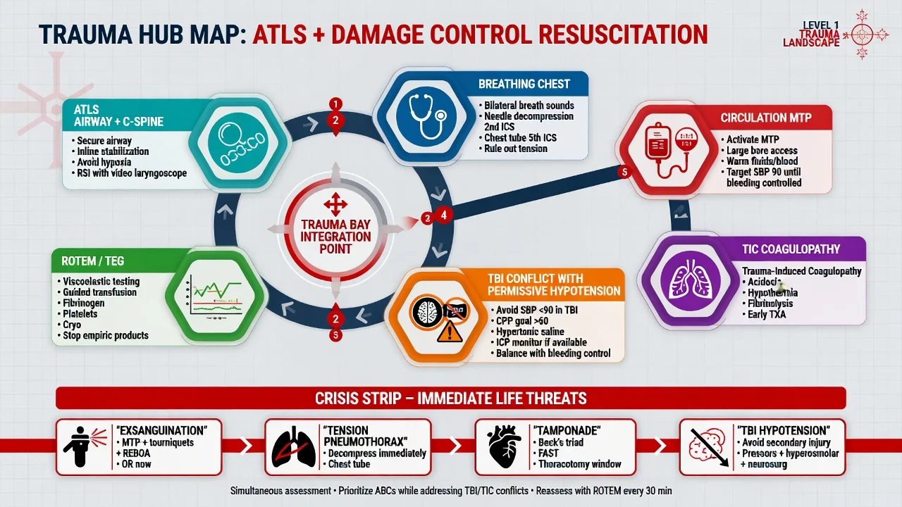
This page is the trauma and massive haemorrhage hub. Use it for the ATLS–DCR spine, lethal triad, MTP, and trial names. Deeper leaves (when built) own isolated crises; cross-links include airway difficult RSI, massive transfusion viscoelastic practice, and TBI under neuroanaesthesia. [1]
| Domain | Examiner focus |
|---|---|
| Trauma airway + C-spine | MILS, RSI, VL, FONA backup |
| Breathing emergencies | Tension, massive haemothorax, tamponade |
| DCR pillars | Permissive BP, 1:1:1, TXA, MTP, DCS |
| TIC + ROTEM/TEG | Mechanism, product choice |
| TBI conflict | No permissive hypotension if brain injured |
| Special scenarios | Pelvis, pregnancy, blast, burns crossover |
Crisis bank
- Exsanguinating torso haemorrhage — MTP + theatre/IR.
- Tension pneumothorax on induction — decompress.
- Cardiac tamponade — thoracotomy pathway.
- TBI + bleeding — CPP defence overrides permissive hypotension.
- Pelvic binder + MTP + packing/IR.
- Citrate hypocalcaemia during MTP — replace calcium.
- Hyperkalaemic arrest during massive transfusion. [1]
Regional practice deltas
ANZ. Local MTP pack composition varies by blood service; state your hospital pack contents and activation phrase. Metaraminol/noradrenaline common; TXA pre-hospital programmes expanding.
SAQ / viva scaffolds
SAQ: "Motorbike vs car, BP 70, open-book pelvis. Outline first 15 minutes of anaesthetic care."
- Team, C-spine, oxygen, primary survey simultaneous.
- Pelvic binder, large-bore access, activate MTP, TXA if within 3 h.
- Airway plan with MILS if needed; ketamine-based RSI when indicated.
- Permissive hypotension unless TBI; warm; calcium; ROTEM.
- To CT only if stable enough, else theatre/IR. [1]
Viva openers: CRASH-2 dose and window; PROPPR result; lethal triad components; why crystalloid harms; TBI MAP target vs permissive hypotension. [1]
Summary
The trauma and massive haemorrhage topic at fellowship level rewards a structured, values-rich answer that links the physiology to the action. The scaffold is: the ATLS primary survey (A with the cervical spine, B, C with the haemorrhage control, D, E); the damage control resuscitation (the permissive hypotension until the haemostasis, the haemostatic 1:1:1 resuscitation, the early tranexamic acid within 3 hours, the activated MTP, the damage control surgery); the lethal triad (the acidosis, the hypothermia, the coagulopathy) and its prevention; the trauma-induced coagulopathy (the thrombomodulin-protein C pathway, the hyperfibrinolysis, the viscoelastic-guided replacement); the traumatic brain injury (the normoxia, the normocapnia, the MAP of 80 mmHg or above, the early intubation and the CT); the specific scenarios (the pelvic binder, the resuscitative thoracotomy, the pregnant patient's left lateral tilt); and the anaesthetic technique (the haemodynamically-tolerated RSI with the ketamine, the titrated maintenance, the multimodal analgesia once stable). The candidate who can quote the trials (the CRASH-2, the CRASH-3, the PROPPR) with the doses and the windows, and who can defend the permissive hypotension and the 1:1:1 ratio with the mechanism and the evidence, passes comfortably. [1]
References
- [1]CRASH-2 trial collaborators; Shakur H, Roberts I, et al. Effects of tranexamic acid on death, vascular occlusive events, and blood transfusion in trauma patients with significant haemorrhage (CRASH-2): a randomised, placebo-controlled trial Lancet, 2010.PMID 20554319
- [2]Holcomb JB, Tilley BC, Baraniuk S, et al.; PROPPR Study Group. Transfusion of plasma, platelets, and red blood cells in a 1:1:1 vs a 1:1:2 ratio and mortality in patients with severe trauma: the PROPPR randomized clinical trial JAMA, 2015.PMID 25647203
- [3]Spahn DR, Bouillon B, Cerny V, et al.; Rossaint R. The European guideline on management of major bleeding and coagulopathy following trauma: fifth edition Crit Care, 2019.PMID 30917843
- [4]Holcomb JB, Jenkins D, Rhee P, et al. Damage control resuscitation: directly addressing the early coagulopathy of trauma J Trauma, 2007.PMID 17297317
- [5]Brohi K, Cohen MJ, Ganter MT, et al. Acute traumatic coagulopathy: initiated by hypoperfusion: modulated through the protein C pathway? Ann Surg, 2007.PMID 17457176
- [6]CRASH-3 trial collaborators. Effects of tranexamic acid on death, disability, vascular occlusive events and other morbidities in patients with acute traumatic brain injury (CRASH-3): a randomised, placebo-controlled trial Lancet, 2019.PMID 31623894