Anaes · Cardiac anaesthesia
Anaesthesia for valve surgery
Also known as Anaesthesia for valve surgery · Aortic stenosis anaesthesia · Mitral repair SAM
Haemodynamic goals for AS, AR, MS, MR; TOE assessment; CPB and cardioplegia nuances; prosthesis checks and post-repair SAM risk.
On this page & tools
Your progress
Saved locally on this device.
10 MCQs with explanations
Target exams
Red flags
Why this is examined / the one-line answer
Valve surgery is classic SS_CS Final material because examiners can walk the candidate from lesion-specific haemodynamics through induction, CPB nuances, post-bypass TOE, and the canonical crisis of systolic anterior motion (SAM) with left ventricular outflow tract obstruction (LVOTO) after mitral repair. Pass-level answers state exact haemodynamic goals for AS, AR, MS, and MR, quote heparin and ACT numbers without hesitation, and describe SAM management as the opposite of pure pump-failure treatment.
One-line opener: I match preload, afterload, and rate to the lesion, prove the ACT before bypass, survey the prosthesis and ventricle with TOE after CPB, and for SAM I fill, squeeze, slow, and stop pure inotropes.
[2]Preoperative assessment and risk stratification
Lesion inventory. Severity and mechanism of each valve lesion; mixed disease (e.g. AS plus MR); left and right ventricular size and function; pulmonary artery pressures; left atrial size and thrombus risk in mitral disease/AF; coronary anatomy if CABG may be combined; aortic dimensions if aortopathy accompanies bicuspid AS; endocarditis stigmata (emboli, heart block, abscess, sepsis physiology).
[2]Functional status and comorbidities. NYHA class, syncope or angina (critical AS red flags), frailty, renal function, COPD, prior sternotomy, anticoagulation plan (mechanical valves, AF), and anaemia (TRICS III context for transfusion thresholds once on the cardiac pathway).[1]
Airway and TOE feasibility. Standard airway assessment; absolute/relative TOE contraindications (oesophageal pathology, recent upper GI surgery, severe dysphagia). Plan alternative imaging if TOE impossible.
[1]Critical aortic stenosis — special induction plan. Fixed stroke volume physiology: cardiac output depends heavily on heart rate and systemic vascular resistance. Hypotension from induction agents or neuraxial blockade collapses coronary perfusion pressure (CPP ≈ DBP − LVEDP) against high LV wall tension and hypertrophied myocardium. Avoid elective major regional sympathectomy in isolation for critical AS without full monitoring and vasopressor readiness.
[1]Investigations to voice. ECG (LVH, AF, conduction disease), echocardiography report with gradients/areas/regurgitant severity, catheterisation or CT as indicated, bloods including haemoglobin, platelets, coagulation, creatinine, and group and screen/crossmatch per unit protocol.
[2]Applied pathophysiology (lesion-specific)
Aortic stenosis (AS)
Obstruction to LV ejection → pressure overload → concentric hypertrophy → diastolic dysfunction and high LVEDP. Stroke volume is relatively fixed; tachycardia shortens diastole (coronary filling time) and may reduce filling; profound bradycardia reduces output because stroke volume cannot rise enough. Vasodilation drops aortic diastolic pressure and coronary perfusion. Atrial kick matters — new AF can cause abrupt decompensation.
[2]Aortic regurgitation (AR)
Volume overload of the LV; regurgitant fraction rises with high SVR and long diastole (bradycardia). Modest afterload reduction and a modestly higher heart rate shorten diastole and may reduce regurgitant volume. Cardioplegia delivery is complicated because antegrade aortic root cardioplegia may wash into the LV rather than coronary ostia when AR is severe — ostial or retrograde cardioplegia often required.
[2]Mitral stenosis (MS)
Obstruction to LV filling; left atrial hypertension; pulmonary hypertension and right ventricular strain in advanced disease. Cardiac output is rate-dependent in a dangerous way: tachycardia shortens diastolic filling time across the stenotic valve and raises left atrial pressure. Maintain sinus rhythm when possible; treat new AF urgently. Avoid aggressive vasodilation that drops systemic pressure while pulmonary pressures remain high.
[1]Mitral regurgitation (MR)
Part of LV stroke volume goes backwards into the LA. High afterload worsens regurgitant fraction; modest afterload reduction can improve forward stroke volume. Bradycardia lengthens systole and may worsen regurgitation; sinus rhythm preferred. After repair, geometry of the LVOT and anterior mitral leaflet can produce SAM.
[2]Mixed and multi-valve disease
Prioritise the dominant lesion for induction goals, then reassess after each surgical stage with TOE. Combined AS and AR, or mitral disease with tricuspid regurgitation and RV failure, requires explicit discussion of which haemodynamic compromise is least tolerable.
[2]Anaesthetic goals (by lesion)
Cross-cutting goals
- Preserve coronary perfusion pressure, especially in AS and LVH.
- Maintain oxygen delivery (haemoglobin strategy per TRICS III context and bleeding).[1]
- Avoid air embolism after open-chamber surgery (de-airing discipline).
- Enable high-quality TOE before and after CPB.[2]
- Plan for conduction block after aortic surgery and for pulmonary hypertension/RV failure in advanced mitral disease.
Technique options and decision matrix
Monitoring. Arterial line before or immediately after induction as unit practice for critical lesions; large-bore IV access; central venous access for drugs and CVP context; TOE probe after induction when safe; urinary catheter; temperature; ACT capability; defibrillator pads for redo or arrhythmia risk; pulmonary artery catheter selectively (severe PHT, complex RV failure, transplant/VAD pathways).
[2]Induction. Opioid-based balanced cardiac induction is common (e.g. fentanyl or equivalent dosing per practice, propofol or etomidate carefully titrated, neuromuscular blockade for intubation). Have vasopressors drawn (metaraminol or phenylephrine boluses; noradrenaline infusion ready) before induction in critical AS. Avoid large histamine-releasing boluses and uncontrolled vasodilation.
[1]Maintenance. Volatile or TIVA both used; many units use volatile into the oxygenator on CPB. Depth monitoring adjuncts as available. Antifibrinolytic therapy (tranexamic acid) per institutional cardiac protocol.
[1]Heparin and CPB. Unfractionated heparin 300–400 IU/kg IV before arterial cannulation/bypass. Confirm ACT ≥400 s (many units ≥480 s) before full CPB. Redose heparin on CPB as ACT falls or per heparin concentration systems. Cardioplegia route: antegrade, retrograde, or combined — explicitly plan for severe AR.
[2]Protamine after stable wean. Approximately 1 mg protamine per 100 IU heparin administered, given slowly, with readiness for reaction phenotypes (hypotension, anaphylactoid, catastrophic pulmonary hypertension/RV failure).[3]
Monitoring and TOE essentials
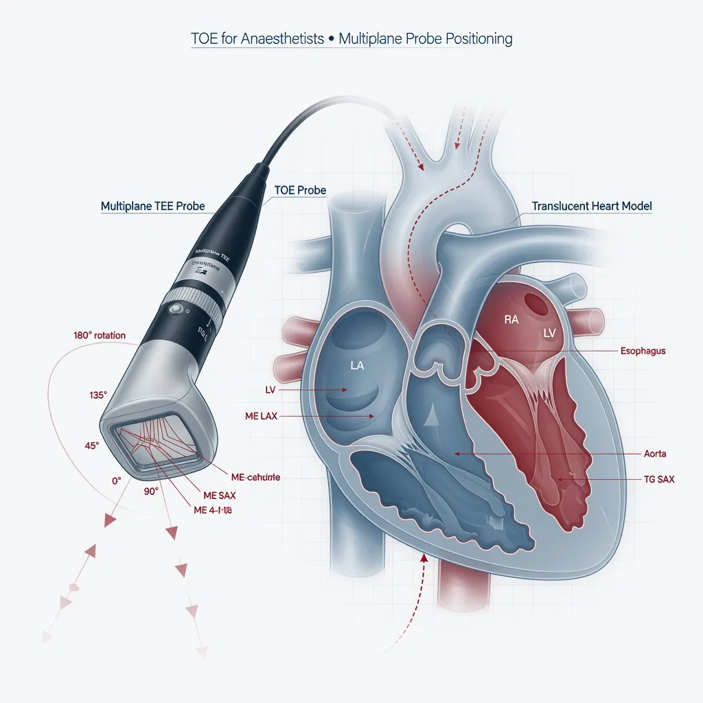
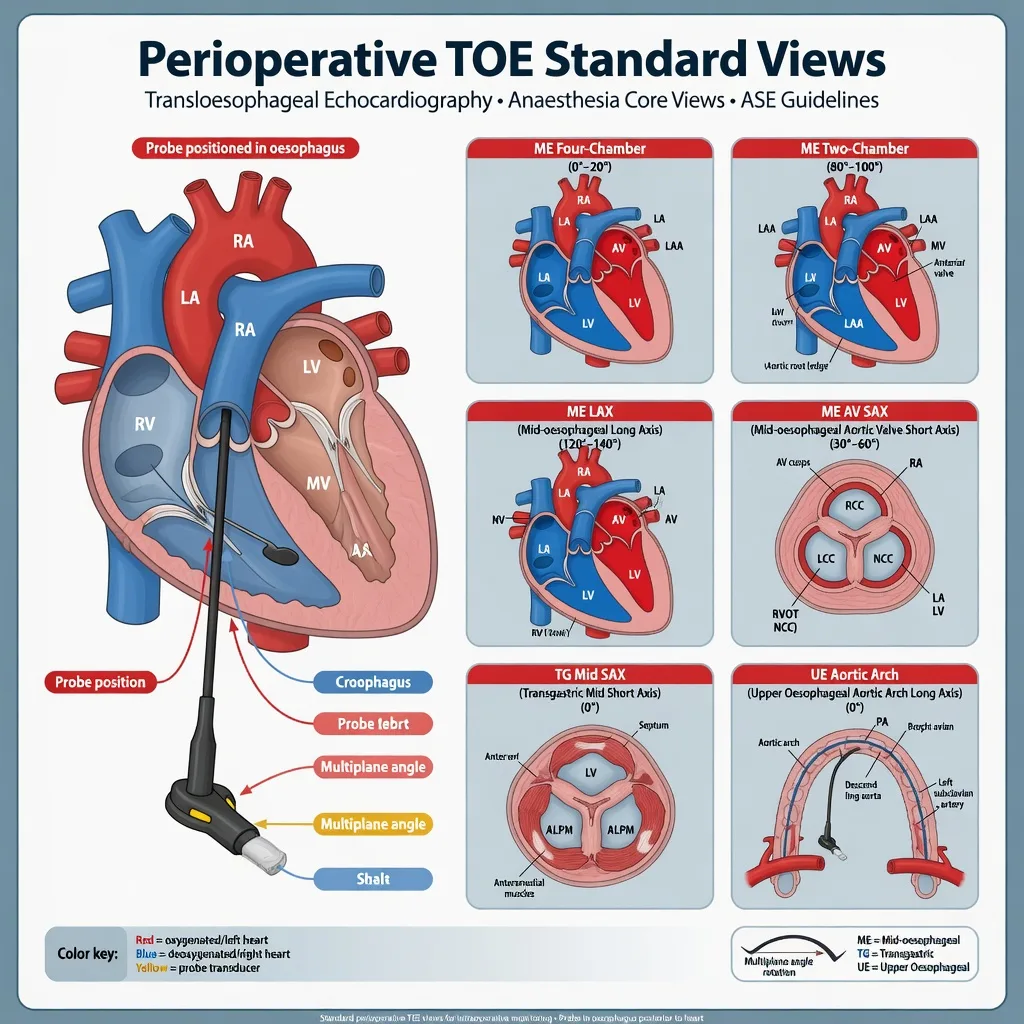
ASE/SCA comprehensive TOE examination standards frame the peri-CPB survey for valve surgery.[2]
Pre-CPB TOE checklist (exam list)
- Confirm lesion severity and mechanism; look for unexpected additional lesions
- LV and RV size/function; regional wall motion if CAD
- Estimate pulmonary pressures where feasible
- Baseline SAM risk anatomy: small hyperdynamic LV, anterior leaflet redundancy, narrow aorto-mitral angle, sigmoid septum
- Aortic atheroma before cannulation when relevant
- Baseline prosthetic or native valve gradients for comparison
Post-CPB TOE checklist
- Prosthesis seating and leaflet motion (mechanical: both leaflets open/close; bioprosthetic: leaflet excursion)
- Transvalvular gradients (compare with expected)
- Paravalvular leak (trace vs significant)
- Residual MR after repair; SAM/LVOTO with or without MR
- Intracardiac air (especially LA roof, LV apex, pulmonary veins)
- Ventricular function and new regional wall motion abnormalities
- Dynamic LVOTO after AVR in small hypertrophied ventricles (not only after mitral repair)

Intraoperative management by phase
Pre-bypass
Stabilise haemodynamics to lesion goals; treat new AF; avoid prolonged hypotension during lines and TOE; heparinise and prove ACT; coordinate cannulation and venting with surgeon; discuss cardioplegia route for AR.
[2]On bypass
Anaesthetic depth via circuit; MAP typically 50–80 mmHg and indexed flows about 2.2–2.5 L/min/m² as unit targets; correct potassium after cardioplegia; watch urine, temperature strategy, haematocrit, glucose; plan de-airing early for open-chamber cases.
[3]Weaning
Use RRRAC (rate, rhythm, preload, afterload, contractility) with TOE. Epicardial pacing wires often essential after aortic valve surgery (AV block risk). For mitral disease with PHT, prepare RV support (inodilators such as milrinone, pulmonary vasodilators per availability, optimise ventilation to avoid hypoxia/hypercarbia/high PEEP that raise PVR).
[1]After wean
Protamine only when surgically ready and haemodynamics acceptable; reassess TOE after chest closure physiology changes; plan ICU vasoactive support and bleeding thresholds.
[3]Crisis pivots — what changes the plan
SAM / LVOTO after mitral repair (canonical crisis)
Recognition. Hypotension, high inotrope requirement that paradoxically worsens obstruction, TOE showing anterior leaflet SAM into the LVOT, dagger-shaped Doppler profile, often posteriorly directed MR jet.
[2]Immediate management (opposite of pure pump failure):
[2]- Stop or reduce pure inotropes and chronotropes (dobutamine, high-dose adrenaline may worsen obstruction).
- Volume load from the reservoir under TOE guidance (increase LV cavity size).
- Increase systemic afterload (vasopressors such as noradrenaline/phenylephrine) to raise LVOT size relative to stroke volume dynamics.
- Slow the heart rate (beta-blockade carefully if not contraindicated; pacing rate reduction if over-paced).
- If refractory: discuss surgical revision (slideplasty, further ring adjustment, or even valve replacement as surgical judgment).
Model phrase: Fill, squeeze, slow, stop inotropes — then revise if still severe.
[2]Other crises
- New severe paravalvular leak or stuck leaflet: return-to-bypass conversation, not observation alone in theatre.
- RV failure on wean (mitral/PHT): pulmonary vasodilators, inodilators, optimise ventilation, avoid pure systemic vasoconstriction that ignores PVR, rest on CPB if needed.
- AV block after AVR: epicardial pacing; temporary pacing plan to ICU.
- Air lock / residual air: de-airing manoeuvres, Trendelenburg strategies as agreed, TOE-guided removal of air.
- Protamine reaction: stop drug, support SVR and RV, consider re-heparinisation and return to CPB for catastrophic pulmonary hypertension.[3]
Postoperative / ICU / PACU plan
- Continuous arterial pressure, ECG with pacing capability, urine output, chest drain losses
- Anticoagulation timing for mechanical valves (unit protocol; balance bleeding vs thrombosis)
- Pulmonary hypertension and RV support weaning plan
- Conduction surveillance after aortic surgery
- Restrictive red-cell transfusion when clinically appropriate (TRICS III non-inferiority context)[1]
- Analgesia that preserves haemodynamic stability; early physiotherapy when stable
Special populations and comorbidities
- Endocarditis: septic vasoplegia, abscess-related heart block, embolic risk, prolonged antibiotics, fragile tissues and bleeding
- Redo sternotomy: external defibrillator pads, massive haemorrhage readiness, prolonged dissection time
- Pregnancy with valve disease: lesion-specific physiology plus aortocaval compression and higher cardiac output demand — multidisciplinary plan
- TAVI / percutaneous pathways: hybrid theatre, rapid pacing for deployment, vascular access complications, conversion readiness
- Hypertrophic cardiomyopathy physiology overlapping AS: dynamic obstruction risk post-AVR
SAQ answer scaffold
- State lesion haemodynamic goals in a four-row table (AS/AR/MS/MR).
- Induction plan for critical AS with vasopressors ready.
- Heparin 300–400 IU/kg, ACT ≥400–480 s, protamine ~1 mg/100 IU slowly.[3]
- Pre- and post-CPB TOE checklists for valves.[2]
- SAM: fill, raise SVR, reduce inotropy/chronotropy, surgical revision if refractory.
- ICU priorities: pacing, bleeding, anticoagulation timing, RV/PHT, TRICS III context.[1]
Viva stem bank and model phrases
- “How do you anaesthetise critical aortic stenosis?”
- “Cardioplegia will not arrest the heart in severe AR — why, and what next?”
- “After mitral repair the blood pressure is 70 mmHg on high-dose adrenaline — your thoughts?”
- “Show me your post-CPB TOE checklist for a mechanical AVR.”
Model phrases:
[2]- “I will not go on full bypass without a confirmed therapeutic ACT.”
- “For SAM I will fill, raise afterload, and reduce inotropes before asking for surgical revision.”
- “New severe prosthetic dysfunction is a return-to-bypass conversation.”
Common traps
- Treating SAM like primary pump failure with pure inotropes and empty LV
- Ignoring AR when planning antegrade aortic root cardioplegia
- Missing stuck leaflet or significant paravalvular leak on TOE
- Neuraxial hypotension in critical AS without monitoring and support
- Starting protamine during an unstable failed wean
- Forgetting pacing wires after AVR when conduction tissue is at risk
Extended lesion haemodynamics (exam depth)
Critical AS numbers and coronary logic
Stroke volume is relatively fixed by the stenotic orifice. Mean gradient and aortic valve area stratify severity on echocardiography (severe AS commonly discussed when AVA is less than about 1.0 cm² with high gradients, acknowledging low-flow low-gradient variants). Coronary perfusion pressure approximates aortic diastolic pressure minus LVEDP. Hypertrophied myocardium has high oxygen demand and reduced coronary reserve; even brief hypotension can cause subendocardial ischaemia, ventricular arrhythmias, and spiral shock. Phenylephrine or metaraminol boluses restore SVR quickly while longer-term noradrenaline infusions stabilise diastolic pressure. Avoid pure inodilators as first-line for induction hypotension in critical AS.
[2]AR and cardioplegia logistics
Severe AR produces LV volume overload and a wide pulse pressure. On CPB, antegrade aortic root cardioplegia may fail to enter the coronary ostia because the aortic valve is incompetent; the LV may distend unless vented. Options: direct ostial cardioplegia after aortotomy, retrograde cardioplegia via the coronary sinus, or combined strategies. Communicate this before cross-clamp. After AVR for pure AR, the LV faces a sudden increase in afterload relative to the regurgitant state — support contractility and avoid acute hypertensive spikes on the fresh suture line.
[2]Mitral stenosis and pulmonary hypertension
MS elevates left atrial pressure; chronic cases remodel the pulmonary vasculature and strain the RV. Intraoperative tachycardia (pain, light anaesthesia, inotropes, AF with rapid ventricular response) is poorly tolerated. Treat rate with beta-blockade carefully if SVR allows, amiodarone or cardioversion for new AF with instability, and maintain systemic pressure for RV coronary perfusion. After mitral valve replacement for MS, the LA and pulmonary pressures may fall, but RV dysfunction can persist — wean with RV-aware pharmacology.
[2]Mitral regurgitation repair versus replacement
Repair preserves subvalvular apparatus and is preferred when feasible for degenerative MR. After repair, TOE grades residual MR and screens for SAM. Ischaemic MR may behave differently with LV remodelling. Replacement eliminates SAM risk from leaflet geometry but removes apparatus unless chordal-sparing techniques are used; prosthesis type dictates anticoagulation later.
[2]Induction scripts you can speak
Critical AS: arterial line if time; draw-up metaraminol 0.5 mg/mL or phenylephrine 100 microg/mL boluses; noradrenaline infusion primed; opioid-first induction; carefully titrated hypnotic; early intubation to control airway without prolonged mask hypotension; treat any MAP fall immediately; maintain sinus rhythm.
[2]Severe MR with low EF: avoid pure vasoconstriction without supporting forward flow; modest afterload reduction once coronary perfusion is safe; prepare for inotropic support post-CPB.
[2]Mixed AS/AR: maintain SVR near baseline; avoid extremes of rate; plan cardioplegia route for the AR component.
[2]De-airing after open-chamber surgery
Air exits poorly from pulmonary veins, LA roof, LV apex, and left atrial appendage. Trendelenburg, surgical venting, TOE-guided detection, and sustained ventilation/recruitment manoeuvres help. Coronary air presents as sudden RWMA or VT/VF on weaning — support, raise perfusion pressure, and allow time while surgeons continue de-airing. Do not attribute all new RWMA to graft failure without considering air in AVR/MVR contexts.
[1]Prosthetic valve assessment phrases
- Mechanical bileaflet: both leaflets mobile, physiological washing jets, no large paravalvular jet
- Bioprosthetic: leaflets open without restriction, expected gradients for size
- Patient–prosthesis mismatch: unexpectedly high gradients for body size — surgical discussion
- Stuck leaflet: urgent return to bypass
Anticoagulation after mechanical valves (exam framing)
Balance postoperative bleeding against valve thrombosis. Timing of heparin bridge and warfarin initiation is unit- and patient-specific (bleeding risk, valve position — mitral higher thrombosis risk than aortic in many protocols). Do not invent a single universal hour mark; state multidisciplinary protocol.
[2]Regional exam deltas
- ANZCA Final: lesion tables, SAM crisis, TOE PS46 culture, exact heparin/ACT
- FRCA: similar cardiac viva; emphasis on pathophysiology explanations
- ABA: TEE examination culture heavily weighted
- EDAIC/FCAI: structured goals and crisis algorithms
Worked mental example
A 78-year-old with critical AS becomes hypotensive to MAP 50 mmHg after induction: stop volatile if running high, give phenylephrine 50–100 microg boluses, start noradrenaline if recurrent, check rhythm (not new AF?), reduce surgical stimulus, deepen carefully once pressure restored, avoid spinal for hip fracture narratives without full AS plan — the same physiology applies outside cardiac theatres when AS is incidental.
[1]Full perioperative timeline (consultant narrative)
Night before and preassessment
Identify the dominant lesion and secondary lesions on the most recent echocardiogram. Critical AS with syncope or heart failure symptoms warrants careful scheduling and ICU bed planning. Review coronary angiography if performed; combined CABG changes ischaemia risk and cross-clamp time. Stop or bridge anticoagulants per protocol; continue beta-blockers usually; discuss ACE inhibitor / ARB holding practices for vasoplegia risk on CPB. Optimise anaemia when time allows because transfusion decisions later sit in the TRICS III evidence context. [1]
On arrival in the anaesthetic room
Large-bore IV access, arterial line timing based on lesion severity (before induction for critical AS is defensible), five-lead ECG, defibrillator pads for reoperations. Draw-up vasopressors before induction drugs. TOE probe check, machine check, perfusion briefing: expected procedure, cardioplegia route if AR, circulatory arrest unlikely vs possible for root work. [1]
Induction to cannulation
Match haemodynamics to the lesion table. After intubation, place central line and TOE. Pre-CPB TOE confirms the surgical plan and looks for unexpected findings (LA thrombus in MS/AF, additional valve lesions, aortic atheroma). Heparin 300–400 IU/kg; ACT ≥400–480 s; only then full bypass. [1]
Cross-clamp and valve intervention
Myocardial protection quality determines ease of wean. For AR, confirm ostial/retrograde delivery. For mitral repair, the anaesthetist anticipates SAM screening immediately after separation. Communicate any difficulty coming off partial bypass early. [1]
Separation and post-CPB
RRRAC, TOE prosthesis survey, treat SAM if present, pacing for AV block after AVR, protamine only when stable (~1 mg per 100 IU heparin slowly), assess bleeding, plan ICU vasoactives. Re-TOE after chest closed if haemodynamics change. [1]
Dynamic LVOTO after AVR (not only mitral)
Small hypertrophied LV cavities after AVR for AS can develop dynamic obstruction. Phenotype mimics SAM management: volume, vasoconstriction, avoid pure inotropes and tachycardia. Beta-blockade may help once blood pressure allows. This is a high-yield discriminator when the candidate only associates LVOTO with mitral repair. [1]
Pulmonary vasodilators in mitral disease (exam agents)
When RV fails with high PVR after mitral surgery, options (availability varies): inhaled nitric oxide (commonly 5–20 ppm ranges titrated), inhaled epoprostenol/prostacyclin analogues per protocol, milrinone as inodilator, optimise ventilation (avoid hypoxia, hypercarbia, excessive PEEP). Systemic hypotension from inodilators requires noradrenaline co-infusion to protect RV coronary perfusion. [1]
Endocarditis valve surgery specifics
Sepsis physiology, vasoplegia, abscess-related conduction block, embolic stroke risk, friable annular tissue, prolonged antibiotics, and sometimes tricuspid involvement in IVDU. TOE is diagnostic gold standard preoperatively for vegetations and complications. Blood conservation still matters but active infection and long CPB predict coagulopathy. [1]
Hybrid and percutaneous overlap
TAVI lists test rapid pacing for deployment, vascular access bleeding, conversion to sternotomy readiness, and post-deploy AR/paravalvular leak assessment. MitraClip-type procedures need TOE guidance and different haemodynamic profiles. Know when to abandon percutaneous plans for open surgery. [1]
Examiner SAQ timing guide
Spend the first third on lesion goals, the middle third on TOE and CPB numbers, the last third on SAM and complications. Always include exact heparin/ACT/protamine figures once. [1]
[2] [2] [1]References
- [1]Mazer CD, Whitlock RP, Fergusson DA, et al. Restrictive or Liberal Red-Cell Transfusion for Cardiac Surgery N Engl J Med, 2017.PMID 29130845
- [2]Hahn RT, Abraham T, Adams MS, et al. Guidelines for performing a comprehensive transesophageal echocardiographic examination: recommendations from the American Society of Echocardiography and the Society of Cardiovascular Anesthesiologists J Am Soc Echocardiogr, 2013.PMID 23998692
- [3]Levy JH, et al. What's fishy about protamine? Clinical use, adverse reactions, and potential alternatives J Thromb Haemost, 2023.PMID 37062523