EM · Primary survey
The primary survey (ABCDE) — the trauma assessment framework
Also known as ABCDE · Primary trauma survey · Trauma assessment · ATLS primary survey
The primary survey — the ABCDE framework for the systematic, prioritised assessment of the trauma patient (Airway with cervical-spine control, Breathing with ventilation, Circulation with haemorrhage control, Disability, Exposure), the immediately life-threatening conditions sought and treated at each step before moving to the next, and the transition to the secondary survey once the patient is stabilised. The foundational framework of all emergency and trauma medicine. ACEM-primary, globally tagged.
On this page & tools
Your progress
Saved locally on this device.
Target exams
Red flags
The primary survey is the single most important framework in all of emergency and trauma medicine — the systematic, prioritised assessment that treats the most immediately fatal derangement before moving to the next. The Fellowship candidate must know the ABCDE in depth, the immediately life-threatening conditions sought at each letter, and the specific interventions that treat them, because this framework governs every emergency from the road traffic collision to the septic collapse. The principle is simple and absolute: treat first what kills first.[1][2]
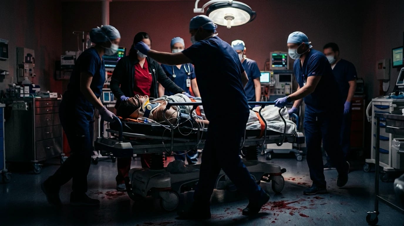
The principle — treat first what kills first
Three principles govern the whole approach. First, the problems are treated in the order in which they kill — a blocked airway kills before a failing circulation, which kills before a raised intracranial pressure — so the airway is secured before the circulation is assessed.[1] Second, the resuscitation and the investigation proceed simultaneously — the intravenous access, the bloods, the gas and the ECG are obtained while the airway and the breathing are secured, not deferred until afterwards. Third, the framework is iterative — after every intervention, the clinician returns to the airway and reassesses; the primary survey is a cycle, not a single pass.[2]
The evolution — from ABCDE to <C>ABCDE
The original ATLS sequence, codified in 1980 from the Nebraska light-aircraft crash that nearly killed the orthopaedic surgeon who designed it, began at the airway — because in the blunt civilian trauma of the era, airway loss was the dominant early killer. Forty years of military experience (the Iraq and Afghanistan campaigns) and the registry data of the civilian penetrating-trauma centres have refined that picture: the patient who is exsanguinating from a limb or a junctional wound dies of haemorrhage before the airway is threatened, and so the modern doctrine inserts a catastrophic-haemorrhage step BEFORE the airway — the <C>ABCDE (read "C-ABCDE"), or its military cousin MARCH (Massive haemorrhage, Airway, Respiration, Circulation, Head/Hypothermia). The civilian consensus is identical in spirit: a 5-second scan from the end of the bed for the exsanguinating limb wound, controlled by the tourniquet or the direct pressure, before the formal airway assessment begins. The <C> is set in the angle brackets precisely because it is a rapid, opportunistic step — performed only if the catastrophic external haemorrhage is present — and the rest of the ABCDE proceeds unchanged once it is controlled.[1]
Kragh et al. — the US military tourniquet registry
Journal of Trauma, 2009
Registry of 499 combat casualties with 749 tourniquets applied prehospital; survival analysed by indication, limb and timing.
Key finding
Prehospital tourniquet use on the exsanguinating limb was associated with a clear survival benefit when applied before the onset of shock; no patient in whom a tourniquet was applied before the onset of shock died of the limb injury itself. Morbidity from the limb was low within the 2-hour ischaemic window.
Practice change
Tourniquets moved from a "last resort" to a first-line prehospital intervention; the early limb tourniquet is now standard ATLS / TECC / civilian-mass-casualty doctrine.
ATLS-1 (1980)
- ABCDE — Airway first
- Civilian blunt trauma
- Airway loss was the dominant early killer
- Crystalloid-first resuscitation
`<C>`ABCDE / MARCH
- Catastrophic haemorrhage first
- Born from Iraq / Afghanistan
- Limb & junctional exsanguination kills first
- Haemostatic resuscitation (1:1:1, TXA)
TECC / Civ-MCI
- Hot / warm / cold zones
- Stop the bleed — public tourniquets
- Hypothermia & head injury tracked
- Triangulates with the hospital ATLS
Differential diagnosis — the immediately life-threatening conditions by letter
The primary survey is the systematic search for the immediately life-threatening conditions. The differential at each letter is the set of conditions that kill fastest, and each is treated before moving to the next.[1][2]
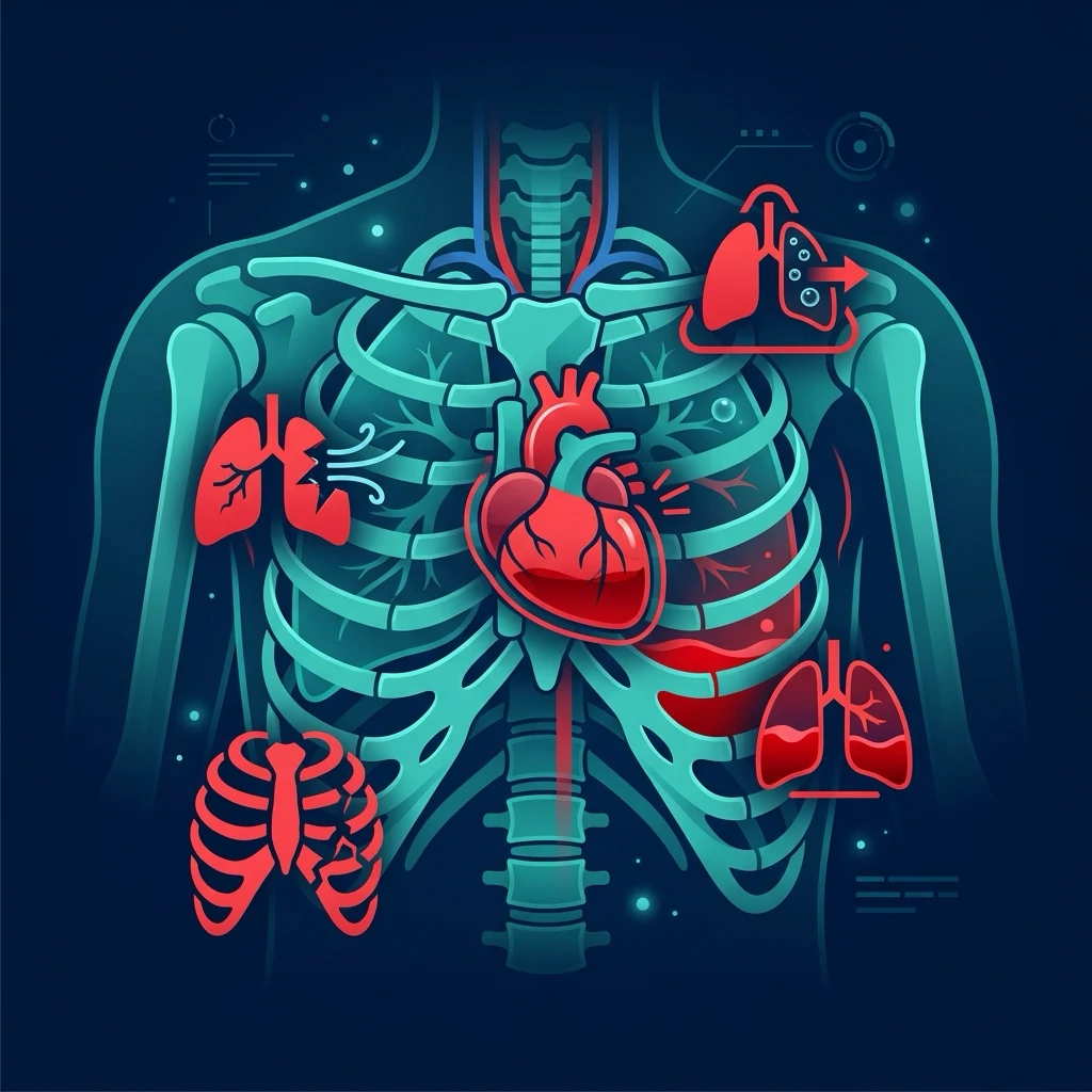
A — Airway
- Airway obstruction (blood, vomit, facial fracture, swelling)
- The C-spine is immobilised from the start
- The definitive airway (RSI) if the airway is threatened
- Surgical airway (FONA) for the can't-intubate
B — Breathing
- Tension pneumothorax → decompress before imaging
- Massive haemothorax → drain (thoracotomy if >1500 mL)
- Open pneumothorax → seal and drain
- Flail chest + contusion → NIV or ventilation
C — Circulation
- Haemorrhagic shock (external, chest, abdomen, pelvis)
- Cardiac tamponade → FAST or echo; thoracotomy if traumatic
- The massive haemorrhage protocol (1:1:1)
- Pelvic binder, tourniquet, direct pressure
D & E
- GCS, pupils, the bedside glucose (hypoglycaemia)
- Hypothermia — warm the patient (the lethal triad)
- The complete exposure for the full assessment
- The secondary survey after the stabilisation
<C> — Catastrophic haemorrhage control
The <C> step is not the full circulation assessment — that is the C of ABCDE. It is the rapid, targeted control of the catastrophic EXTERNAL haemorrhage that will kill the patient in the next 3 to 5 minutes. Three tools carry almost the entire burden.[1]
The limb tourniquet
A catastrophic limb bleed (a partial amputation, a femoral arterial laceration, a deep knife wound to the thigh) is controlled with a windlass tourniquet (the combat application tourniquet — the CAT — or the equivalent) placed 5 to 8 cm proximal to the wound, never over a joint, tightened until the bleeding stops and the distal pulse disappears. The time of application is written on the patient (the forehead is the military convention) — the tourniquet is left on until the theatre, and the safe ischaemic time in the awake patient is at least 2 hours with very low risk, rising to a limb-threatening concern after 6 hours. Improvised tourniquets (a belt, a blood-pressure cuff inflated above the systolic) are inferior and reserved for the austere setting. The tourniquet is NOT applied to a venous ooze or a non-compressible trunk bleed — it is reserved for the limb that is going to kill the patient.[1]
The pelvic binder
A mechanically unstable pelvic fracture (the open-book, the lateral-compression fracture with disruption) can bleed catastrophically from the venous plexus and the internal iliac arteries into the retroperitoneum — a volume that is invisible externally and that kills within the first hour. The pelvic binder (a circumferential sheet sling, applied at the level of the greater trochanters, NOT the iliac crests) closes the pelvic ring, reduces the pelvic volume, and tamponades the bleeding by approximating the bony surfaces. It is applied on the CLINICAL suspicion (a deformed pelvis, the mechanism, the shock) BEFORE the imaging — and it is left in place until the definitive fixation or the angiographic embolisation. A sheet wrapped firmly around the greater trochanters is the austere equivalent and works near-as-well.[1]
Direct and packing pressure
For the junctional bleed (the neck, the axilla, the groin — where the tourniquet cannot reach) and for the trunk, the control is direct pressure with a haemostatic dressing (the chitosan-based Celox or the kaolin-based QuikClot gauze) packed firmly into the wound and held for at least 3 minutes. Plain gauze packing is the fallback. The deep-packing principle is that the cavity is filled under pressure; a superficial dressing over a deep cavity fails.[1]
[1] [1]Limb bleed
- CAT windlass tourniquet
- 5–8 cm proximal to the wound
- Tighten until distal pulse lost
- Note the application time
Pelvic fracture
- Pelvic binder at the greater trochanters
- Apply before the imaging
- Closes the ring, reduces the volume
- Leave in place to theatre or IR
Junctional/trunk
- Direct pressure + haemostatic gauze
- Pack the cavity firmly
- Celox / QuikClot
- Hold ≥ 3 min before re-check
A — Airway with the cervical-spine control
The first question is whether the airway is patent, protected and maintainable, because an obstructed airway kills before any other problem. The cervical spine is immobilised from the first contact in any patient with a mechanism compatible with a spinal injury — a rigid collar, head blocks and tape, and the manual in-line stabilisation during the intubation. The assessment is rapid and clinical: look for the chest movement and the abdominal breathing, listen for the obstructive noises (the stridor, the gurgling, the snoring), and feel for the airflow at the mouth. The airway is cleared (the suction, the head tilt and the chin lift, or the jaw thrust if the C-spine is injured) and the adjuncts are inserted (the oropharyngeal airway for the deeply unconscious, the nasopharyngeal for the semi-conscious). If the airway cannot be maintained or protected, the definitive airway — the rapid-sequence intubation — is performed by the most skilled operator, with the difficult-airway equipment and the surgical airway kit prepared from the start. The drugs for the RSI: the induction (the ketamine 1 to 2 mg per kilogram, or the propofol 1 to 2 mg per kilogram, or the thiopental 3 to 5 mg per kilogram) and the muscle relaxant (the suxamethonium 1.5 mg per kilogram, or the rocuronium 1 mg per kilogram). The difficult airway (the can't-intubate, the can't-oxygenate) is rescued by the scalpel-bougie-tube cricothyroidotomy (the front-of-neck access).[1]
The trauma RSI — the high-risk airway
The trauma RSI is a high-risk procedure — the patient may be hypovolaemic (the induction agents drop the pressure), may have a full stomach (aspiration risk), may have a cervical-spine injury (the in-line stabilisation impairs the view), and may have blood or swelling in the airway (the view is poor). The sequence:[2]
The trauma RSI
Prepare
Suction on, oxygen on, two operators, the difficult-airway trolley open, the surgical-airway kit on the tray. Drugs drawn up: ketamine 1–2 mg/kg, suxamethonium 1.5 mg/kg, rocuronium 1 mg/kg if sugammadex is available.
Pre-oxygenate
3 minutes of high-flow oxygen via a non-rebreather, or 8 vital-capacity breaths. Denitrogenate the functional residual capacity.
Pre-treatment
Fentanyl 1–3 µg/kg for the sympathetically responsive hypertensive patient; the routine use of atropine in adults is no longer recommended.
Induction + paralysis
Ketamine is the agent of choice in trauma (preserves the blood pressure); thiopental and propofol are avoided in the shocked. Suxamethonium is the default relaxant; rocuronium 1 mg/kg is the alternative when the sugammadex reversal is available.
Apnoeic oxygenation
Nasal cannulae at 15 L/min throughout the apnoeic period — the THRIVE technique — prolongs the safe apnoea time.
Bougie first, in-line stabilisation
Manual in-line stabilisation by an assistant (NOT the head-tilt). The bougie-led, video-laryngoscope-aided intubation is the first attempt.
Confirm and secure
Waveform capnography — the gold standard. Secure the tube; check the chest for the bilateral air entry.
Rescue — FONA
The can't-intubate, can't-oxygenate situation is rescued by the scalpel-bougie-tube cricothyroidotomy (the ATLS-10 / DAS-2015 standard). The needle cricothyroidotomy has been de-emphasised — it fails.
The surgical airway — the front-of-neck access (FONA)
The can't-intubate, can't-oxygenate situation in the adult is rescued by the scalpel-bougie-tube cricothyroidotomy: a transverse incision through the cricothyroid membrane, a bougie passed into the trachea, and a size 6.0 cuffed tube railroaded over the bougie. The technique is muscle-memory, taught on the manikin, and the kit must be at the bedside before the RSI begins. The needle cricothyroidotomy (the large-bore cannula through the membrane) has been de-emphasised in the adult because of the high failure rate (kinking, malposition, the inadequate ventilation); it remains a temporising option in the child under 8 years (where the cricoid is too small for the tube).[1]
B — Breathing with the ventilation
The breathing is assessed for the work, the effectiveness and the symmetry. The respiratory rate is counted over a full minute; the effort is assessed by the accessory-muscle use, the tracheal tug and the ability to speak in full sentences; and the chest is examined for the symmetry of the movement, the expansion, the percussion and the auscultation. The oxygen saturation is recorded and the capillary refill bridges the breathing and the circulation assessments. The immediately life-threatening breathing problems are treated before the examination is completed: a tension pneumothorax is decompressed (the 5th intercostal space, anterior axillary line); a massive haemothorax is drained (a large-bore chest drain at the 4th or 5th intercostal space, anterior axillary — a thoracotomy if the immediate output exceeds 1500 mL or the ongoing rate exceeds 200 mL per hour); and a flail chest with a pulmonary contusion is managed with the oxygen, the analgesia and, if failing, the non-invasive or the invasive ventilation. The oxygen is given at a high concentration via the non-rebreather mask (10 to 15 L per minute, up to 90 per cent), titrated to a target saturation.[1]
Tension pneumothorax — the clinical diagnosis and the decompression
The tension pneumothorax is a CLINICAL diagnosis made at the bedside — the triad of respiratory distress, hypoxia, and shock, with the hemithorax signs of the tracheal deviation AWAY from the affected side (a late sign), the unilateral hyper-resonance, the absent breath sounds, and the distended neck veins. The chest radiograph is NOT awaited. The immediate decompression is by needle thoracostomy — a 14-gauge (or larger) cannula in the 2nd intercostal space, mid-clavicular line, OR (the modern preference) the 5th intercostal space, anterior axillary line — the same site as the chest drain, which is the definitive treatment. The traditional 2nd-ICS site has a high failure rate in the muscular or the obese patient (the cannula is too short to reach the pleural space) and is increasingly replaced by the 5th-ICS approach, or by the finger thoracostomy in the intubated patient (the blunt dissection down to the pleura, the finger sweep to release the tension, and the chest drain through the tract).[1]
Open pneumothorax — the three-sided dressing
An open (sucking) chest wound is sealed with a chest seal or a three-sided occlusive dressing (the open side allows the air to escape on expiration, preventing the conversion to a tension pneumothorax). A formal chest drain is placed at a SEPARATE site (NOT through the wound). If the patient deteriorates after the dressing, the seal is briefly lifted to release the trapped air — the "burping" of the dressing.[1]
Massive haemothorax — drain, then thoracotomy
A massive haemothorax is the accumulation of more than 1500 mL of blood in the pleural cavity (or the ongoing loss of more than 200 mL per hour). The patient is in shock with the absent breath sounds and the dullness to percussion on the affected side. A large-bore (28 to 36 French) chest drain is placed at the 5th intercostal space, anterior axillary line, and the output is measured in a graduated collector. The autotransfusion of the drained blood is available in many centres. If the immediate output exceeds 1500 mL OR the ongoing rate exceeds 200 mL per hour, the patient goes to the resuscitative thoracotomy (or the theatre) for the source control. The indications for the ED thoracotomy are the witnessed cardiac arrest with the penetrating chest trauma (the salvage rate up to 30%), and the persistent shock despite the drainage in the blunt trauma (the salvage rate much lower).[2]
Flail chest and the pulmonary contusion
A flail segment — three or more ribs fractured in two or more places — produces a paradoxical (inward on inspiration) segment of the chest wall. The flail itself is rarely the killer; the underlying pulmonary contusion is, with the progressive hypoxia and the shunt over the first 24 to 48 hours. The management is the aggressive analgesia (the regional blocks — the serratus anterior plane, the erector spinae plane — are first-line, with the systemic opioids as the adjunct), the oxygen as required, and the early non-invasive ventilation (the CPAP or the BiPAP) for the failing oxygenation or the rising work of breathing. The intubation and the ventilation are reserved for the patient who fails the NIV or who has the other indication.[1]
The surgical stabilisation of the flail chest — observational cohort
Chest / Annals of Thoracic Surgery, 2017
Multicentre observational cohort of severe blunt chest injury; the surgical rib fixation compared with the non-operative management.
Key finding
The severe flail chest with the surgical stabilisation reduced the pneumonia, the ventilator days and the ICU length of stay in the selected cohort; the benefit was greatest in the marked displacement and the respiratory failure.
Practice change
The surgical rib fixation is now a recognised option for the severe, displaced flail segment; the analgesic-first, NIV-supported approach remains the default for most.
C — Circulation with the haemorrhage control
The circulatory assessment determines whether the tissue perfusion is adequate and, if not, why. The heart rate, the blood pressure, the capillary refill, the peripheral temperature and the pulse character are measured. The skin is examined for the pallor and the sweating. Any external haemorrhage is controlled with the direct pressure, the tourniquet (for the exsanguinating limb — note the time of application), and the pelvic binder (for the suspected unstable pelvic fracture — applied before the imaging). Two large-bore intravenous cannulae are sited and the blood is drawn for the group and crossmatch, the full blood count, the coagulation, the urea and electrolytes, the lactate and the venous gas. An ECG and a rhythm strip are obtained. A balanced crystalloid is given to the hypotensive or the poorly perfused patient (but the crystalloid is minimised — the blood-product-first strategy of the damage-control resuscitation), and the response is assessed. The shock is classified by which determinant fails: the hypovolaemic (the cold peripheries, the narrow pulse pressure — haemorrhage or the fluid loss), the distributive (the warm early, the bounding pulse — the sepsis or the anaphylaxis), the cardiogenic (the cold, the raised JVP — the infarction or the arrhythmia), and the obstructive (the raised JVP with the clear lungs — the tamponade or the tension pneumothorax or the massive PE). The intravenous fluids, the blood products, the vasopressor (the noradrenaline) and the tranexamic acid (1 gram intravenously within 3 hours) are given by the cause and the response. The calcium chloride 10 mL of 10 per cent is given after every four units of the blood products for the citrate-induced hypocalcaemia.[1]
Damage-control resuscitation and the permissive hypotension
The modern circulation management of the trauma patient is damage-control resuscitation (DCR): the permissive hypotension, the haemostatic (1:1:1) ratio of the plasma to the platelets to the red cells, the minimisation of the crystalloid, and the early use of the tranexamic acid. The principle is to avoid the three limbs of the lethal triad — the hypothermia, the acidosis, and the coagulopathy — that, once established, take the patient into an unstoppable cycle of bleeding.[1]
The permissive hypotension permits a systolic blood pressure of 80 to 90 mmHg (or a MAP of 65 mmHg) in the actively bleeding trauma patient without a traumatic brain injury, on the logic that a higher pressure blows out the fresh clots and increases the bleeding. The crystalloid is minimised because it dilutes the clotting factors, causes the acidosis (the normal saline has a pH of ~5.5 and a high chloride that induces a hyperchloraemic acidosis), and contributes to the hypothermia. The 1:1:1 ratio is the plasma to the platelets to the packed red cells in the equal units, which approximates the loss of the whole blood.[2]
The PROPPR trial (Pragmatic, Randomised Optimal Platelet and Plasma Ratios)
JAMA, 2015
Multicentre RCT of 1:1:1 vs 1:1:2 (plasma:platelet:RBC) transfusion ratios in 680 patients with the severe trauma and the major bleeding.
Key finding
No difference in the 24-hour or 30-day mortality overall; the 1:1:1 group achieved the earlier haemostasis and fewer deaths from the exsanguination within 24 hours.
Practice change
The 1:1:1 ratio became the default target for the massive haemorrhage protocol; the early empiric plasma is now standard.
The CRASH-2 trial (tranexamic acid in trauma)
Lancet, 2010
International, placebo-controlled RCT of tranexamic acid (1 g IV loading dose over 10 min, then 1 g over 8 h) vs placebo in 20,211 trauma patients with, or at risk of, the major bleeding.
Key finding
Tranexamic acid reduced the all-cause mortality (relative risk 0.91, p=0.003) with no increase in the vascular occlusive events. The benefit was greatest when given within 1 hour; the benefit was lost and the harm emerged if given after 3 hours.
Practice change
Tranexamic acid 1 g IV within 3 hours (ideally within 1 hour) of injury became the global standard for the trauma patient with the significant bleeding.
The shock classification — the four types by the determinant
| Type | Peripheries | Pulse pressure | JVP | Examples |
|---|---|---|---|---|
| Hypovolaemic | Cold, clammy | Narrow | Low | Haemorrhage, the GI loss, the burns |
| Distributive | Warm early | Wide | Normal / low | The sepsis, the anaphylaxis, the spinal |
| Cardiogenic | Cold | Narrow | Raised | The MI, the arrhythmia, the cardiomyopathy |
| Obstructive | Cold | Narrow | Raised (distended) | The tamponade, the tension PTX, the massive PE |
The trauma patient is most often in the hypovolaemic shock (haemorrhage) but may also have the obstructive shock (the tension pneumothorax, the cardiac tamponade) — and the recognition of these at B and C, with the raised JVP and the clear lungs, is the prompt to the specific treatment rather than the fluid bolus.[2]
D — Disability
The disability assessment evaluates the central neurological function and several rapidly reversible causes of the altered consciousness. The Glasgow Coma Score is recorded (the eye response 1 to 4, the verbal 1 to 5, the motor 1 to 6 — the best motor response is the most informative). The pupils are examined for the size, the symmetry and the reactivity — a unilateral fixed dilated pupil suggests the uncal herniation. The bedside glucose is checked on every obtunded, the agitated or the fitting patient (the hypoglycaemia is immediately reversible). The AVPU (the Alert, the Voice, the Pain, the Unresponsive) is the rapid alternative. The quick SOFA (the altered mentation, the systolic under 100, the respiratory rate over 22) is the prompt to consider the sepsis. The reversible contributors: the hypoglycaemia, the hypoxia, the opioid toxicity (the pin-point pupils and the respiratory depression — reversed by the naloxone 400 micrograms), and the alcohol.[1]
The Glasgow Coma Score — the components and the pitfalls
The GCS is composed of three responses: the eye (1 to 4), the verbal (1 to 5), and the motor (1 to 6). The best motor response is the most informative single value — the localising response (the patient localises the central pain, e.g. reaches above the clavicle to a supraorbital pressure) implies an intact motor pathway; the withdrawing response (the patient withdraws from a peripheral nailbed pressure but does not localise the central pain) is one step down; the abnormal flexion (the decorticate) and the abnormal extension (the decerebrate) are the signs of the deep hemisphere and the brainstem dysfunction respectively.[1]
| Score | Eye opening | Verbal response | Motor response |
|---|---|---|---|
| 6 | — | — | Obeys commands |
| 5 | — | Oriented | Localises pain |
| 4 | Spontaneous | Confused | Withdraws to pain |
| 3 | To voice | Inappropriate words | Abnormal flexion (decorticate) |
| 2 | To pain | Incomprehensible sounds | Abnormal extension (decerebrate) |
| 1 | None | None | None |
The GCS is recorded as the best response in each component (the best eye, the best verbal, the best motor — not summed). The summed "GCS 8" (E1 V2 M5, for example) loses the information that the motor is localising. The GCS of 8 or below is the traditional threshold for the intubation — the airway is no longer protected. The pupils are examined for the size, the symmetry and the reactivity: a unilateral fixed dilated pupil in the patient with a depressed conscious level suggests the uncal herniation (the third-nerve compression at the tentorial hiatus) and is a neurosurgical emergency. The bedside glucose is checked on every obtunded, fitting or agitated patient — the hypoglycaemia is the immediately reversible mimic of every brain injury and is treated with 50 mL of 50% dextrose (or the equivalent).[2]
[1] [1] [1]E — Exposure with the temperature control
The full exposure allows a complete head-to-toe examination while preserving the dignity and preventing the heat loss — a cold patient deteriorates across every system (the coagulopathy, the arrhythmia, the slowed drug metabolism). The examination seeks the clue that explains the presentation: a petechial or a purpuric rash in the meningococcal disease, a surgical emphysema in the pneumothorax or the oesophageal rupture, a hidden external haemorrhage, an obvious source of the infection, or the surgical scars and the indwelling devices of the comorbidity. The core temperature is measured: the hypothermia is corrected actively (the forced-air warmer, the warmed fluids) and the hyperthermia is treated. The log-roll (for the spinal and the back examination) is performed once the patient is stable.[1]
The log-roll and the back examination
The log-roll is the manoeuvre that turns the supine trauma patient through 90 degrees onto the side, with the spinal alignment maintained throughout by at least four staff (the leader at the head controlling the C-spine, two at the chest and pelvis, one at the legs, and one to examine the back). The log-roll is performed AFTER the patient is stabilised on the primary survey — turning a shocked patient onto the side causes a cardiovascular decompensation, and the log-roll is delayed until the circulation is secured. The examination during the log-roll: the whole of the back is inspected for the bruising, the swelling, the deformity, the wounds and the tenderness; the pelvic spring test is gently applied (the lateral compression, not the AP — the AP squeeze on an unstable fracture opens the book and worsens the bleed); the spine is palpated for the step-off, the swelling and the tenderness; the rectal examination is performed for the tone (the spinal-cord injury), the blood (the rectal or the bowel injury), the high-riding prostate (the urethral injury) and the sacral sensation.[2]
The log-roll — the procedure
Stabilise first
The patient is stabilised on the primary survey before the log-roll — the airway secured, the breathing managed, the circulation at least partly addressed. The log-roll is delayed until the patient can tolerate the turn.
Brief the team
The leader at the head controls the C-spine in-line. Three staff line up on the side to which the patient will be turned — at the chest, the pelvis, and the legs.
Command on "three"
The team lifts the patient towards themselves as a single unit, maintaining the spinal alignment, then rolls the patient 90° onto the side. The leader at the head maintains the in-line stabilisation throughout.
Examine the back
Inspect the whole back for the bruising, swelling, wounds and deformity. Palpate the spine for the step-off, the tenderness and the swelling. Examine the pelvis with the gentle lateral compression.
Rectal examination
Check the anal tone, the blood in the rectum, the high-riding prostate and the sacral sensation. A flaccid tone and the absence of the sacral sensation are the signs of the spinal-cord injury.
Roll back and secure
The patient is rolled back supine, the spinal alignment maintained throughout. The findings are documented and the secondary survey continues.
Temperature — the lethal triad revisited
The core temperature is measured in every trauma patient on arrival. A temperature below 35°C is the hypothermia of the lethal triad — the coagulopathy worsens (the clotting enzymes are temperature-dependent), the fibrinolysis increases, the platelet function falls, and the mortality rises exponentially. The active warming is the standard: the forced-air warmer (the Bair Hugger) over the trunk and the limbs, the warmed intravenous fluids (the crystalloid and the blood products in the 37°C blood warmer), the heated humidified inspired gases if intubated. The patient is kept warm throughout the resuscitation, the theatre and the ICU.[2]
[1] [1] [1]The adjuncts to the primary survey
The adjuncts are performed during or immediately after the primary survey. The FAST (the focused assessment with sonography in trauma) screens for the intra-abdominal and the pericardial free fluid. The chest and the pelvic radiographs screen for the pneumothorax, the haemothorax, the widened mediastinum and the pelvic fracture. The blood tests (the group and crossmatch, the coagulation, the venous gas, the lactate) and the urinary and the gastric catheters are placed if not contraindicated (check for the blood at the urethral meatus, the perineal haematoma and the high-riding prostate before the urinary catheter — a urethral injury is a contraindication). The ECG screens for the arrhythmia and the ischaemia.[2]
Management adjuncts — the key drug doses
The key pharmacological adjuncts used during the primary survey, summarised: the RSI induction (the ketamine 1 to 2 mg per kilogram, or the propofol 1 to 2 mg per kilogram) and the muscle relaxant (the suxamethonium 1.5 mg per kilogram, or the rocuronium 1 mg per kilogram); the tranexamic acid 1 g intravenously within 3 hours for the haemorrhagic shock; the calcium chloride 10 mL of 10 per cent intravenously after every four units of the blood products; the analgesia (the morphine 5 to 10 mg intravenously, or the fentanyl 50 to 100 micrograms, titrated); and the naloxone 400 micrograms intravenously for the suspected opioid-induced respiratory depression. The fluid is a balanced crystalloid in 250 mL aliquots (minimised — the blood-product-first strategy), and the vasopressor (the noradrenaline 0.05 to 0.5 micrograms per kilogram per minute) is reserved for the non-responder.[1]
The transition to the secondary survey
The secondary survey — the complete head-to-toe examination, the AMPLE history and the full imaging — is performed AFTER the primary survey has identified and treated the immediately life-threatening conditions and the patient is stabilised. It does NOT precede the primary survey and it does NOT delay the management of a life-threatening condition. If the patient deteriorates at any point during the secondary survey, the clinician returns to the primary survey (the ABCDE) and repeats the cycle.[1]
Complications and pitfalls
The complications of the missed primary-survey condition are the preventable death — the untreated tension pneumothorax, the uncontrolled haemorrhage, the missed airway, the uncorrected hypoglycaemia. The pitfalls are the violation of the framework: performing the CT before the primary survey; not immobilising the cervical spine; missing a tension pneumothorax behind a "shocked trauma patient"; giving large-volume crystalloid to the actively bleeding patient; not checking the bedside glucose in the obtunded patient; allowing the hypothermia to persist; and forgetting the secondary survey after the stabilisation.[1]
Prognosis and disposition
The primary survey stabilises the patient for the definitive imaging and the management. The patient who stabilises on the primary survey goes to the CT scanner for the full trauma series; the patient who does not stabilise goes directly to the theatre. The primary survey is repeated whenever the patient deteriorates, and it is the last assessment before the disposition.[2]
Special populations
The paediatric primary survey uses the weight-based drug doses and the age-specific vital signs (the paediatric assessment triangle — the appearance, the work of breathing, the circulation to the skin).[2] The elderly trauma patient may be seriously injured from a low-energy mechanism, and the comorbidity and the anticoagulation lower the threshold for the full trauma assessment. The pregnant patient is managed with the left-lateral tilt (the aortocaval decompression) and the fetal monitoring after 20 weeks. The intoxicated patient has a GCS that is masked by the alcohol or the drug — the CT excludes the mass.[1]
Evidence and regional guidelines
The contemporary framework is the ATLS (the advanced trauma life support) and the local trauma protocol. The primary survey and the secondary survey are the global ATLS standard. The drug doses, the fluid strategy and the imaging pathway follow the local trauma and the transfusion-medicine policy.[1]
ANZ practice note. The ABCDE primary survey follows the ATLS/EMST framework via the local trauma protocol; the FAST is the standard bedside screen, the chest and the pelvic radiographs are the primary-survey adjuncts, and the patient who stabilises goes to the CT for the full trauma series.[1]
Exam pearls
- Treat first what kills first — A before B before C before D before E.
- The C-spine is immobilised from the first contact.
- A tension pneumothorax is a clinical diagnosis → decompress before imaging.
- The bedside glucose is checked on every altered-conscious-level patient.
- Minimise the crystalloid; use the blood products (1:1:1) for the haemorrhagic shock.
- The secondary survey is AFTER the primary survey stabilisation — never before.
- If the patient deteriorates at any point, return to the ABCDE.
- The
<C>step is a 5-second scan from the end of the bed for the catastrophic limb bleed. - Tourniquet time on the forehead; ischaemic clock starts at application, not arrival.
- Pelvic binder at the greater trochanters — apply before the imaging, on the clinical suspicion.
- Ketamine is the trauma induction agent of choice — halve the dose in the profoundly shocked.
- Tension PTX — the 5th ICS, anterior axillary, is the modern preferred site (the 2nd ICS fails).
- Permissive hypotension does NOT apply to the traumatic brain injury — target SBP ≥ 110.
- Tranexamic acid 1 g IV within 3 hours (ideally 1 hour) — the CRASH-2 trial.
- The 1:1:1 ratio of plasma:platelets:RBC for the massive haemorrhage — the PROPPR trial.
- Record the GCS components (E V M), not just the sum — the components carry the meaning.
- The lethal triad — hypothermia, acidosis, coagulopathy — keep the patient warm, the pH up.
- The can't-intubate, can't-oxygenate is rescued by the scalpel-bougie-tube cricothyroidotomy (FONA).
- The AP pelvic squeeze opens the book — use the lateral compression only, or skip and apply the binder.
- Massive haemothorax — drain at the 5th ICS, anterior axillary; thoracotomy if > 1500 mL or > 200 mL/h.
- The needle thoracostomy is a bridge to the chest drain, not the treatment.[2]
Exam practice
SAQ — Multisystem blunt trauma in haemorrhagic shock
10 minutes · 10 marks
A 24-year-old unrestrained motorcyclist is brought in after a high-speed crash. He is agitated and cyanosed, BP 78/50 palpable only at the femorals, HR 138, RR 32, GCS 12. There is a large scalp laceration, a visibly deformed and swollen left thigh, a tender pelvis on the limited exam, and a seatbelt/handlebar contusion across the abdomen.
SAQ — The trauma rapid-sequence intubation in the head-injured, shocked patient
10 minutes · 10 marks
A 45-year-old man is brought in after a fall from a height. GCS 7, he responds to pain with abnormal flexion, BP 88/60, SpO2 90 per cent on 15 L oxygen. There is blood in the mouth, noisy breathing, and an obvious facial injury. The cervical spine is immobilised.
Red flags
[2]References
- [1]Polmear MM, Moed BR, Mousavi-Ideh M, et al. Early care of polytraumatized patients: a framework for orthopaedic surgeons. Journal of the American Academy of Orthopaedic Surgeons, 2025.PMID 39739953
- [2]Hannon MM, Lee-Plew K, Bose S. The initial approach to the multisystem pediatric trauma patient. Pediatric Emergency Care, 2022.PMID 35639432