EM · Pulmonary oedema
Pulmonary oedema
The pulmonary oedema from the fluid in the alveolar interstitium through the CARDIOGENIC (the raised pulmonary capillary pressure from the left ventricular failure — the ischaemia and the MI, the hypertensive crisis, the arrhythmia, the valve, the cardiomyopathy) distinguished from the NON-CARDIOGENIC (the ARDS, the increased permeability), the flash pulmonary oedema, the presentation (the dyspnoea, the orthopnoea, the frothy pink sputum, the bilateral crackles, the cardiac asthma), and the management (the upright position, the high-flow oxygen and the CPAP, the IV GTN, the IV furosemide, and the treat-the-cause).
On this page & tools
Your progress
Saved locally on this device.
Target exams
Red flags
The pulmonary oedema is the fluid in the alveolar interstitium that impairs the gas exchange. The Fellowship candidate must distinguish the cardiogenic (the raised pulmonary capillary pressure from the left ventricular failure) from the non-cardiogenic (the ARDS, the increased permeability), recognise the flash presentation, and apply the early intervention — the upright position, the oxygen and the CPAP, the GTN, and the furosemide.[1][3]
The cardiogenic versus the non-cardiogenic
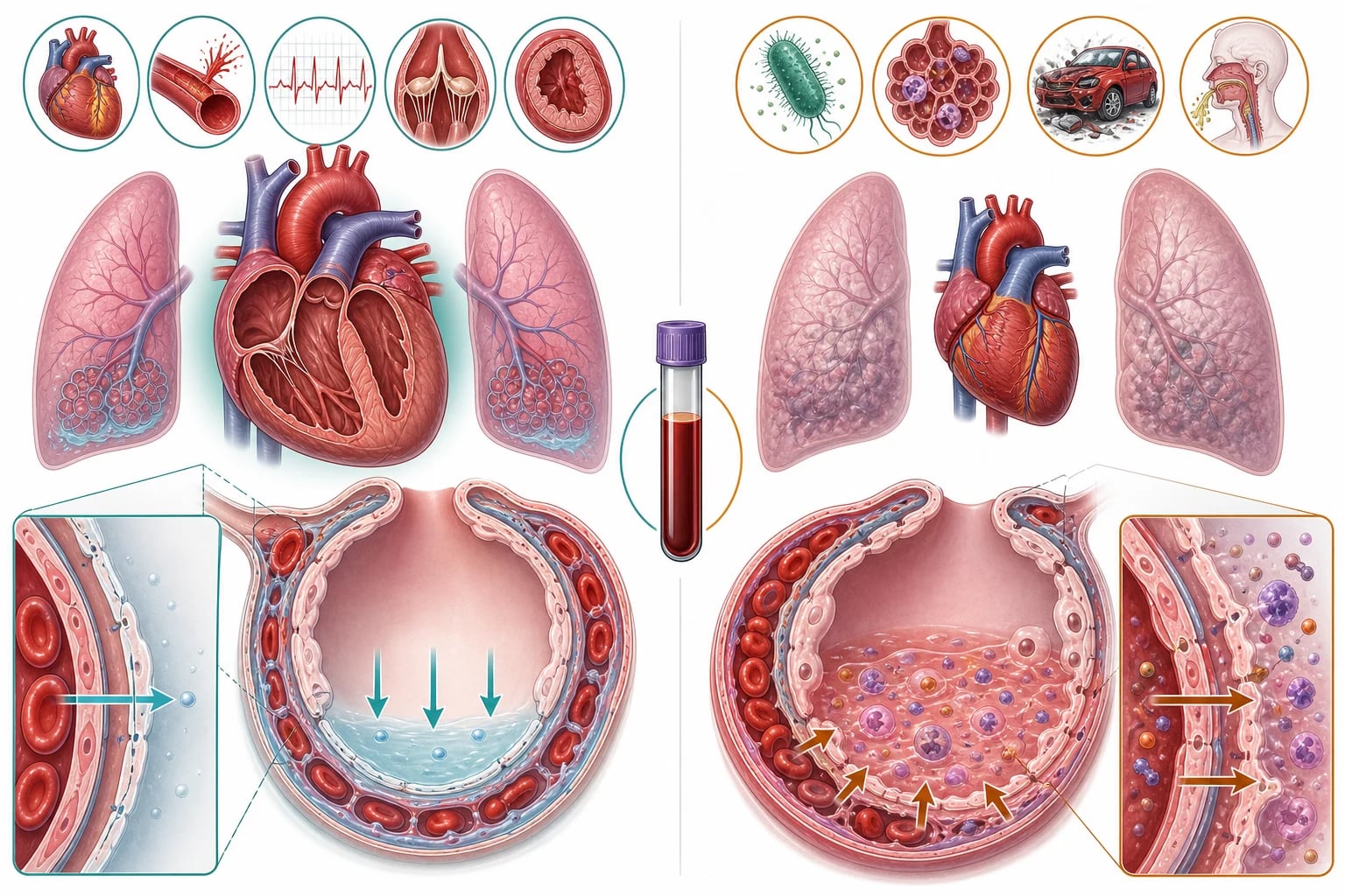
The cardiogenic pulmonary oedema arises from the raised pulmonary capillary pressure (the hydrostatic) from the left ventricular failure. The causes are the ischaemia and the myocardial infarction, the hypertensive crisis, the arrhythmia (the atrial fibrillation with the rapid ventricular response, the bradycardia), the valve failure (the acute mitral regurgitation, the critical aortic stenosis), and the cardiomyopathy. The fluid is the protein-poor, and the heart is the enlarged on the chest radiograph.[1][2]
The non-cardiogenic pulmonary oedema (the ARDS) arises from the increased alveolar-capillary permeability (the inflammatory, the leak). The causes are the sepsis, the pneumonia, the trauma, the aspiration, the pancreatitis, and the transfusion. The fluid is the protein-rich, the heart is the normal size, and the criteria are the ARDS (the timing, the chest imaging, the origin of the oedema, and the hypoxaemia).[1]
Cardiogenic
hydrostatic — the raised pulmonary capillary pressure
- The mechanism: the raised LV end-diastolic pressure transmitted to the pulmonary capillaries (the wedge pressure typically above 18 to 25 mmHg)
- The causes: the acute MI, the ischaemia, the AF with RVR, the hypertensive emergency, the acute mitral or aortic valve failure, the cardiomyopathy, the fluid overload
- The fluid: the protein-poor transudate
- The heart: the enlarged on the chest radiograph, the raised cardiothoracic ratio
- The chest radiograph: the perihilar bat-wing, the Kerley B lines, the pleural effusion, the upper-lobe venous diversion
- The BNP and the NT-proBNP: the markedly raised
- The echo: the reduced ejection fraction, the regional wall motion abnormality, the valve lesion
- The management: the preload and the afterload reduction — the NIV, the GTN, the furosemide
Non-cardiogenic (ARDS)
permeability — the leak at the normal pressure
- The mechanism: the inflammatory alveolar-capillary damage, the glycocalyx shedding, the tight-junction opening at a NORMAL hydrostatic pressure
- The causes: the sepsis, the pneumonia, the aspiration, the trauma, the pancreatitis, the transfusion (the TRALI), the near-drowning, the toxin
- The fluid: the protein-rich exudate
- The heart: the normal size on the chest radiograph
- The chest radiograph: the diffuse bilateral opacities, no cardiomegaly, no dominant pleural effusion
- The BNP and the NT-proBNP: the low or the normal
- The echo: the normal LV function
- The Berlin criteria: within 1 week, the bilateral opacities not explained by the effusion or the nodules, not fully cardiac, the PaO2 to FiO2 ratio (the mild 200 to 300, the moderate 100 to 200, the severe under 100 with the PEEP or CPAP of 5 or more)
- The management: the lung-protective ventilation and the treat-the-cause — the diuretic does NOT help the primary lesion
Acute MI / ischaemia
the systolic and the diastolic failure
- The commonest single trigger — the loss of the contractile function raises the LVEDP
- The regional wall motion abnormality on the echo, the raised troponin, the ECG changes
- Open the artery — the PCI pathway, the dual antiplatelet, the anticoagulation
AF with RVR
the loss of the atrial kick and the short filling
- The tachycardia shortens the diastole, the atrial kick is lost (15 to 30 per cent of the filling), the mitral stenosis patient decompensates the fastest
- The rate control (the IV metoprolol, cautious, or the digoxin in the low-output), or the synchronised cardioversion if unstable
- Treat the trigger (the sepsis, the thyroid, the ischaemia)
Hypertensive emergency
the pressure overload and the diastolic failure
- The afterload spike worsens the LV ejection, the diastolic dysfunction raises the LVEDP, the flash pulmonary oedema
- The controlled reduction (the GTN, the labetalol — 10 to 20 per cent in the first hour, NOT the rapid drop)
- Search for the end-organ damage — the brain, the kidneys, the retina, the heart
Acute valve failure
the mechanical catastrophe
- The acute mitral regurgitation (the papillary muscle rupture post-MI, the endocarditis) — the new pansystolic murmur
- The critical aortic stenosis — the slow-rising pulse, the ejection systolic murmur, the flash oedema
- The surgical or the transcatheter urgency — the echo is the priority
Cardiomyopathy / overload
the pump or the volume
- The decompensated chronic heart failure — the non-adherence, the NSAID, the ischaemia, the infection
- The fluid overload — the renal failure, the iatrogenic fluid, the post-operative
- The high-output failure — the anaemia, the thyrotoxicosis, the AV fistula, the beriberi
The Berlin Definition of ARDS — the Ranieri and the ARDS Definition Task Force
JAMA, 2012
A consensus panel redefining the acute respiratory distress syndrome, replacing the 1994 American-European Consensus Conference criteria.
Key finding
The ARDS is defined by the timing (within 1 week of the clinical insult), the chest imaging (the bilateral opacities not explained by the effusion, the atelectasis or the nodules), the origin of the oedema (the respiratory failure not fully explained by the cardiac failure or the fluid overload), and the hypoxaemia (the PaO2 to FiO2 ratio with the PEEP or the CPAP of 5 cmH2O or more — the mild 200 to 300, the moderate 100 to 200, the severe under 100).
Practice change
The standard diagnostic framework for the ARDS worldwide — the basis for the trial entry and the ventilatory strategy.
The pathophysiology
The fluid movement across the pulmonary capillary is governed by the Starling forces — the hydrostatic pressure (the outward push) against the oncotic pressure (the inward pull), with the capillary membrane integrity as the barrier. In the cardiogenic pulmonary oedema, the raised left ventricular end-diastolic pressure is transmitted back through the left atrium to the pulmonary veins and capillaries, raising the hydrostatic pressure above the oncotic threshold (typically above 25 mmHg), and the protein-poor fluid is pushed into the interstitium and then into the alveoli, first as the interstitial oedema (the Kerley B lines, the thickened bronchovascular markings) and then as the alveolar flooding (the bilateral fluffy infiltrates, the bat-wing pattern, the frothy pink sputum). In the non-cardiogenic oedema (the ARDS), the capillary membrane is damaged by the inflammatory mediators, the glycocalyx is shed, the tight junctions open, and the protein-rich fluid leaks into the alveoli at a normal hydrostatic pressure. The ARDS lung is the stiff, the heavy, and the oedematous, with the surfactant depletion and the micro-atelectasis, producing the severe shunt and the refractory hypoxaemia. The distinction is the foundation of the management — the cardiogenic oedema is treated with the preload and the afterload reduction (the NIV, the GTN, the furosemide), the non-cardiogenic oedema is treated with the lung-protective ventilation and the treatment of the underlying cause. [1]
The bedside ultrasound
The lung ultrasound is the rapid bedside tool that distinguishes the cardiogenic from the non-cardiogenic and the pulmonary oedema from the COPD or the pneumonia. The B-lines (the vertical comet-tail artefacts arising from the pleural line) indicate the interstitial fluid — multiple bilateral B-lines confirm the pulmonary oedema, and their absence points to the COPD or the pulmonary embolism. The focal B-lines suggest the pneumonia, while the diffuse bilateral pattern suggests the cardiogenic oedema. The reduced B-line clearance and the pleural effusions (seen as the anechoic spaces above the diaphragm) support the cardiogenic diagnosis. The cardiac ultrasound (the focused echo) evaluates the left ventricular function (the global hypokinesia in the cardiomyopathy, the regional wall motion abnormality in the MI), the valve (the mitral regurgitation, the aortic stenosis), and the right ventricular size (the right-heart strain suggests the PE). The IVC (the plethoric, the non-collapsible IVC supports the right-heart failure and the fluid overload). The lung-plus-heart ultrasound is the modern standard for the rapid aetiological diagnosis of the acute dyspnoea in the emergency department. [1]
The presentation
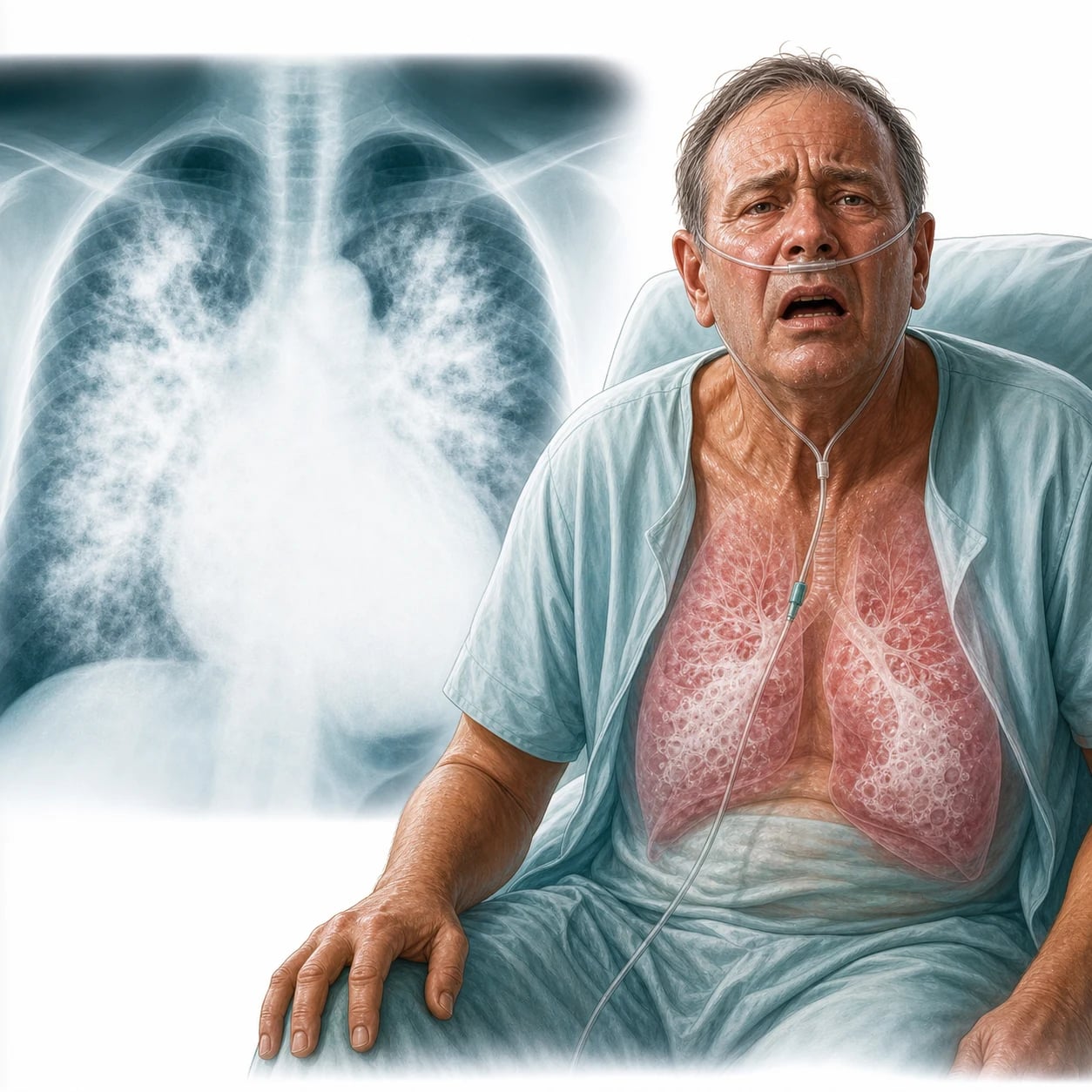
The presentation is the acute dyspnoea, the orthopnoea, the paroxysmal nocturnal dyspnoea, the frothy and the pink sputum, the bilateral crackles, the wheeze (the cardiac asthma), the hypoxia, the tachypnoea, the diaphoresis, and the raised jugular venous pressure. The flash pulmonary oedema is the sudden, the severe, the rapid onset — often from the hypertensive crisis or the acute ischaemia. The hypoxia and the respiratory distress dominate.[1][2]
The investigations
The chest radiograph shows the bilateral fluffy infiltrates, the perihilar bat-wing pattern, the cardiomegaly, the pleural effusion, and the Kerley B lines (the interstitial). The BNP and the NT-proBNP are the raised in the cardiogenic (the low level favours the non-cardiogenic). The troponin (the ischaemia), the electrocardiogram (the MI, the arrhythmia), the echocardiography (the left ventricular function, the valve, the regional wall motion), the renal function and the electrolytes, and the blood gas (the hypoxia, the acidosis, and the lactic acid). The pulmonary oedema is distinguished from the pneumonia (the fever, the purulent sputum, the focal consolidation) and the COPD exacerbation (the history, the hypercapnia).[1][3]
The management
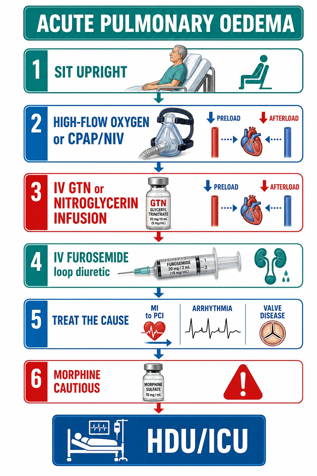
The management relieves the pulmonary congestion and treats the cause.[1][2][3]
- The ABCDE, the sit upright (the pooling of the venous blood, the reduced preload).
- The high-flow oxygen to the target saturation of 94 to 98 per cent; the CPAP or the NIV for the moderate-to-severe — the CPAP reduces the preload and the afterload, the splints the alveoli, the recruits the collapsed lung, and the reduces the work of the breathing (the Cochrane confirms the benefit of the CPAP and the BiPAP in the cardiogenic oedema).[1][2]
- The IV GTN or the nitroglycerin infusion — the venodilatation and the arteriolar dilatation, the reduced preload and the afterload, the reduced pulmonary capillary pressure.
- The IV furosemide — the loop diuretic, the diuresis and the venodilation (the immediate effect before the diuresis). The dose by the chronic use and the renal function.[3]
- The morphine — the cautious (the analgesia and the anxiolysis, the mild venodilation); the concern for the respiratory depression and the hypotension limits the use in the modern practice.
- The treat-the-cause — the acute coronary syndrome to the PCI pathway; the arrhythmia to the rate or the rhythm control; the valve to the surgery; the hypertensive crisis to the controlled blood-pressure reduction; the fluid overload to the diuresis and the avoidance of the further fluid.
- The disposition — the high-dependency or the intensive care for the severe and the refractory, the monitoring of the response. The specific drug doses: furosemide 40 to 80 mg IV (double the oral dose in the chronic user, up to 200 mg IV); glyceryl trinitrate infusion 10 to 200 mcg/min (start low, titrate to the BP and the symptom relief); morphine 2.5 to 5 mg IV (use cautiously, or omit); CPAP at 5 to 10 cm H2O; and the target oxygen saturation 94 to 98 per cent. The non-invasive ventilation reduces the intubation rate and the mortality in the cardiogenic pulmonary oedema.
The 3-minute, the 30-minute, the 3-hour approach
The acute cardiogenic pulmonary oedema is the time-critical emergency that the Fellowship candidate manages in the three nested horizons — the immediate stabilisation, the diagnostic and the therapeutic hour, and the disposition and the cause-resolution phase.[7]
The 3-minute approach — the immediate stabilisation at the bedside
The airway, the breathing, the saturations
The patient is sat upright (the pooling of the venous return, the reduced preload, the improved vital capacity). The high-flow oxygen (15 L/min via the non-rebreather mask) to the target saturation of 94 to 98 per cent. The continuous waveform capnography and the ECG monitor from the first minute.
The NIV — the CPAP or the BiPAP at once
The non-invasive ventilation is the first-line for the respiratory distress. The CPAP at 5 to 10 cm H2O (start at 5, titrate up). The BiPAP (the IPAP 10 to 15, the EPAP 5 to 8) if the hypercapnia coexists or the CPAP is not tolerated. The NIV reduces the preload (the intrathoracic pressure) and the afterload (the reduced LV transmural pressure), the recruits the alveoli, and the reduces the work of the breathing.<Cite id="1" /><Cite id="4" />
The IV access and the bloods
The two large-bore cannulae. The venous gas, the troponin, the BNP or the NT-proBNP, the U&E, the full blood count, the coagulation, the glucose, the lactate. The arterial gas if the severe distress or the SpO2 below 92 per cent.
The sublingual GTN and the nitrates
The sublingual glyceryl trinitrate 400 mcg (one to two sprays) for the immediate venodilation while the IV access is established. The IV GTN infusion if the SBP is above 110 mmHg (start at 10 to 20 mcg/min, titrate every 5 minutes to the response and the BP).
The IV furosemide
The furosemide 40 to 80 mg IV (the double of the oral dose in the chronic loop-diuretic user, up to the 200 mg). The immediate venodilation precedes the diuresis by 5 to 15 minutes — the symptom relief is the early signal.<Cite id="3" />
The ECG and the cause hunt
The 12-lead ECG within the 10 minutes — the STEMI pathway triggers the catheter-lab activation. The bedside echo and the lung ultrasound for the LV function, the valve, the B-lines, the effusion.
The 30-minute approach — the diagnostic and the therapeutic hour
Reassess the response — the clinical and the saturations
The respiratory rate, the SpO2, the heart rate, the blood pressure, and the work of the breathing at the 15- and the 30-minute marks. The falling respiratory rate and the rising SpO2 with the reduced accessory-muscle use is the response. The persistent distress escalates the NIV pressure or moves to the intubation.
Titrate the GTN and the furosemide
The GTN infusion titrated every 5 minutes to the SBP of 100 to 120 mmHg (the systolic above 110 to start, the cautious in the borderline). The second dose of the furosemide at the 30 to 60 minutes if the inadequate response (the 1 to 2.5 times the home oral dose is the total IV dose). The urinary catheter for the accurate output.<Cite id="3" />
The chest radiograph and the formal echo
The portable CXR (the bat-wing, the cardiomegaly, the Kerley B lines, the effusion) and the formal echocardiography (the EF, the regional wall motion, the valve, the right heart). The CT pulmonary angiogram if the PE is the differential (the right-heart strain, the normal BNP).
The treat-the-cause in parallel
The STEMI to the PCI; the AF with RVR to the rate control (the IV metoprolol 5 mg, cautious, or the amiodarone 300 mg if the impaired); the hypertensive emergency to the controlled reduction; the acute mitral regurgitation to the cardiology and the surgery; the sepsis to the antibiotic.<Cite id="7" /><Cite id="8" />
The disposition decision
The high-dependency or the intensive care for the severe, the intubated, the vasopressor-dependent, or the NIV-dependent beyond the 1 to 2 hours. The monitored bed for the responder. The catheter-lab or the CCU for the ischaemic.
The 3-hour approach — the cause resolution and the safe disposition
The weaning of the NIV
The NIV weaned as the saturations, the respiratory rate and the work of the breathing improve — the pressure reduced, the mask breaks increased, the transition to the standard oxygen. The failed wean prompts the reassessment (the residual oedema, the new cause, the diuretic under-dosing) and the intensive-care admission.
The ongoing diuresis and the electrolyte
The conversion to the oral furosemide when the stable. The potassium and the magnesium replacement (the loop diuretic wastes both). The renal function monitoring (the AKI from the low output and the diuresis). The ACE inhibitor or the ARB started cautiously once the stable.
The cause defined and the definitive treatment
The angiography for the ischaemia; the rate or the rhythm strategy for the AF; the valve repair or the replacement for the mechanical lesion; the renal replacement therapy for the fluid overload; the treatment of the sepsis, the thyroid, the anaemia.
The secondary prevention and the heart-failure bundle
The four pillars of the chronic heart failure (the ACE inhibitor or the ARNI, the beta-blocker, the MRA, the SGLT2 inhibitor) started or optimised. The education on the weight, the fluid and the symptom monitoring. The cardiology and the heart-failure nurse follow-up.<Cite id="7" /><Cite id="8" />
The safe discharge criteria
The euovolaemic, the off the NIV and the stable on the standard oxygen or the room air, the cause addressed, the medications optimised, the follow-up arranged, the patient educated. The early clinic review at the 7 to 14 days.
CPAP
the continuous positive airway pressure — one pressure
- A single continuous pressure (5 to 10 cm H2O, start at 5) — no inspiratory assistance
- Increases the mean airway pressure, the recruits the alveoli, the reduces the work of the breathing
- Reduces the LV preload (the intrathoracic pressure) and the afterload (the reduced transmural pressure) — the haemodynamic benefit
- The preferred first-line in the pure cardiogenic pulmonary oedema
- Contraindicated in the profound hypotension, the vomiting, the facial trauma, the inability to protect the airway
BiPAP
the bilevel positive airway pressure — the IPAP and the EPAP
- Two pressures — the IPAP (inspiratory, 10 to 15) and the EPAP (expiratory or PEEP, 5 to 8)
- Adds the inspiratory pressure support to the EPAP — the ventilatory assistance
- Preferred where the hypercapnia coexists (the COPD overlap, the tiring patient, the rising PaCO2)
- The pressure support reduces the work of the breathing more than the CPAP alone
- The same contraindications as the CPAP
The 3CPO trial — the Gray, the Goodacre and the Newby (NEJM 2008)
New England Journal of Medicine, 2008
A multicentre randomised controlled trial of 1,069 patients with the acute cardiogenic pulmonary oedema, comparing the continuous positive airway pressure (CPAP), the bilevel non-invasive positive-pressure ventilation (NIPPV), and the standard oxygen therapy, in the three arms.
Key finding
No significant difference in the 7-day mortality between the NIV (the CPAP or the BiPAP) and the standard oxygen (around 9.5 per cent), but a significant reduction in the need for the intubation with both the NIV modalities. The breathlessness and the physiological markers improved faster with the NIV.
Practice change
The NIV (the CPAP or the BiPAP) is the first-line for the acute cardiogenic pulmonary oedema — the intubation rate is reduced, the symptom relief is faster, and the mortality benefit is at most modest.
The morphine in the acute cardiogenic pulmonary oedema — the controversy (the Gil et al, Curr Heart Fail Rep 2019)
Current Heart Failure Reports, 2019
A narrative review and the pooled analysis of the observational data and the post-hoc analyses of the NIV trials, examining the association of the morphine with the outcomes in the acute cardiogenic pulmonary oedema.
Key finding
The morphine provides the anxiolysis, the analgesia and the mild venodilation, but the observational data associate its use with the higher intubation rate, the ICU admission, and the mortality — a confounded signal, but a consistent one. The mechanism is the respiratory depression, the reduced sympathetic drive, and the hypotension.
Practice change
The morphine is the cautious, the low-dose (2.5 to 5 mg IV), and the reserved for the patient with the genuine pain or the distressing anxiety that the NIV and the anxiolysis do not control — it is NOT the routine, and the modern ESC and the NICE guidance de-emphasise it.
The NIV escalation — when the mask is not enough
Start the NIV early and correctly
The CPAP at 5 cm H2O via the full-face mask, titrated up to 10 cm H2O as the tolerated. The BiPAP if the hypercapnia or the CPAP intolerance. The mask fit and the patient comfort are the determinants of the success — the explanation and the reassurance.
Reassess at the 15 to 30 minutes — the response or the failure
The response: the falling respiratory rate, the rising SpO2, the reduced accessory-muscle use, the falling heart rate. The failure: the persisting or the worsening distress, the rising PaCO2, the falling GCS, the hypotension.
The failed NIV — escalate to the intubation
The NIV failure (around 10 to 15 per cent of the cardiogenic oedema) is the indication for the prompt intubation — the delay worsens the outcome. The full stomach, the pulmonary oedema, and the reduced reserve make the rapid-sequence intubation the high-risk — the experienced operator, the haemodynamic optimisation, and the vasopressor ready.
The NIV in the hypotensive — the caution
The NIV reduces the venous return and the cardiac output — the cardiogenic shock with the SBP below 90 is the relative contraindication. The judicious vasopressor (the noradrenaline) and the low EPAP (5 cm H2O) if the NIV is essential. The intubation if the shock and the respiratory failure coexist.
The intubated and the ventilated — the lung-protective strategy
The tidal volume of 6 mL per kg of the ideal body weight, the plateau pressure below 30 cm H2O, the PEEP titrated to the oedema. The diuresis and the vasodilatation continue. The cause treated definitively.
Differential diagnosis — the acute dyspnoea with the bilateral infiltrates
- Cardiogenic pulmonary oedema — the LV failure, the MI, the arrhythmia; the raised BNP, the enlarged heart, the protein-poor fluid; responds to the NIV and the GTN.
- Non-cardiogenic pulmonary oedema (ARDS) — the sepsis, the pneumonia, the aspiration; the normal heart size, the protein-rich fluid; treated with the lung-protective ventilation, not the diuretic.
- Bilateral pneumonia — the fever, the purulent sputum, the focal consolidation; treated with the antibiotic, not the diuretic.
- COPD exacerbation — the history, the hypercapnia, the wheeze; treated with the bronchodilator and the steroid.
- Pulmonary embolism — the risk factors, the pleuritic pain, the raised D-dimer; treated with the anticoagulation, not the diuretic. [1]
The complications and the prognosis
The complications are the respiratory failure (the progressive hypoxaemia from the alveolar flooding, the need for the intubation and the mechanical ventilation), the cardiogenic shock (the end-stage pump failure, the hypotension, the need for the inotrope), the arrhythmia (the atrial fibrillation as the trigger or the consequence), the acute kidney injury (the low cardiac output, the diuretic), the myocardial infarction (the cause or the consequence of the oedema), and the death. The prognosis depends on the cause and the speed of the relief — the early CPAP and the cause treatment (the PCI for the MI, the rate control for the AF) improve the outcome. The in-hospital mortality of the acute cardiogenic pulmonary oedema is 5 to 10 per cent; the one-year mortality after the admission is 20 to 30 per cent, reflecting the underlying heart disease. The patient with the flash pulmonary oedema from the critical aortic stenosis or the acute mitral regurgitation has the worse prognosis unless the valve is corrected. [1]
Special populations
The renal-failure patient with the fluid overload is treated with the haemodialysis or the haemofiltration if the diuretic fails — the pulmonary oedema in the anuric patient is the dialysis indication. The pregnant patient with the pulmonary oedema (the pre-eclampsia, the peripartum cardiomyopathy) is managed with the cautious diuresis, the hydralazine or the labetalol for the blood pressure, and the early obstetric involvement. The elderly patient with the multiple comorbidities and the polypharmacy (the NSAID, the calcium-channel-blocker) may present with the gradual fluid overload that tips into the acute oedema — the precipitant is identified and removed. The post-operative patient develops the pulmonary oedema from the fluid overload, the transfusion (the TRALI), or the postoperative atrial fibrillation. [1]
Evidence and regional guidelines
The contemporary framework is the ESC heart failure guideline and the NICE acute heart failure guideline for the cardiogenic oedema, and the Berlin definition for the ARDS. The CPAP or the BiPAP is the first-line for the cardiogenic oedema with the respiratory distress, the IV GTN and the furosemide are the standard, and the morphine is used sparingly. The treat-the-cause is the simultaneous priority. [1]
ANZ practice note. The pulmonary oedema management follows the local ED and the cardiology protocol; the CPAP or the BiPAP is the first-line non-invasive ventilation; the GTN infusion and the furosemide are the standard; the treat-the-cause (the PCI, the rate control) is the simultaneous priority; and the high-dependency admission for the severe or the refractory case is the standard. [1]
Common pitfalls
The recurring errors are: the failure to distinguish the cardiogenic from the non-cardiogenic (the ARDS treated as the LV failure); the under-use of the CPAP or the NIV in the cardiogenic oedema; the over-reliance on the morphine (the respiratory depression, the hypotension); the failure to identify and to treat the trigger (the MI, the arrhythmia); the under-dosing of the furosemide in the chronic user; and the under-recognition of the flash pulmonary oedema.
Exam practice
SAQ — Acute cardiogenic pulmonary oedema with atrial fibrillation and ischaemia
10 minutes · 10 marks
A 78-year-old man presents with acute dyspnoea, orthopnoea and frothy pink sputum. RR 34, SpO2 86 per cent on room air, BP 168/96, HR 126 in atrial fibrillation. There are bilateral crackles and a wheeze. The ECG shows the AF with a rapid ventricular response and 2 mm of ST depression in V3 to V4. The bedside lung ultrasound shows the diffuse bilateral B-lines.
SAQ — Flash pulmonary oedema in critical aortic stenosis with shock
10 minutes · 10 marks
A 70-year-old woman with known severe aortic stenosis presents with the flash pulmonary oedema. SpO2 84 per cent on 15 L oxygen, BP 88/56 (a cardiogenic-shock overlap), HR 110, with bilateral crackles and a slow-rising pulse. An ejection systolic murmur is audible at the right upper sternal edge.
Red flags
The following features identify the cardiogenic oedema or the dangerous situation, in which the CPAP and the cause treatment are the priority: [1]
[1]Clinical pearls — the exam-exhaustive deep dive
[1] [1] [1] [1] [1] [1] [1] [1] [1] [1] [1] [1] [1] [1] [1]References
- [1]Berbenetz N, Brown E, Nour A, et al. Non-invasive positive pressure ventilation (CPAP or bilevel NPPV) for cardiogenic pulmonary oedema Cochrane Database Syst Rev, 2019.PMID 30950507
- [2]Liu C, Liu XL. Non-invasive ventilation in cardiogenic pulmonary edema Ann Transl Med, 2018.PMID 30370282
- [3]Pourafshar N, Kansal S, Davis JS, Sobotka PA. Diuretic Treatment in Heart Failure: A Practical Guide for Clinicians J Clin Med, 2024.PMID 39124738
- [4]Gray A, Goodacre S, Newby DE, et al. Noninvasive ventilation in acute cardiogenic pulmonary edema N Engl J Med, 2008.PMID 18614781
- [5]ARDS Definition Task Force, Ranieri VM, Rubenfeld GD, et al. Acute respiratory distress syndrome: the Berlin Definition JAMA, 2012.PMID 22797452
- [6]Gil V, Dominguez-Rodriguez A, Masip J, et al. Morphine Use in the Treatment of Acute Cardiogenic Pulmonary Edema and Its Effects on Patient Outcome: A Systematic Review Curr Heart Fail Rep, 2019.PMID 31183779
- [7]McDonagh TA, Metra M, Adamo M, et al. 2021 ESC Guidelines for the diagnosis and treatment of acute and chronic heart failure Eur Heart J, 2021.PMID 34447992
- [8]McDonagh TA, Metra M, Adamo M, et al. 2023 Focused Update of the 2021 ESC Guidelines for the diagnosis and treatment of acute and chronic heart failure Eur Heart J, 2023.PMID 37622666